. 2023 Dec:54:211-222.
doi: 10.1016/j.jare.2023.01.016. Epub 2023 Jan 24.

Ferroptosis in acute kidney injury following crush syndrome: A novel target for treatment

Affiliations

Ferroptosis in acute kidney injury following crush syndrome: A novel target for treatment

Ou Qiao et al. J Adv Res. 2023 Dec.

Abstract

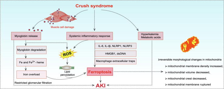
Background: Crush syndrome (CS) is a kind of traumatic and ischemic injury that seriously threatens life after prolonged compression. It is characterized by systemic inflammatory reaction, myoglobinuria, hyperkalemia and acute kidney injury (AKI). Especially AKI, it is the leading cause of death from CS. There are various cell death forms in AKI, among which ferroptosis is a typical form of cell death. However, the role of ferroptosis has not been fully revealed in CS-AKI.

Aim of review: This review aimed to summarize the evidence of ferroptosis in CS-AKI and its related molecular mechanism, discuss the therapeutic significance of ferroptosis in CS-AKI, and open up new ideas for the treatment of CS-AKI.

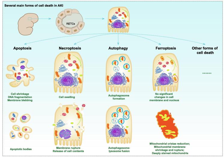
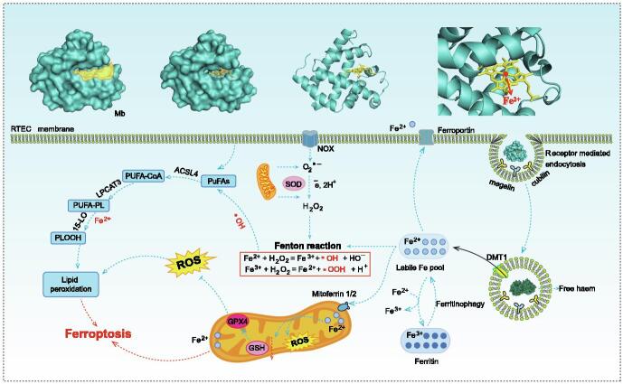
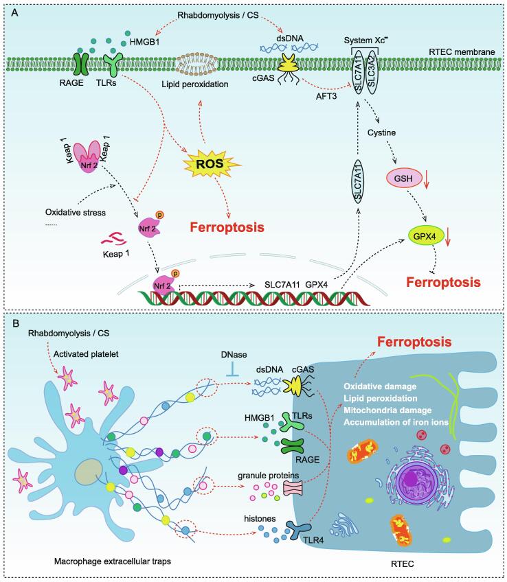
Key scientific concepts of review: One of the main pathological manifestations of CS-AKI is renal tubular epithelial cell dysfunction and cell death, which has been attributed to massive deposition of myoglobin. Large amounts of myoglobin released from damaged muscle deposited in the renal tubules, impeding the normal renal tubules function and directly damaging the tubules with oxidative stress and elevated iron levels. Lipid peroxidation damage and iron overload are the distinguishing features of ferroptosis. Moreover, high levels of pro-inflammatory cytokines and damage-associated molecule pattern molecules (HMGB1, double-strand DNA, and macrophage extracellular trap) in renal tissue have been shown to promote ferroptosis. However, how ferroptosis occurs in CS-AKI and whether it can be a therapeutic target remains unclear. In our current work, we systematically reviewed the occurrence and underlying mechanism of ferroptosis in CS-AKI.

Keywords: Acute kidney injury; Crush syndrome; Drug development; Ferroptosis; Myoglobin; Systemic inflammatory responses.


Conflict of interest statement

Declaration of Competing Interest The authors declare that they have no known competing financial interests or personal relationships that could have appeared to influence the work reported in this paper.

Figures

None
Graphical abstract
Fig. 1
Fig. 1
Several main forms of cell death in AKI. The forms of cell death that have been revealed in AKI include apoptosis, necroptosis, autophagy, ferroptosis, and so on. Apoptosis: cells contracted, nuclear chromatin condensed, DNA broken, and cells decomposed into several apoptotic bodies. Necroptosis: cells and organelles swelled and disintegrated, cell membranes broken, and cell contents leaked. Autophagy: autophagosomes were formed and transported to lysosomes to form autophagosomes. Subsequently, aging and damaged organelles in autophagosomes were degraded. Ferrotosis: the volume and cristae of mitochondria decreased, the density of mitochondrial membrane increased, and the mitochondrial membrane ruptured.
Fig. 2
Fig. 2
Potential mechanisms of ferroptosis caused by Mb. Excessive Mb released from damaged muscle cells accumulated in renal tubules and was taken up by RTECs in receptor-mediated endocytosis (megalin and cubilin). Mb was subsequently acidified and degraded in the lysosomes to produce Fe2+. Accumulated Fe2+ could generate hydroxyl radicals through Fenton reaction. Both Fe2+ and hydroxyl radicals could aggravate the oxidation process of PUFAs in cell membrane, resulting in lipid peroxidation damage, and eventually ferroptosis. Fe2+ could be transported by mitoferrin 2 to accumulate in the mitochondria, thus causing oxidative stress in the mitochondria. The chemical and molecular mechanisms in this process remains to be investigated. DMT1, divalent metal transporter 1; Mitoferrin 2, also known as mitochondrial RNA-splicing protein 3/4 homolog (MRS3/4) or solute carrier family 25 member 28 (SLC25A28); PUFAs, polyunsaturated fatty acids; ACSL4, acyl-CoA synthetase long-chain family member 4; FA-CoA, fatty acyl coenzyme A; LPCAT3, lysophosphatidylcholine acyltransferase 3; 15-LO, 15-lipoxygenase; PLOOH, phospholipid hydroperoxides.
Fig. 3
Fig. 3
Molecular mechanisms of ferroptosis caused by several identified DAMP molecules in CS (rhabdomyolysis) –AKI (A) Damaged muscle cells released a large amount of HMGB1, which was recognized by RAGE and TLRs in RTECs, causing ROS accumulation and lipid peroxidation damage by blocking Nrf2 pathway. dsDNA could activate cGAS to up-regulate AFT and directly inhibit SLC7A11 expression, resulting in GSH depletion. System Xc was an important intracellular antioxidant system, which consisted of two subunits, SLC7A11 and SLC3A2. SLC7A11 was responsible for the major transport activity and was highly specific for cystine and glutamate, whereas SLC3A2 acted as a chaperone protein. Inhibition of System Xc activity would inhibit the cystine uptake and affected the synthesis of GSH, leading to reduced GPX4 activity and reduced cellular antioxidant capacity, thereby promoting ferroptosis. (B) Activated platelets induced macrophages to form ETs during rhabdomyocytolysis, which have been shown to promote ferroptosis via dsDNA, HMGB1, and so on. HMGB1, high mobility group box 1; RAGE, the receptor for advanced glycation end products; TLRs, toll-like receptors; Nrf2, nuclear factor erythroid 2-related factor 2; dsDNA, double-strand DNA; cGAS, cyclic GMP-AMP synthase; AFT3, activating transcription factor 3; SLC3A2, solute carrier family 3 member 2; SLC7A11, solute carrier family 7 member 11; GSH, glutathione; GPX4, glutathione peroxidase 4.
Fig. 4
Fig. 4
Chemical structure of Fer-1, Lip-1 and their analogues for inhibiting ferroptosis. (A) Chemical structure of Fer-1. (B-H) Chemical structure of Fer-1 analogues. The aromatic primary amines and N-cyclohexyl portion were the key functional groups of Fer-1 antioxidant activity. Moreover, the N-cyclohexyl portion aslo acted as a lipophilic anchor within the cellular biofilm. The SRS 16–86 had better plasma stability and stronger inhibition of ferroptosis compared to Fer-1. The amide group and sulfonamide subunit provide Lip-1 with good stability and drug absorption distribution. Both Fer-1 and Lip-1 are monoarylamines. In contrast, diarylamines such as phenoxazines and phenothiazines have stronger pharmacodynamic activity. The underlying chemical mechanisms need to be further explored.

Similar articles

Cited by

References

    1. Liu Y., Yu M., Chen L., Liu J., Li X., Zhang C., et al. Systemic review of animal models used in the study of crush syndrome. Shock. 2022;57:469–478. doi: 10.1097/SHK.0000000000001911. - DOI - PubMed
    1. Sever M.S., Vanholder R., Lameire N. Management of crush-related injuries after disasters. N Engl J Med. 2006;354:1052–1063. doi: 10.1056/NEJMra054329. - DOI - PubMed
    1. Wang P.T., Li N., Wang X.Y., Chen J.L., Geng C.H., Liu Z.Q., et al. RIG-I, a novel DAMPs sensor for myoglobin activates NF-kappaB/caspase-3 signaling in CS-AKI model. Mil Med Res. 2021;8:37. doi: 10.1186/s40779-021-00333-4. - DOI - PMC - PubMed
    1. Li N., Chen J., Geng C., Wang X., Wang Y., Sun N., et al. Myoglobin promotes macrophage polarization to M1 type and pyroptosis via the RIG-I/Caspase1/GSDMD signaling pathway in CS-AKI. Cell Death Discov. 2022;8:90. doi: 10.1038/s41420-022-00894-w. - DOI - PMC - PubMed
    1. Jin H., Lin X., Liu Z., Wang J., Wang J., Zhang Y., et al. Remote ischemic postconditioning protects against crush-induced acute kidney injury via down-regulation of apoptosis and senescence. Eur J Trauma Emerg Surg. 2022;48:4585–4593. doi: 10.1007/s00068-022-01910-5. - DOI - PMC - PubMed

Publication types