Review
. 2023 May;48(5):463-476.
doi: 10.1016/j.tibs.2022.12.006. Epub 2023 Jan 24.

Coenzyme Q biochemistry and biosynthesis

Affiliations
Review

Coenzyme Q biochemistry and biosynthesis

Rachel M Guerra et al. Trends Biochem Sci. 2023 May.

Abstract

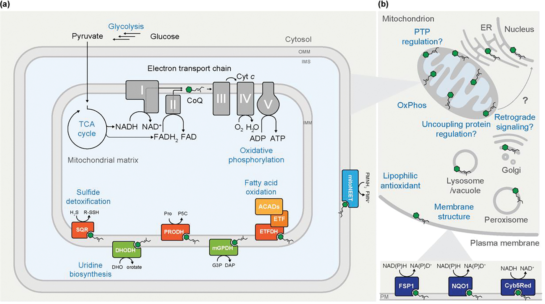
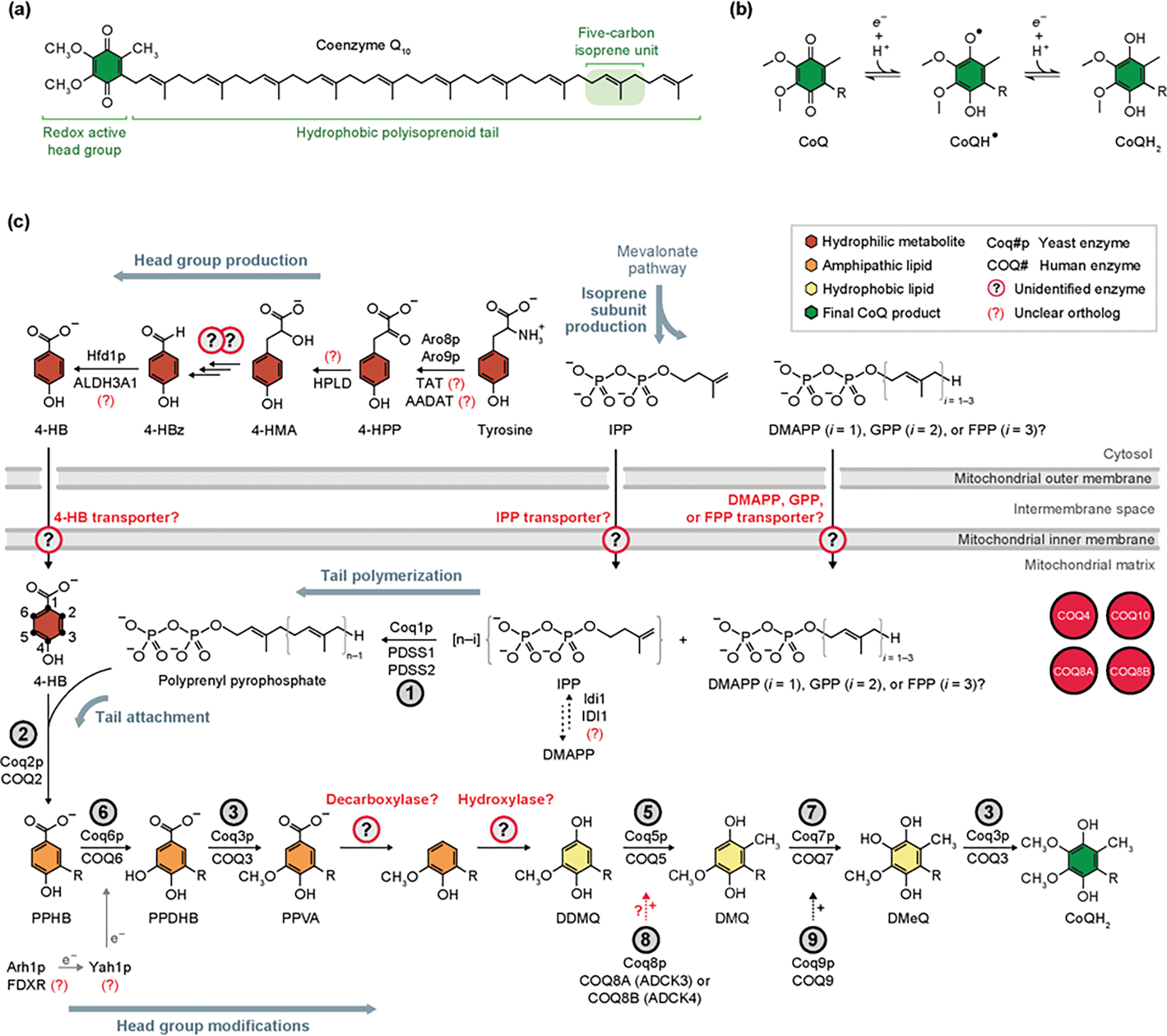
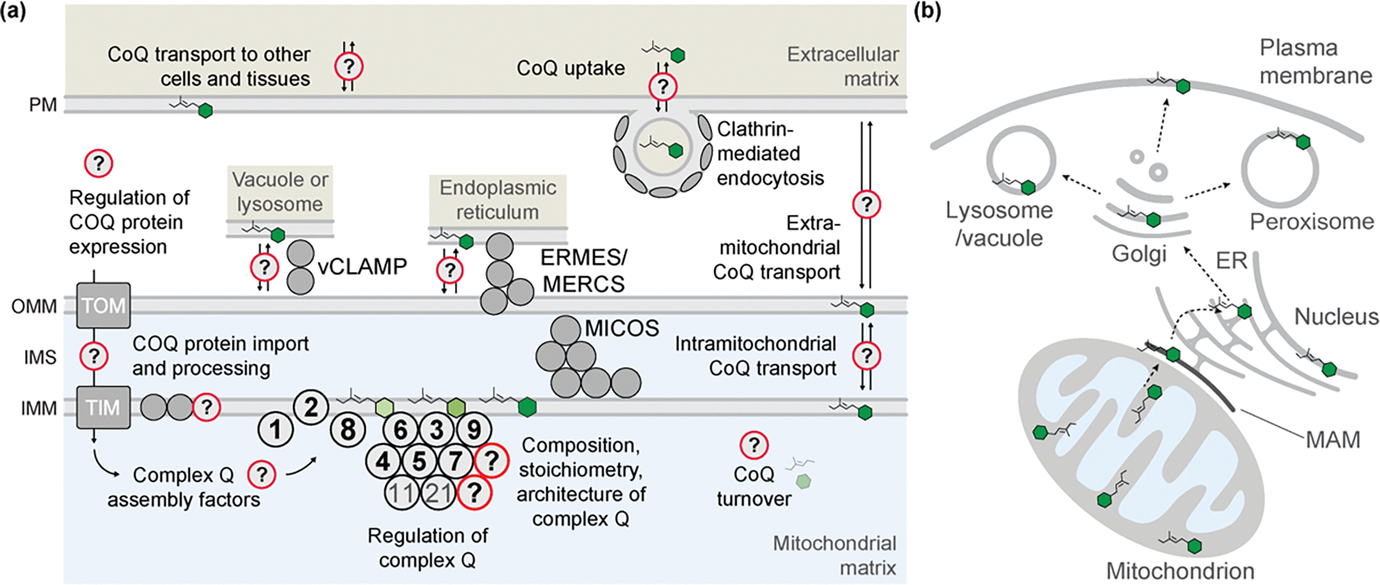
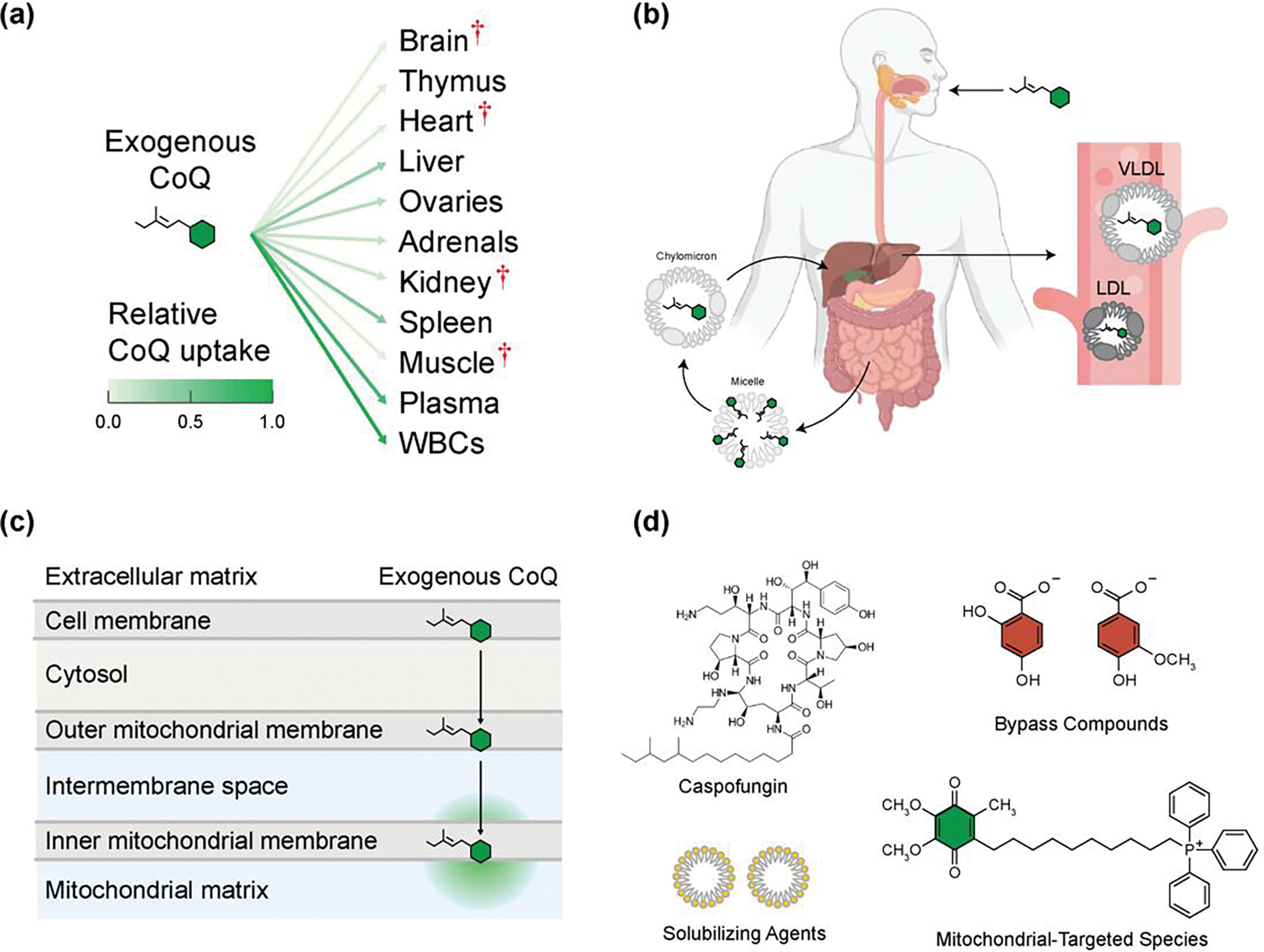
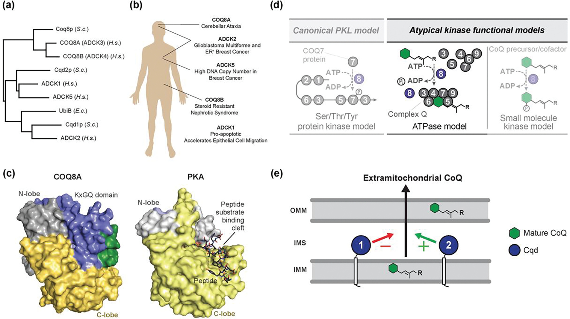
Coenzyme Q (CoQ) is a remarkably hydrophobic, redox-active lipid that empowers diverse cellular processes. Although most known for shuttling electrons between mitochondrial electron transport chain (ETC) complexes, the roles for CoQ are far more wide-reaching and ever-expanding. CoQ serves as a conduit for electrons from myriad pathways to enter the ETC, acts as a cofactor for biosynthetic and catabolic reactions, detoxifies damaging lipid species, and engages in cellular signaling and oxygen sensing. Many open questions remain regarding the biosynthesis, transport, and metabolism of CoQ, which hinders our ability to treat human CoQ deficiency. Here, we recount progress in filling these knowledge gaps, highlight unanswered questions, and underscore the need for novel tools to enable discoveries and improve the treatment of CoQ-related diseases.

Keywords: coenzyme Q; complex Q; lipids; mitochondria; oxidative phosphorylation; ubiquinone.


Conflict of interest statement

Declaration of interests None are declared by the authors.

Figures

Figure 1.
Figure 1.. Cellular Roles of Coenzyme Q.
(A) Although coenzyme Q (CoQ) is typically regarded for its central role in the mitochondrial electron transport chain and oxidative phosphorylation (OxPhos), it additionally acts as a cofactor for multiple mitochondrial enzymes. As part of central energy metabolism, the tricarboxylic acid (TCA) cycle provides reducing equivalents that enable the reduction of CoQ at OxPhos complexes I and II. Reduction of CoQ by enzymes involved in uridine biosynthesis, fatty acid oxidation, and sulfide detoxification, among others, provides additional routes of electron entry to complex III of the ETC, which ultimately drives ATP synthesis. (B) An overview of the widespread cellular functions of CoQ. CoQ is present in nearly all cellular membranes. Some extramitochondrial roles of CoQ are depicted, including modulating membrane structure dynamics and regulating the uncoupling protein, though many of the roles are incompletely characterized. At the plasma membrane (PM), reduced CoQH2 acts as lipophilic antioxidant where it can be reduced by various oxidoreductases to combat lipid peroxidation and oxidative damage.
Figure 2.
Figure 2.. Coenzyme Q Structure and Eukaryotic Biosynthesis.
(A) The chemical structure of coenzyme Q10 (CoQ10). (B) Single electron transfer redox reactions of the CoQ head group, whereby CoQ can cycle through the oxidized (CoQ), radical (CoQH), and fully reduced (CoQH2) forms. This redox activity allows CoQ function as a cofactor for numerous enzymes, relay electrons in the ETC, and act as an antioxidant. ‘R’ indicates the polyisoprenoid tail. (C) The primary eukaryotic pathway, conserved from S. cerevisiae to humans, of CoQ biosynthesis is depicted. CoQ production begins with the head group precursor 4-HB, derived from tyrosine, and tail subunit IPP, derived from the mevalonate pathway, which are both transported into the mitochondrial matrix by unknown means. Following tail polymerization and head group attachment, CoQ intermediates are processed through a series of head group modifications to yield mature CoQ. Biosynthetic enzymes that catalyze each reaction are denoted by the circled number above each arrow. Unidentified enzymes and transporters indicated by circled question marks. Auxiliary proteins with some unclear involvement in CoQ biosynthesis are depicted in red circles. ‘+’ symbol near dashed arrows designates a hypothesized supportive role for the indicated reaction. Abbreviations: 4-HBz, 4-hydroxybenzaldehyde; 4-HMA, 4-hydroxymandelate; 4-HPP, 4-hydroxyphenylpyruvate; AADAT, mitochondrial alpha-aminoadipate aminotransferase; ALDH3A1, aldehyde dehydrogenase 3A1; DDMQ, demethoxy-demethyl-coenzyme Q; DMQ, demethoxy-coenzyme Q; DMeQ, demethyl-coenzyme Q; DMAPP, dimethylallyl pyrophosphate; FDX2 (FDX1L), mitochondrial ferredoxin 2 (ferredoxin 1-like); FDXR, adrenodoxin reductase; FPP, farnesyl pyrophosphate; GPP, geranyl pyrophosphate; HPLD, hydroxyphenylpyruvate dioxygenase-like protein; IDI1, isopentenyl-diphosphate delta isomerase 1; IPP, isopentenyl pyrophosphate; PDSS1, prenyl (decaprenyl) diphosphate synthase subunit 1; PPDHB, polyprenyl-dihydroxybenzoate; PPHB, polyprenyl-hydroxybenzoate; PPVA, polyprenyl-vanillic acid; TAT, tyrosine aminotransferase.
Figure 3.
Figure 3.. Models Framing Knowledge Gaps in Complex Q Organization and CoQ Distribution.
(A) A depiction of outstanding questions regarding complex Q composition and assembly, along with possible mechanisms contributing to CoQ transport and cellular distribution. Although there is much evidence to support the existence and identify core components of complex Q, its full composition, stoichiometry, and architecture are undefined, and how its assembly is regulated is unknown. All COQ genes are nuclearly encoded, but many questions exist regarding factors that regulate their gene expression and how they are processed and imported into mitochondria via TIM and TOM complexes. As complex Q has been observed in proximity to MICOS and ERMES, it is hypothesized that these complexes could facilitate import of CoQ precursors to mitochondria or export of mature CoQ to other organelles. Studies involving administration of exogenous CoQ has suggested that clathrin-mediated endocytosis machinery may be involved in CoQ assimilation. (B) Depiction of a putative transport pathway of CoQ throughout the cell. Following synthesis at the IMM, CoQ passes through mitochondrial-associated membranes (MAM) to the ER. There, CoQ encounters the endomembrane system and is packaged in the Golgi for widespread mobilization to different organelles and cellular membranes. Abbreviations: ERMES, ER-mitochondria encounter structure; MERCS, mitochondria-ER contact sites; MICOS, mitochondrial contact site and cristae organization system; TIM, translocase of the inner mitochondrial membrane; TOM, translocase of the outer mitochondrial membrane; vCLAMP, vacuolar and mitochondrial patch.
Figure 4.
Figure 4.. Challenges in Uptake of Exogenous CoQ and Strategies to Overcome Them to Treat CoQ Deficiency.
(A) Exogenous CoQ is taken up poorly by most rat tissues after intraperitoneal injection. Red cross denotes organs most commonly affected in CoQ deficiency. Data from [Bentinger et al.] [112]. (B) Following oral intake of exogenous CoQ, it is absorbed in the small intestine and packaged into chylomicrons, then taken up by the liver and repackaged into low-density lipoproteins (LDL) and very low-density lipoproteins (VLDL), which can then enter blood circulation. Created with BioRender.com. (C) Once exogenous CoQ is in the bloodstream, it then must be transported across multiple lipid membranes and aqueous compartments to reach the mitochondrial inner membrane. (D) Depicted are examples of strategies to increase the solubility and uptake of exogenous CoQ by packaging with solubilizing agents like micelles, in novel formulations such as with caspofungin, or increasing organellar localization with the attachment of mitochondrial-targeting species like TPP. Alternative, CoQ deficiencies could by treated with bypass compounds that supersede the need for enzymes in the biosynthetic pathway that may be mutant or defective.
Figure I.
Figure I.. UbiB Protein Structure and Function.
(A) UbiB proteins are conserved across all domains of life, from humans to bacteria. (B) Mutations in human UbiB family proteins have been linked to numerous diseases. (C) Surface representations of COQ8ANΔ254 (PDB=4PED) and Protein Kinase A (PDB=1ATP) showing occlusion of the canonical peptide binding site by the highly conserved and UbiB-specific KxGQ domain (adapted from Stefely et al. [101]. (D) Potential models for the role of COQ8 in supporting CoQ biosynthesis. COQ8 could act as a canonical protein kinase to phosphorylate an enzyme in the biosynthetic pathway and modulate its activity, but this is not supported by current data. Alternatively, COQ8 could leverage its ATPase activity to extract hydrophobic substrates from the membrane and seed the formation of complex Q. Finally, COQ8 may act as a small-molecule kinase to support CoQ production, but no such substrate has been identified (adapted from Stefely et al. [40]. (E) Model of UbiB protein function in CoQ cellular distribution. Localized at the IMM, yeast Cqd1p and Cqd2p are proposed to have opposing roles in the extramitochondrial transport of mature CoQ, though the mechanism has yet to be defined. Abbreviations: S.c., Saccharomyces cerevisiae; H.s., Homo sapiens; E.c., Escherichia coli.

Similar articles

Cited by

References

    1. Crane FL et al. (1957) Isolation of a quinone from beef heart mitochondria. Biochim Biophys Acta 25, 220–221 - PubMed
    1. Morton RA (1958) Ubiquinone. Nature 182, 1764–1767 - PubMed
    1. Stefely JA and Pagliarini DJ (2017) Biochemistry of Mitochondrial Coenzyme Q Biosynthesis. Trends Biochem Sci 42, 824–843 - PMC - PubMed
    1. Baschiera E et al. (2021) The multiple roles of coenzyme Q in cellular homeostasis and their relevance for the pathogenesis of coenzyme Q deficiency. Free Radical Bio Med 166, 277–286 - PubMed
    1. Martínez-Reyes I et al. (2020) Mitochondrial ubiquinol oxidation is necessary for tumour growth. Nature 585, 288–292 - PMC - PubMed

Publication types

LinkOut - more resources