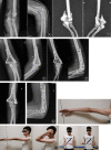
Combined Medial and Lateral Approach Versus Paratricipital Approach in Open Reduction and Internal Fixation for Type C Distal Humerus Fracture: A Randomized Controlled Study
- PMID: 36702763
- PMCID: PMC10432446
- DOI: 10.1111/os.13658
Combined Medial and Lateral Approach Versus Paratricipital Approach in Open Reduction and Internal Fixation for Type C Distal Humerus Fracture: A Randomized Controlled Study
Abstract
Objective: Olecranon osteotomy and paratricipital approaches were widely used in the treatment of type C distal humerus fracture but some disadvantages exist, so a combined medial and lateral approach was designed. The objective of this study was to investigate and compare the clinical outcomes of combined medial and lateral approach with the paratricipital approach in open reduction and internal fixation of type C distal humerus fractures.
Methods: From May 2018 to April 2020, 37 patients with type C distal humerus fracture who accepted open reduction and internal fixation in our hospital were enrolled in this study. All cases were randomly divided into two groups according to the surgical approach: combined medial and lateral approach group (19 cases), paratricipital approach group (18 cases). All of the patients received open reduction and double vertical plates fixation. The operation and follow-up indexes, including operation time, blood loss, incision length, triceps muscle strength, flexion-extension arc of elbow and forearm rotation arc, were recorded and compared. Caja score was used to assess the quality of fractures reduction. Mayo Elbow Performance Score (MEPS) was used to evaluate the elbow function in the follow-up. Complications such as incision infection, ulnar nerve injury, degenerative osteoarthritis, and heterotopic ossification were analyzed.
Results: The differences in age, gender, and AO classification of fractures between two groups were not statistically significant (p > 0.05). The sum of medial and lateral incision length of combined approach group was longer than the midline incision of paratricipital approach group (15.4 ± 0.8 vs. 14.6 ± 0.8, p < 0.05), but there was no significant difference in operation time (103.5 ± 10.2 vs. 106.0 ± 8.8, p > 0.05), blood loss (71.3 ± 24.5 vs. 72.8 ± 24.6, p > 0.05), and Caja score (16.05 ± 5.67 vs. 15.56 ± 5.66, p > 0.05). During the follow-up, the MEPS of combined approach group was higher than that of paratricipital approach group at 3 months postoperatively (80.5 ± 5.7 vs. 68.9 ± 8.1, p < 0.05), but there was no significant difference in MEPS at 6 months postoperatively (83.9 ± 6.6 vs. 79.7 ± 7.0, p > 0.05) and at the last follow-up (86.8 ± 7.1 vs. 86.9 ± 7.7, p > 0.05) between the two groups. There was no significant difference in triceps muscle strength (p > 0.05), flexion-extension arc (126.8 ± 5.3 vs. 128.9 ± 6.0, p > 0.05), and forearm rotation arc (163.2 ± 5.3 vs. 163.6 ± 4.8, p > 0.05) at the last follow-up. Although the incidence of complication of combined approach group (15.8%) was lower than that of paratricipital approach group (22.2%), the difference was not statistically significant (p > 0.05).
Conclusions: The combined medial and lateral approach was an effective and safe way of open reduction and internal fixation for type C distal humerus fractures. Compared with the paratricipital approach, the combined medial and lateral approach could restore the elbow function more quickly postoperatively, and the long-term results were comparable.
Keywords: Combined Medial and Lateral Approach; Distal Humerus Fractures; Open Reduction and Internal Fixation; Paratricipital Approach; Type C Fractures.
© 2023 The Authors. Orthopaedic Surgery published by Tianjin Hospital and John Wiley & Sons Australia, Ltd.
Conflict of interest statement
All authors declare that they have no conflict of interest.
Figures


Similar articles
-
[Effectiveness comparison between the paratricipital approach and the chevron olecranon V osteotomy approach in the treatment of type C3 distal humeral fractures].Zhongguo Xiu Fu Chong Jian Wai Ke Za Zhi. 2018 Oct 15;32(10):1321-1325. doi: 10.7507/1002-1892.201803036. Zhongguo Xiu Fu Chong Jian Wai Ke Za Zhi. 2018. PMID: 30215496 Free PMC article. Chinese.
-
[Operative effect and treatment strategies for the low distal humerus fracture].Zhonghua Wai Ke Za Zhi. 2020 Mar 1;58(3):213-219. doi: 10.3760/cma.j.issn.0529-5815.2020.03.009. Zhonghua Wai Ke Za Zhi. 2020. PMID: 32187925 Chinese.
-
Modified paratricipital approach without mobilization of the ulnar nerve prevents postoperative ulnar neuropathy in distal humerus fractures.J Orthop Sci. 2023 Sep;28(5):1113-1117. doi: 10.1016/j.jos.2022.06.018. Epub 2022 Jul 31. J Orthop Sci. 2023. PMID: 35922365
-
Comparison of olecranon osteotomy and paratricipital approach in distal humerus intra-articular fracture: A systematic review and meta-analysis.Medicine (Baltimore). 2022 Aug 26;101(34):e30216. doi: 10.1097/MD.0000000000030289. Medicine (Baltimore). 2022. PMID: 36042686 Free PMC article.
-
Surgical approaches for open reduction and internal fixation of intra-articular distal humerus fractures in adults: A systematic review and meta-analysis.Injury. 2018 Aug;49(8):1381-1391. doi: 10.1016/j.injury.2018.06.018. Epub 2018 Jun 19. Injury. 2018. PMID: 29934098
Cited by
-
Shoulder and Elbow Surgery Special Issue.Orthop Surg. 2023 Aug;15(8):1929-1930. doi: 10.1111/os.13861. Orthop Surg. 2023. PMID: 37587546 Free PMC article. No abstract available.
-
Superior outcome with anterolateral approach in treating O'Driscoll type II ulnar coronoid process fractures: a retrospective analysis.Am J Transl Res. 2025 Jan 15;17(1):440-449. doi: 10.62347/OUWK4004. eCollection 2025. Am J Transl Res. 2025. PMID: 39959191 Free PMC article.
References
-
- Sharma S, John R, Dhillon MS, Kishore K. Surgical approaches for open reduction and internal fixation of intra‐articular distal humerus fractures in adults: a systematic review and meta‐analysis. Injury. 2018;49:1381–91. - PubMed
-
- Nauth A, McKee MD, Ristevski B, Hall J, Schemitsch EH. Distal humeral fractures in adults. J Bone J Surg. 2011;93:686–700. - PubMed
Publication types
MeSH terms
LinkOut - more resources
Full Text Sources
Medical
Miscellaneous

