. 2023 May 26;224(2):iyad010.
doi: 10.1093/genetics/iyad010.

Hotspot of de novo telomere addition stabilizes linear amplicons in yeast grown in sulfate-limiting conditions

Affiliations

Hotspot of de novo telomere addition stabilizes linear amplicons in yeast grown in sulfate-limiting conditions

Remington E Hoerr et al. Genetics. .

Abstract

Evolution is driven by the accumulation of competing mutations that influence survival. A broad form of genetic variation is the amplification or deletion of DNA (≥50 bp) referred to as copy number variation (CNV). In humans, CNV may be inconsequential, contribute to minor phenotypic differences, or cause conditions such as birth defects, neurodevelopmental disorders, and cancers. To identify mechanisms that drive CNV, we monitored the experimental evolution of Saccharomyces cerevisiae populations grown under sulfate-limiting conditions. Cells with increased copy number of the gene SUL1, which encodes a primary sulfate transporter, exhibit a fitness advantage. Previously, we reported interstitial inverted triplications of SUL1 as the dominant rearrangement in a haploid population. Here, in a diploid population, we find instead that small linear fragments containing SUL1 form and are sustained over several generations. Many of the linear fragments are stabilized by de novo telomere addition within a telomere-like sequence near SUL1 (within the SNF5 gene). Using an assay that monitors telomerase action following an induced chromosome break, we show that this region acts as a hotspot of de novo telomere addition and that required sequences map to a region of <250 base pairs. Consistent with previous work showing that association of the telomere-binding protein Cdc13 with internal sequences stimulates telomerase recruitment, mutation of a four-nucleotide motif predicted to associate with Cdc13 abolishes de novo telomere addition. Our study suggests that internal telomere-like sequences that stimulate de novo telomere addition can contribute to adaptation by promoting genomic plasticity.

Keywords: Saccharomyces cerevisiae; Cdc13; DNA repair; copy number variation; de novo telomere addition; evolutionary genomics; gene amplification.


Conflict of interest statement

Conflicts of interest The authors declare no conflict of interest.

Figures
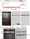
Fig. 1.
Fig. 1.
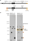
Amplification of SUL1 chromosomal fragments in sulfate-limited chemostats. a) Array Comparative Genomic Hybridization (aCGH) of the population of a diploid yeast strain (Chemostat run S712) 33 days after continuous growth in a sulfate-limited chemostat. The locations of SNF5, SUL1, and ARS228 are indicated in the diagram at the top. The Log2 hybridization ratio of day 33/day 0 indicates that the right end of chromosome II, containing the gene for the primary sulfur transporter SUL1, is present in 8 or more copies in excess of the genomic copies. The presence of two discontinuities at 782 and 788 kb suggests two discrete amplicons within the cell population. b) Contour-clamped homogeneous electric field (CHEF) gel electrophoresis of a single population (S712) sampled across the 33 days of continuous growth in sulfate-limited medium. The ethidium bromide-stained gel displays the total yeast karyotype. The CHEF gel was Southern blotted and probed sequentially for SUL1 and CEN2 as indicated. One extrachromosomal band of low molecular weight was detected by the SUL1 probe at day 17 that persists in the population onward. A second, faster migrating band appears at day 23 (more clearly resolved as two bands in c) and persists in the population onward. c) CHEF gel electrophoresis, under conditions to determine the sizes of extrachromosomal amplicons, from specific days of 15 individual chemostat runs. The ethidium bromide-stained gel and Southern blot hybridized with SUL1 reveal multiple small extrachromosomal fragments. The sizes of extrachromosomal amplicons vary from 26 to 40 kb with a recurring event producing a fragment of approximately 33 kb (arrow).
Fig. 2.
Fig. 2.
Linear fragments are capped by new telomeric sequences. a) Top: Positions of the EcoNI restriction sites at the right end of chromosome II (coordinates 770 to 813 kb). Chromosome II-R telomeric sequences are indicated with gold triangles. Bottom: Structure of linear fragments. Proposed telomeric sequences at variable positions at the left margin of the amplicons are indicated by the orange triangles. Positions of two probes [C(1-3)A/G(1-3)T and SUL1 (indicated by the horizontal bars)]. b) Linear fragments purified from the plug supernatant (see Supplementary Fig. 3) for five cultures (S708-S712) were digested with EcoNI and separated on a CHEF gel. Southern blots were sequentially hybridized with a SUL1 probe and a telomere-specific probe (C(1-3)A/G(1-3)T). Cultures S710 and S712 contain two species of extrachromosomal molecules. Triangles indicate the fragments with de novo telomeres. Boxed fragments correspond to the native right telomere.
Fig. 3.
Fig. 3.
Extrachromosomal fragments purified from plug supernatants were prepared for Illumina sequencing. Split reads (red/black sequences) were aligned to chromosome II (top sequence with chromosome II coordinates). The junctions of the left end of the extrachromosomal fragments each contained a new telomeric sequence (red) and potential seed sequences for telomere addition (blue/underline). The number in parentheses to the right of each sequence corresponds to the number of split reads that mapped to that site. One of the 15 sequences (S706) was a junction with an internal portion of a Y’ element. The remaining 14 contained unique junctions with C(1-3)A/G(1-3)T telomere sequences. The four nucleotide 5′-GxGT-3′ motifs (see Fig. 5) are in bold italics in the (top) reference sequence.
Fig. 4.
Fig. 4.
The SNF5 SiRTA is sufficient to stimulate de novo telomere addition at an ectopic location. a) Top schematic: the endogenous right arm of chromosome II; distance between the endogenous SNF5 sequence and SUL1 is shown. Triangles represent the endogenous telomere. Middle schematic: lines representing the relative size of each fragment from SNF5 that was integrated and tested (actual size in bp shown). Black arrows above the 718 bp fragment depict approximate locations of de novo telomere addition events stimulated by HO cleavage. Blue arrows below the 718 bp fragment depict approximate locations of spontaneous de novo telomere addition events from the chemostat experiments. Arrow size is proportional to the number of independent events at that location (see Supplementary Fig. 5 for exact numbers and locations). Bottom schematic: modified left arm of chromosome VII. Distances between the integrated SNF5 sequence and the HO cleavage site or the most distal essential gene (BRR6) on this chromosome arm are shown. b) Chromosome truncation events following HO cleavage preferentially involve de novo telomere addition in the SiRTA. At least 30 clones that survived HO cleavage and lost the URA3 marker were assayed for each experiment. The percentage of clones that incurred telomere addition within the sequence of interest is shown. Bar height is the average of three to four independent experiments; error bars represent standard deviations. A significant difference is indicated (*P < 0.05) by unpaired Student's t-test comparing 226 to 175 bp. See Supplementary File 2 for data used to generate the graph.
Fig. 5.
Fig. 5.
The SNF5 SiRTA requires Cdc13 binding to stimulate de novo telomere addition. a) Top: lines representing 718, 175 bp, and SNF5 SiRTA Stim::2XUAS fragments integrated and tested on chromosome VII. Sites of de novo telomere addition are depicted as in Fig. 4. Bottom: Twenty-nine nucleotides of the TG-rich strand of the SNF5 SiRTA Stim sequence are shown from 3′ to 5′. Note that a double-strand break that occurs to the left of this sequence will expose the TG-rich sequence in single-stranded DNA following 5′-end resection. 5′-GxGT-3′ motifs and corresponding mutations are highlighted in gray. b) Percentage of 5-FOA-resistant clones that incurred de novo telomere addition within the 718 bp SNF5 SiRTA in the presence of the indicated mutation. Wild-type (WT) samples are repeated from Fig. 4 for comparison. Samples with statistically significant values by one-way ANOVA with post hoc Dunnett's are indicated (**P < 0.01). c) Percentage of 5-FOA-resistant clones that incurred de novo telomere addition within the 175 bp SNF5 fragment when two copies of the Gal4 UAS are added (Stim::2xUAS) or when the 175 bp fragment is present alone. Cells were transformed with pRS414 (empty vector) or pRS414 expressing either Gal4 DNA binding domain (GBD) or GBD fused to the N-terminus of Cdc13 as indicated. For b and c, at least 30 colonies were assayed for each experiment. Bar height is the average of three to four independent experiments; error bars represent standard deviations. Samples with statistically significant values by one-way ANOVA with post hoc Tukey’s are indicated (*P < 0.01; ***P < 0.001). See Supplementary File 2 for data used to generate the graph.
Fig. 6.
Fig. 6.
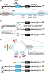
A model to explain how an ODIRA event may generate a substrate for de novo telomere addition at the SNF5 SiRTA. a) The endogenous right arm of chromosome II contains interrupted inverted repeats (IR; arrows) flanking the SNF5 SiRTA (star) and the TG-rich strand of the right telomere (triangles). In the expanded schematic the relevant sequences are shown for one possible pair of inverted repeats (arrows), the start codon of SNF5 (ATG), the Core of the SNF5 SiRTA, and the two 5′-GxGT-3′ motifs from Fig. 5 (labeled Stim 1 and 2). b) Ligation of the leading strand to the lagging strand following fork reversal at the leftward moving fork and template switching at the inverted repeats results in a “closed” fork with the rightward moving fork completing replication through the right chromosomal telomere. When a replication fork from ARS227, proceeding toward the telomere, encounters the closed fork structure, a hairpin-capped linear is displaced (curved arrow). c) In the displaced hairpin-capped linear, the inverted repeats form the base of the terminal single-stranded loop that contains the SiRTA Stim and Core sequences. Cleavage of the hairpin to the 3′ side of the SiRTA (1) and resection at the site of the break (dashed lines) exposes a 3′ single-stranded tail containing SiRTA sequences which can be bound by Cdc13 (2) and telomerase (3) to form a de novo telomere. d) In this model, after a single round of replication, the chromosome that experienced the closed fork produces two unaltered sister chromatids and the linear fragment containing SUL1 which is now protected by a de novo telomere addition at the SNF5 SiRTA. (Normal replication of the homolog in this diploid is not shown.) Under sulfate-limiting conditions, the cells containing the linear fragment are retained in the population due to the growth advantage provided by one or more extra copies of SUL1.

Similar articles

Cited by

References

    1. Alkan C, Coe BP, Eichler EE. Genome structural variation discovery and genotyping. Nat Rev Genet. 2011;12(5):363–376. doi:10.1038/nrg2958. - DOI - PMC - PubMed
    1. Anand R, Memisoglu G, Haber J. Cas9-mediated gene editing in Saccharomyces cerevisiae. Protocol Exchange. 2017. doi:10.1038/protex.2017.021a. - DOI
    1. Anderson EM, Halsey WA, Wuttke DS. Site-directed mutagenesis reveals the thermodynamic requirements for single-stranded DNA recognition by the telomere-binding protein Cdc13. Biochemistry. 2003;42(13):3751–3758. doi:10.1021/bi027047c. - DOI - PubMed
    1. Araya CL, Payen C, Dunham MJ, Fields S. Whole-genome sequencing of a laboratory-evolved yeast strain. BMC Genomics. 2010;11(1):88. doi:10.1186/1471-2164-11-88. - DOI - PMC - PubMed
    1. Bianchi A, Negrini S, Shore D. Delivery of yeast telomerase to a DNA break depends on the recruitment functions of Cdc13 and Est1. Mol Cell. 2004;16(1):139–146. doi:10.1016/j.molcel.2004.09.009. - DOI - PubMed

Publication types

MeSH terms