. 2023 Jan 26;6(1):102.
doi: 10.1038/s42003-023-04485-y.

The HDAC inhibitor zabadinostat is a systemic regulator of adaptive immunity

Affiliations

The HDAC inhibitor zabadinostat is a systemic regulator of adaptive immunity

Geng Liu et al. Commun Biol. .

Abstract

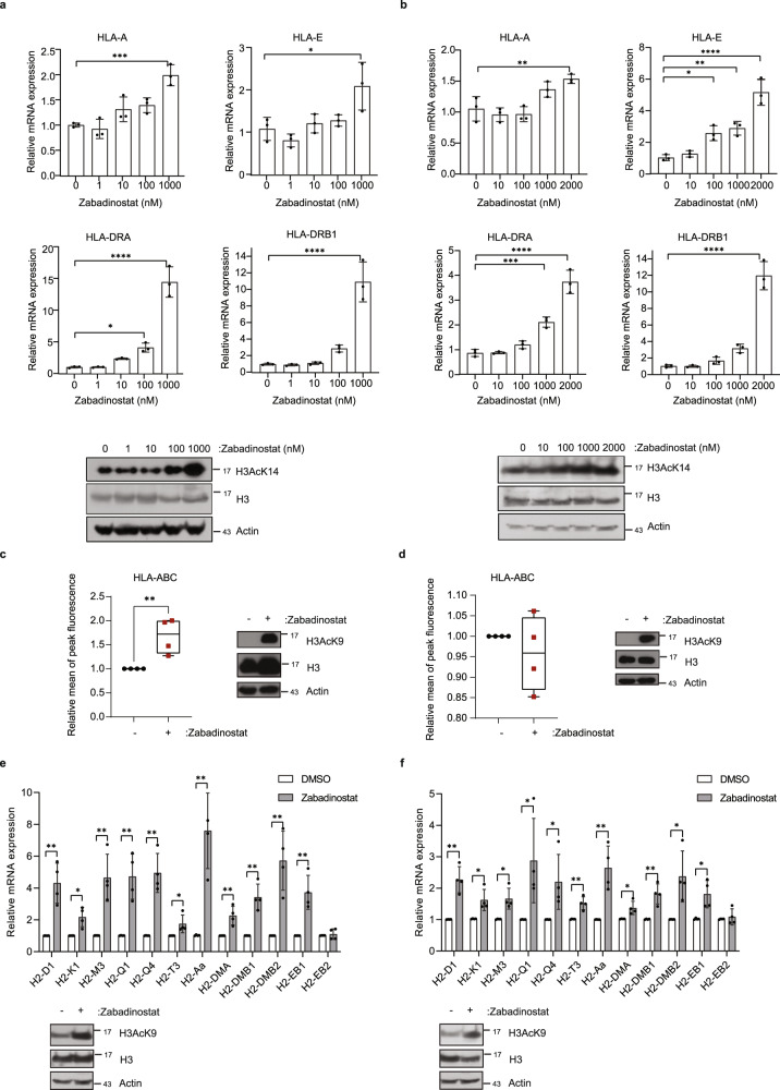
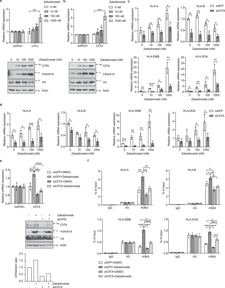
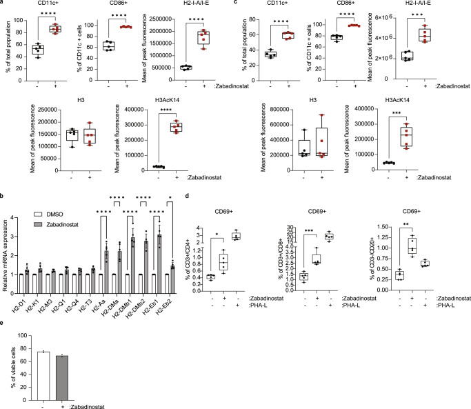
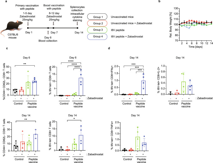
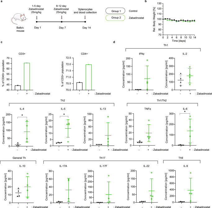
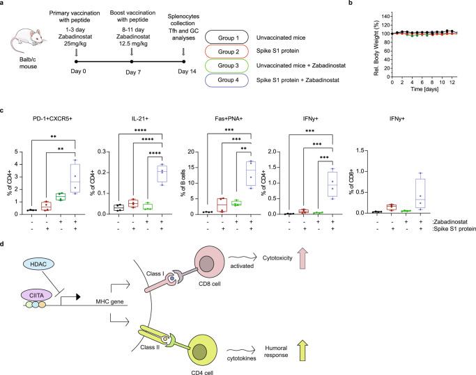
Protein acetylation plays a key role in regulating cellular processes and is subject to aberrant control in diverse pathologies. Although histone deacetylase (HDAC) inhibitors are approved drugs for certain cancers, it is not known whether they can be deployed in other therapeutic contexts. We have explored the clinical HDAC inhibitor, zabadinostat/CXD101, and found that it is a stand-alone regulator of the adaptive immune response. Zabadinostat treatment increased expression of MHC class I and II genes in a variety of cells, including dendritic cells (DCs) and healthy tissue. Remarkably, zabadinostat enhanced the activity of DCs, and CD4 and CD8 T lymphocytes. Using an antigenic peptide presented to the immune system by MHC class I, zabadinostat caused an increase in antigen-specific CD8 T lymphocytes. Further, mice immunised with covid19 spike protein and treated with zabadinostat exhibit enhanced covid19 neutralising antibodies and an increased level of T lymphocytes. The enhanced humoral response reflected increased activity of T follicular helper (Tfh) cells and germinal centre (GC) B cells. Our results argue strongly that zabadinostat has potential to augment diverse therapeutic agents that act through the immune system.


Conflict of interest statement

N.L.T. is a shareholder in Celleron Therapeutics Ltd. The remaining authors declare no competing interests.

Figures

Fig. 1
Fig. 1. Expression of MHC class I and class II genes upon zabadinostat treatment.
a Quantitative reverse transcription PCR (qRT-PCR) of MHC class I and class II genes in BEAS2B cells treated for 3 days with 1, 10, 100, 1000 nM zabadinostat or DMSO control; n = 3; results presented as mean values +/−SD; one-way ANOVA; The acetylation mark (H3AcK14) was detected by immunoblotting. b Quantitative reverse transcription PCR (qRT-PCR) of MHC class I and class II genes in NBE1 cells treated for 3 days with 10, 100, 1000, 2000 nM zabadinostat or DMSO control; n = 3; results presented as mean values +/−SD; one-way ANOVA. The acetylation mark (H3AcK14) was detected by immunoblotting. c Flow cytometry analysis of extracellular HLA class I proteins in BEAS2B cells treated for 2 days with 1 µM zabadinostat or DMSO control, n = 4, results presented as mean values +/−SD, Student’s t test; The acetylation mark (H3AcK9) was detected by immunoblotting in BEAS2B cells. d Flow cytometry analysis of extracellular HLA class I proteins in NBE1 cells treated for 2 days with 1 µM zabadinostat or DMSO control, n = 4, results presented as mean values +/−SD, Student’s t test; The acetylation mark (H3AcK9) was detected by immunoblotting in NBE1 cells; Quantitative reverse transcription PCR (qRT-PCR) of MHC class I and class II genes in lung RNA from Balb/c mice treated for 14 days (e) and 30 days (f) with 10 mg/kg zabadinostat or DMSO control; n = 4; results presented as mean values +/−SD; one-way ANOVA. The acetylation mark (H3AcK9) of representative mouse samples was detected by immunoblotting.
Fig. 2
Fig. 2. The effect of CIITA on the expression of MHC class I and class II genes.
a Quantitative reverse transcription PCR (qRT-PCR) of CIITA in BEAS2B cells treated for 3 days with 10, 100, 1000 nM zabadinostat or DMSO control; n = 3; results presented as mean values +/−SD; one-way ANOVA; The CIITA protein level and acetylation mark (H3AcK14) was detected by immunoblotting. b Quantitative reverse transcription PCR (qRT-PCR) of CIITA in NBE1 cells treated for 3 days with 10, 100, 1000 nM zabadinostat or DMSO control; n = 3; results presented as mean values +/−SD; one-way ANOVA; The CIITA protein level and acetylation mark (H3AcK14) was detected by immunoblotting. c Quantitative reverse transcription PCR (qRT-PCR) of MHC genes in BEAS2B cells treated for 2 days with 50 nM siCIITA and for 3 days with 10, 100, 1000 nM zabadinostat or DMSO control; n = 3; results presented as mean values +/−SD; one-way ANOVA. d Quantitative reverse transcription PCR (qRT-PCR) of MHC class I genes in NBE1 cells treated for 2 days with 50 nM siCIITA and for 3 days with 10, 100, 1000 nM zabadinostat or DMSO control; n = 3; results presented as mean values +/−SD; one-way ANOVA. e Quantitative reverse transcription PCR (qRT-PCR) of CIITA in BEAS2B cells treated for 2 days with 50 nM siCIITA and for 3 days with 10, 100, 1000 nM zabadinostat or DMSO control; n = 3; results presented as mean values +/−SD; one-way ANOVA. The CIITA protein level and acetylation mark (H3AcK14) were detected by immunoblotting and quantified. f Histone H3 and H3AcK9 ChIP on MHC gene promoters in BEAS2B cells treated for 2 days with 50 nM siCIITA and for 3 days with 1000 nM zabadinostat or DMSO control; n = 3; results presented as mean values +/−SD; one-way ANOVA.
Fig. 3
Fig. 3. Zabadinostat augments MHC expression in dendritic cells, and activates T and B lymphocytes.
a Flow cytometry analysis of the extracellular MHC class II protein level in bone marrow (collected from C57BL/6)-derived dendritic cells (CD11c+/CD86+) treated with 1 µM zabadinostat or DMSO control for 48 h; n = 5, Student’s t test; the acetylation mark (H3AcK14) and total histone 3 level were detected by flow cytometry; n = 5, Student’s t test. b Quantitative reverse transcription PCR (qRT-PCR) of MHC class I and II genes was also performed; n = 5; results presented as mean values +/−SD; one-way ANOVA. c Flow cytometry analysis of the extracellular MHC class II protein level in bone marrow (collected from Balb/c)-derived dendritic cells (CD11c+/CD86+) treated with 1 µM zabadinostat or DMSO control for 48 h, n = 5; the acetylation mark (H3AcK14) and total histone 3 level were detected by flow cytometry; n = 5; Student’s t test. d Activation of CD4, CD8 T cells and B cells upon 1 µM zabadinostat treatment or DMSO control was evaluated in splenocytes collected from Balb/c mice; n = 5; one-way ANOVA. e Viability of splenocytes measured by flow cytometry with L/D staining; graph represents pooled results from two animal experiments (see Fig. 5 (n = 4) and Supplementary Fig. 15 (n = 4)); results presented as mean values +/−SD.
Fig. 4
Fig. 4. Zabadinostat enhances the CD8 T-cell response.
a Schematic representation of the immunogenicity experiment with zabadinostat and peptide vaccine. C57BL/6 mice were treated with orally administrated zabadinostat at 25 mg/kg for 14 days (5 days on/2 days off) and intravenous I8V peptide (days 1 and 7) with respect to vehicle-only control; n = 5 per group; b relative body weight representation of treated and non-treated mice; n = 5; c general effector memory T-cell activation (CD8 + CD44 + CD62L− positive T cells) at days 6 (in the blood) and 14 (in splenocytes), I8V-peptide-specific CD8-positive T cells stained ex vivo with MHC tetramers at days 6 (in blood) and 14 (in splenocytes), and intracellular cytokine IFNγ, IL-2, TNFα secretion of I8V antigen-specific CD8 T cells stained ex vivo with tetramers (day 14; in splenocytes) measurements by flow cytometry (d); n = 4 and n = 5; results presented as mean values +/−SD; one-way ANOVA.
Fig. 5
Fig. 5. Zabadinostat enhances the CD4 T-cell response.
a Schematic representation of the experiment with zabadinostat. Balb/c mice were treated with orally administrated zabadinostat at 25 mg/kg for 14 days (5 days on/2 days off) or vehicle only; n = 4 per group; b relative body weight representation of treated and non-treated mice; n = 4; results presented as mean values +/−SD. c General T-cell activation and CD4-positive T cells measured by flow cytometry; measurements were performed in pooled splenocytes from four mice; also, we noticed similar percentage of CD8-positive T cells in control and zabadinostat-treated groups (26.2% and 24.3%). The viability of the splenocytes was similar in zabadinostat treatment compare to untreated control (70.9% and 76.6% viable cells, respectively). d Analysis of panel of Th1, Th2, Th1/Th2, General Th, Th17 and Th9 cytokines in serum collected from mice treated with zabadinostat and the control group; n = 4; results presented as mean values +/−SD; Student’s t test.
Fig. 6
Fig. 6. Zabadinostat enhances the antibody response against the covid19 spike S1 protein.
a Schematic representation of the immunogenicity experiment with zabadinostat and spike S1 protein. Balb/c mice were treated with orally administrated zabadinostat at 25 mg/kg for 14 days (5 days on/2 days off) and intravenous spike S1 protein (days 1 and 7) with respect to vehicle-only control. Blood samples were collected on days 14, 21, and 30; n = 4 per group; b Relative body weight representation of treated and non-treated mice; n = 4; results presented as mean values +/−SD; Analysis of IgG (c) and IgM (d) spike protein antibodies at days 14, 21, and 30 in serum collected from mice treated with spike protein, zabadinostat, the combination and the control group; n = 4; results presented as mean values +/−SD; one-way ANOVA. e Analysis of spike S1-ACE2 neutralising antibodies at days 14, 21, and 30 in serum collected from mice treated with spike protein, zabadinostat, the combination, and the control group; n = 4; results presented as mean values +/−SD; one-way ANOVA.
Fig. 7
Fig. 7. Zabadinostat augments the level of GC B cells through modulating the activity of Tfh cells.
a Schematic representation of the immunogenicity experiment with zabadinostat and spike S1 protein. Balb/c mice were treated with orally administrated zabadinostat at 25 mg/kg (days 1–3) or 12.5 mg/kg (days 8–11), and intravenous spike S1 protein (10 µg) (days 0 and 7) with respect to vehicle-only control; splenocytes were collected at day 14; b relative body weight representation of treated and non-treated mice; n = 4; results presented as mean values +/−SD; c follicular T-cell level (CD4 + PD-1 + CXCR5 + positive T cells), germinal centre B cells (Fas+ PNA+ positive B cells) and intracellular cytokine IL-21 (in CD4-positive T cells), IFNy (in CD4 and CD8-positive T cells) secretion measurements by flow cytometry; n = 4; results presented as mean values +/−SD; one-way ANOVA. d Model describing the effect of zabadinostat in regulating the adaptive immune response. It is proposed that inhibition of histone deacetylase induces both cellular and humoral immune responses by increasing antigen presentation (e.g., by dendritic cells), activating T and B cells and increasing germinal centre B cells (stimulated by follicular T cells), which coincides with elevated the MHC class I and II proteins.

Similar articles

Cited by

References

    1. Shvedunova M, Akhtar A. Modulation of cellular processes by histone and non-histone protein acetylation. Nat. Rev. Mol. Cell Biol. 2022;3:329–349. doi: 10.1038/s41580-021-00441-y. - DOI - PubMed
    1. Provine NM, et al. MAIT cell activation augments adenovirus vector vaccine immunogenicity. Science. 2021;371:521–526. doi: 10.1126/science.aax8819. - DOI - PMC - PubMed
    1. Choudhary C, Weinert BT, Nishida Y, Verdin E, Mann M. The growing landscape of lysine acetylation links metabolism and cell signalling. Nat. Rev. Mol. Cell Biol. 2014;15:536–550. doi: 10.1038/nrm3841. - DOI - PubMed
    1. Kinnaird A, Zhao S, Wellen KE, Michelakis ED. Metabolic control of epigenetics in cancer. Nat. Rev. Cancer. 2016;16:694–707. doi: 10.1038/nrc.2016.82. - DOI - PubMed
    1. Cappellacci L, Perinelli DR, Maggi F, Grifantini M, Petrelli R. Recent progress in histone deacetylase inhibitors as anticancer agents. Curr. Med. Chem. 2020;27:2449–2493. doi: 10.2174/0929867325666181016163110. - DOI - PubMed

Publication types