. 2023 Jan 26;13(1):1477.
doi: 10.1038/s41598-023-28026-z.

Regulatory mechanisms of the dynein-2 motility by post-translational modification revealed by MD simulation

Affiliations

Regulatory mechanisms of the dynein-2 motility by post-translational modification revealed by MD simulation

Shintaroh Kubo et al. Sci Rep. .

Abstract

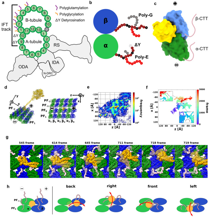
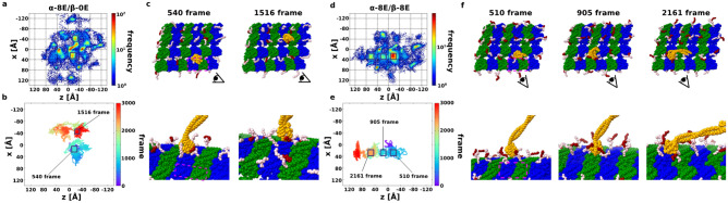
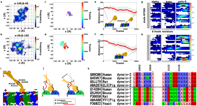
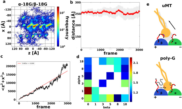
Intraflagellar transport for ciliary assembly and maintenance is driven by dynein and kinesins specific to the cilia. It has been shown that anterograde and retrograde transports run on different regions of the doublet microtubule, i.e., separate train tracks. However, little is known about the regulatory mechanism of this selective process. Since the doublet microtubule is known to display specific post-translational modifications of tubulins, i.e., "tubulin code", for molecular motor regulations, we investigated the motility of ciliary specific dynein-2 under different post-translational modification by coarse-grained molecular dynamics. Our setup allows us to simulate the landing behaviors of dynein-2 on un-modified, detyrosinated, poly-glutamylated and poly-glycylated microtubules in silico. Our study revealed that poly-glutamylation can play an inhibitory effect on dynein-2 motility. Our result indicates that poly-glutamylation of the B-tubule of the doublet microtubule can be used as an efficient means to target retrograde intraflagellar transport onto the A-tubule.


Conflict of interest statement

The authors declare no competing interests.

Figures

Figure 1
Figure 1
Low-affinity state dynein with WT MT. (a) Schematic of the doublet MT and the PTM distribution on the A- and B-tubules. The IFT track indicates the available PF for anterograde and retrograde IFT. (b) Illustration of PTM on CTT of tubulin. (c) Surface models of dynein-MTBD binding (from PDB 6KIQ to tubulin and the position of the flexible CTT (pink). (d) Simulation system of dynein and MT. Dynein, α-tubulin, and β-tubulin colored orange, green, and blue. The minus end of the MT is along the z-axis, and the circumferential direction is x-axis. (e) Heat map of the position of the low-affinity state MTBD on the uMT, with 20 trajectories overlaid. The frequency increases from blue to red. The red area is the stable bound for MTBD, while the blue area is the unstable within the 20 times trajectories. (f) Representative trajectory of MTBD on the MT. The simulation time is 3000 frames, and the trajectory changed a color from purple to red from 0 to 3000 frame. Origin is the initial position of the center of mass of MTBD. (g) Snapshots of a back-landing trajectory. These snapshots are picked up from Fig. 1d bottom. CTTs of α- and β-tubulins colored pink. The semi-transparent model is a previous snapshot. (h) Cartoons for the back, right-side, front, and left-side landing mechanism.
Figure 2
Figure 2
Low-affinity state dynein with detyrosinated MT. (a) Heat map of the position of the low-affinity state MTBD on the ΔY MT, with 20 trajectories overlaid. The coloring method is same with Fig. 1d. (b) Probability of localization of MTBD on the uMT and ΔY MT for each binding lane; PF-1, PF0, PF1, and other percentages are red, green, orange, and blue, respectively. (c) The detyrosinated CTT seems to have an easier contact MTBD than unmodified CTT. The positively charged ARG and LYS are blue, the negatively charged GLU and ASP are red, and the other are gray.
Figure 3
Figure 3
Low-affinity state dynein with short poly-E MT. (a, d) Heat map of the position of the low-affinity state MTBD on the poly-E MT, with 20 trajectories overlaid. The coloring method is same with Fig. 1d. (a) is α-tubulin have 8 length poly-E, and β-tubulin does not have poly-E (simply denoted α-8E/β-0E). (d) is both α- and β-tubulin have 8 length poly-E (α-8E/β-8E). (b, e) Representative trajectory of MTBD on the poly-E MT. The coloring method is same with Fig. 1b. (b) is the representative one in the case α-8E/β-0E, and (e) is the representative one in the case α-8E/β-8E. (c, f) Snapshots picked up from Fig. 3b, e inboxes. The coloring method of dynein and tubulins are same with Fig. 1e, and poly-E colored red.
Figure 4
Figure 4
Low-affinity state dynein with long poly-E MT. (a, b) Heat map of the position of the low-affinity state MTBD on the long poly-E MT, with 20 trajectories overlaid. The coloring method is same with Fig. 1d. (a) is in the α-18E/β-0E case, and (b) is in the α-0E/β-18E case. (c, d) Representative trajectory of MTBD on the poly-E MT. The coloring method is same with Fig. 1b. (c) is in the α-18E/β-0E case, and (d) is in the α-0E/β-18E case. (e, f) The trajectory of the average distance between the tip of the linker to the closest tubulins. The average distance for each frame is red, and 95% confidence interval are gray. (g, h) Residue-by-residue contact maps of CTT and poly-E with ARG or LYS residues on stalk-MTBD. Highly contacted pairs are colored red, lower are blue, and no contact pairs box are white. (g) and (h) is made from 20 trajectories in α-18E/β-0E and α-0E/β-18E setup, respectively. (i) The highest contact residues on the stalk-MTBD region; R2955, R2959, K3122, and R3126. Especially, R2955 and R2959 are specific contact residues in (0, 18) case. (j) Cartoons for understanding poly-E contact features. MTBD, α-tubulin, and β-tubulin colored orange (gray), green, and blue. CTT is black, and poly-E is red. (k) Sequence alignment of dynein-1 and -2 from different species.
Figure 5
Figure 5
Low-affinity state dynein with poly-G MT. (a) Heat map of the position of the low-affinity state MTBD on the poly-G MT, with 20 trajectories overlaid. The coloring method is same with Fig. 1d. This is in the α-18G/β-18G case. (b) The trajectory of the average direction between the tip of the linker to the closest tubulins in the α-18G/β-18G case. The coloring method is same with Fig. 4e. (c) MSD plot of α-18G/β-18G case (black) and its linear approximation line (red). (d) The heatmap of the diffusion coefficient in the various poly-G simulation systems. Blue is small diffusion, red is high diffusion, and the white is not simulated setups. (e) Cartoon for understanding poly-G simulation features. When MT does not have poly-G, CTT directly contacts to MTBD, and CTT induces MTBD flexible motion (above). On the other hand, when poly-G MT prevents CTT from contacting MTBD, thus suppressing the diffusion of MTBD.

Similar articles

Cited by

References

    1. Monroy BY, Tan TC, Oclaman JM, Han JS, Simó S, Niwa S, Ori-McKenney KM. A combinatorial MAP code dictates polarized microtubule transport. Dev. Cell. 2020;53(1):60–72. - PMC - PubMed
    1. Dixit R, et al. Differential regulation of dynein and kinesin motor proteins by tau. Science. 2008;319(5866):1086–1089. - PMC - PubMed
    1. Janke C, Magiera MM. The tubulin code and its role in controlling microtubule properties and functions. Nat. Rev. Mol. Cell Biol. 2020;21(6):307–326. - PubMed
    1. Sirajuddin M, Rice LM, Vale RD. Regulation of microtubule motors by tubulin isotypes and post-translational modifications. Nat Cell Biol. 2014;16(4):335–344. - PMC - PubMed
    1. Gadadhar S, Alvarez Viar G, Hansen JN, Gong A, Kostarev A, Ialy-Radio C, Janke C. Tubulin glycylation controls axonemal dynein activity, flagellar beat, and male fertility. Science. 2021;371(6525):eabd4914. - PMC - PubMed

Publication types

Grants and funding