. 2023 Jan 26;17(1):6.
doi: 10.1186/s13036-023-00325-z.

The synergistic and enhancive effects of IL-6 and M-CSF to expand and differentiate functional dendritic cells from human monocytes under serum-free condition

Affiliations

The synergistic and enhancive effects of IL-6 and M-CSF to expand and differentiate functional dendritic cells from human monocytes under serum-free condition

Chao-Ling Yao et al. J Biol Eng. .

Abstract

Background: Dendritic cells (DCs) are differentiated from monocytes, and have a strong ability to perform phagocytosis, present antigens and activate T cell immune response. Therefore, DCs are one of the key factors in fighting cancer in immunotherapy, and it is an important issue to develop a serum-free system for DC differentiation and expansion in vitro for clinical application.

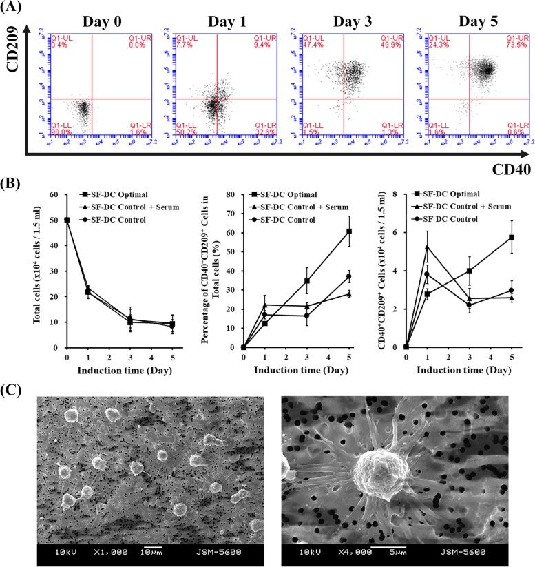
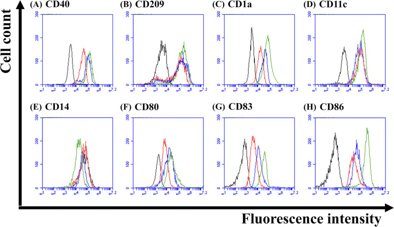
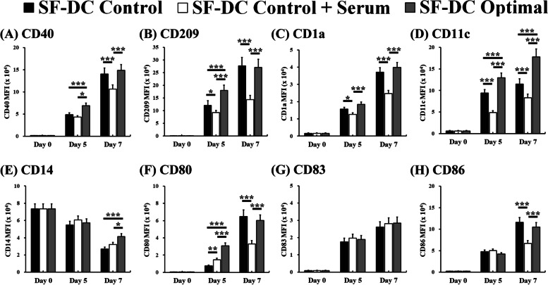
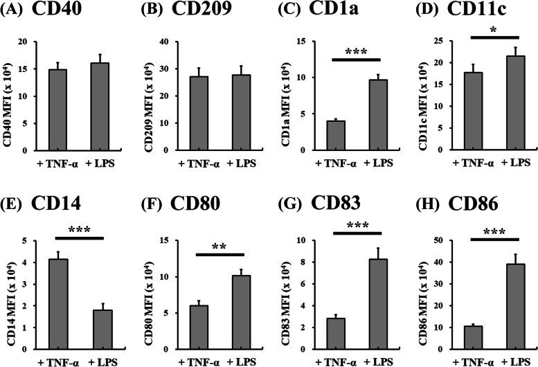
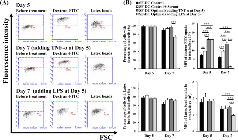
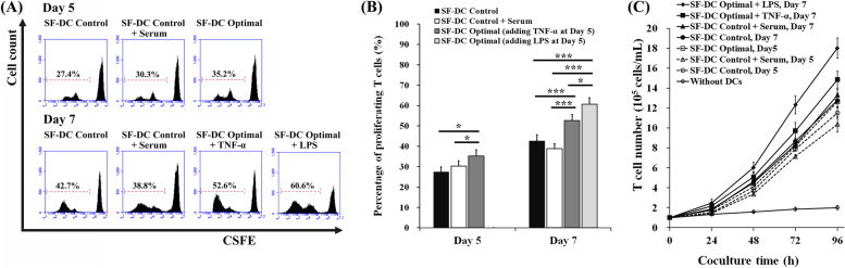
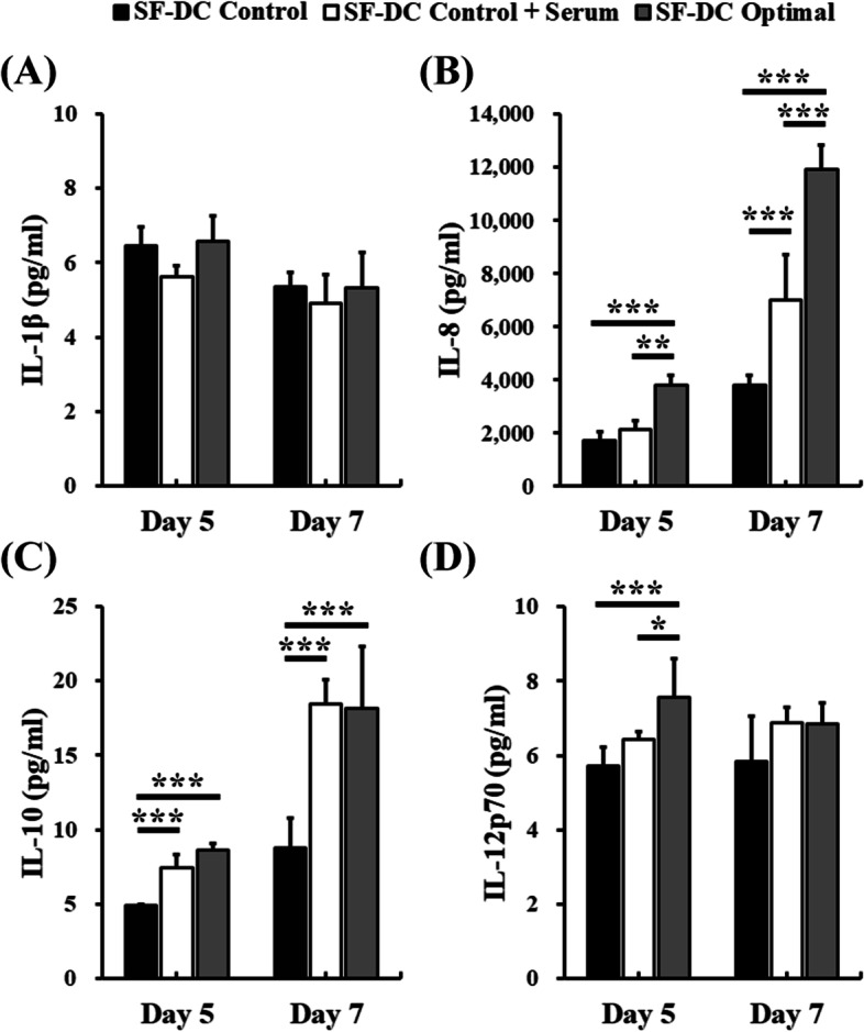
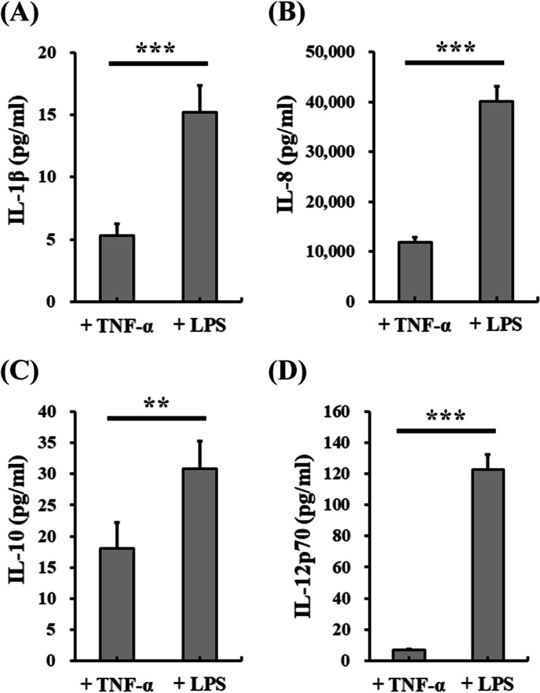
Results: In this study, IL-6 and M-CSF were determined and a concentration combination of cytokines was optimized to develop an optimal DC serum-free differentiation medium (SF-DC Optimal) that can effectively differentiate CD14+ monocytes into CD40+CD209+ DCs. After differentiation, the morphology, growth kinetics, surface antigen expression, phagocytosis ability, cytokine secretion, mixed lymphocyte reaction and stimulation for maturation of the differentiated DCs were checked and confirmed. Importantly, this research is the first report finding that the addition an extra low concentration of IL-6 and M-CSF exhibited a synergistic effect with GM-CSF and IL-4 to generate higher numbers and more fully functional DCs than the addition of GM-CSF and IL-4 only under serum-free condition.

Conclusion: A large number of functional DCs can be generated by using SF-DC Optimal medium and provide an alternative source of DCs for related basic research and clinical applications.

Keywords: Dendritic cell; IL-6; Immunotherapy; M-CSF; Serum-free.


Conflict of interest statement

The authors declare that they have no competing interests.

Figures

Fig. 1
Fig. 1
CD40 and CD209 profiles, growth kinetics and morphologies on DC differentiation from human monocytes. DCs were differentiated from CD14+ monocytes (5 × 105 cells/1.5 mL) in SF-DC Optimal medium, SF-DC Control medium and SF-DC Control + Serum medium for 5 days. A The differentiated cells in SF-DC Optimal medium at the indicated time points were stained with CD40 and CD209-PE. The numbers within the dot plots represent the percentages of the indicated cells in the total cell population by flow cytometry analysis. B The accumulated total cell number, percentage of CD40+CD209+ DCs in the total cells and CD40+CD209+ DC number differentiated from CD14+ monocytes at the indicated time points were determined by flow cytometry analysis (n = 5). (I) Representative morphologies of the differentiated DCs on Day 5 in SF-DC Optimal medium under scanning electron microscopy observation. Scale bars are 10 μm (left figure) and 5 μm (right figure)
Fig. 2
Fig. 2
Representative histograms of DC-related surface marker expressions on differentiated DCs from human monocytes in SF-DC Optimal medium. DCs were differentiated from CD14+ monocytes (5 × 105 cells/1.5 mL) in SF-DC Optimal medium for 5 days (Day 0 to Day 5), and then, 20 ng/ml TNF-α or 1 μg/mL lipopolysaccharide (LPS) was added to stimulate maturation for 2 days (Day 5 to Day 7). After differentiation, the generated cells at the indicated time points (Day 0: black peaks; Day 5: red peaks; Day 7 with TNF-α: blue peaks; Day 7 with LPS: green peaks) were stained with antibodies against (A) CD40, B CD209, C CD1a, D CD11c, E CD14, F CD80, G CD83 and H CD86. The histogram of the indicated surface marker expression in the total cell population was analyzed by flow cytometry
Fig. 3
Fig. 3
Expression of DC-related surface markers on differentiated DCs from human monocytes. DCs were differentiated from CD14+ monocytes (5 × 105 cells/1.5 mL) in SF-DC Optimal medium, SF-DC Control medium and SF-DC Control + Serum medium for 5 days (Day 0 to Day 5), and then, 20 ng/ml TNF-α was added to the corresponding medium to stimulate maturation for 2 days (Day 5 to Day 7). After differentiation, the generated cells at the indicated time points were stained with antibodies against (A) CD40, B CD209, C CD1a, D CD11c, E CD14, F CD80, G CD83 and H CD86. The mean fluorescence intensity (MFI) of the indicated cells in the total cell population was analyzed by flow cytometry. *, ** and *** represent significant differences of p < 0.05, p < 0.01 and p < 0.005, respectively (n = 5)
Fig. 4
Fig. 4
Comparison of DC-related surface marker expression on differentiated DCs from human monocytes in SF-DC Optimal medium with TNF-α and lipopolysaccharide stimulation. DCs were differentiated from CD14+ monocytes (5 × 105 cells/1.5 mL) in SF-DC Optimal medium for 5 days (Day 0 to Day 5), and then, 20 ng/ml TNF-α or 1 μg/mL lipopolysaccharide (LPS) was added to stimulate maturation for 2 days (Day 5 to Day 7). After differentiation, the generated cells on Day 7 were stained with antibodies against (A) CD40, B CD209, C CD1a, D CD11c, E CD14, F CD80, G CD83 and (H) CD86. The mean fluorescence intensity (MFI) of the indicated cells in the total cell population was analyzed by flow cytometry. *, ** and *** represent significant differences of p < 0.05, p < 0.01 and p < 0.005, respectively (n = 5)
Fig. 5
Fig. 5
Endocytosis analysis of the differentiated DCs from monocytes. DCs were differentiated from CD14+ monocytes (5 × 105 cells/1.5 mL) in SF-DC Optimal medium, SF-DC Control medium and SF-DC Control + Serum medium for 5 days (Day 0), and then, 20 ng/ml TNF-α or 1 μg/mL lipopolysaccharide (LPS) was added to the corresponding medium to stimulate maturation for 2 days (Day 5 to Day 7). After differentiation, the cells were treated with 1 mg/mL dextran-fluorescein isothiocyanate (dextran-FITC) or 1 mg/mL fluorescent latex beads and stained with a CD209 fluorescence antibody. The endocytosis ability was determined by the CD209+ cells that expressed the fluorescence of dextran-FITC or latex beads using flow cytometry. A Representative endocytosis analysis of the differentiated DCs in SF-DC Optimal medium before (Day 5) and after (Day 7) TNF-α or LPS stimulation for maturation by flow cytometry. B The percentages and the mean fluorescence intensity (MFI) of the cells with dextran-FITC or fluorescent latex bead uptake in total CD209+ DCs at the indicated time points were analyzed by flow cytometry. ** and *** represent significant differences of p < 0.01 and p < 0.005, respectively (n = 3)
Fig. 6
Fig. 6
Mixed leukocyte reaction of the differentiated DCs from monocytes. DCs were differentiated from CD14+ monocytes (5 × 105 cells/1.5 mL) in SF-DC Optimal medium, SF-DC Control medium and SF-DC Control + Serum medium for 5 days (Day 0 to Day 5), and then, 20 ng/ml TNF-α or 1 μg/mL lipopolysaccharide (LPS) was added to the corresponding medium to stimulate maturation for 2 days (Day 5 to Day 7). After differentiation, CD209+ DCs were isolated from the differentiated cells using anti-CD209 microbeads on a VarioMACS separator. Then, the isolated CD209+ DCs were cocultured with allogenic CFSE-stained CD3+ T cells at a ratio of 1:2 for mixed leukocyte reactions. After 4 days of coculture, total cells were harvested, and proliferating CD3+ T cells were determined using flow cytometry. A Representative analysis of CFSE expression in cocultured cells. The cells in the dotted boundary represent proliferating CD3+ T cells with decayed CFSE expression. B The percentages of proliferating CD3+ T cells among the total CD3+ T cells. * and *** represent a significant difference of p < 0.05 and p < 0.005, respectively (n = 3). C Growth kinetics of CD3+ T-cell expansion stimulated by coculture with various differentiated DCs (n = 3). The initial CD3+ T-cell density was 1 × 105 cells/mL
Fig. 7
Fig. 7
Cytokine secretion of the differentiated DCs from monocytes. DCs were differentiated from CD14+ monocytes (5 × 105 cells/1.5 mL) in SF-DC Optimal medium, SF-DC Control medium and SF-DC Control + Serum medium for 5 days (Day 0 to Day 5), and then, 20 ng/ml TNF-α was added to the corresponding medium to stimulate maturation for 2 days (Day 5 to Day 7). After differentiation, the conditioned media at the indicated time points were collected, and the secreted amounts of (A) IL-1β, B IL-8, C IL-10 and D IL-12p70 were analyzed by flow cytometry. *, ** and *** represent significant differences of p < 0.05, p < 0.01 and p < 0.005, respectively (n = 3)
Fig. 8
Fig. 8
Comparison of cytokine secretion of the differentiated DCs from human monocytes in SF-DC Optimal medium with TNF-α and lipopolysaccharide stimulation. DCs were differentiated from CD14+ monocytes (5 × 105 cells/1.5 mL) in SF-DC Optimal medium for 5 days (Day 0 to Day 5), and then, 20 ng/ml TNF-α or 1 μg/mL lipopolysaccharide (LPS) was added to stimulate maturation for 2 days (Day 5 to Day 7). After differentiation, the conditioned media on Day 7 were collected, and the secreted amounts of (A) IL-1β, B IL-8, C IL-10 and D IL-12p70 were analyzed by flow cytometry. ** and *** represent significant differences of p < 0.01 and p < 0.005, respectively (n = 3)

Similar articles

References

    1. Sharma P, Allison JP. The future of immune checkpoint therapy. Science. 2015;348:56–61. doi: 10.1126/science.aaa8172. - DOI - PubMed
    1. Yu MB, Guerra J, Firek A, Langridge WHR. Extracellular vimentin modulates human dendritic cell activation. Mol Immunol. 2018;104:37–46. doi: 10.1016/j.molimm.2018.09.017. - DOI - PMC - PubMed
    1. Büchler T, Hajek R, Bourkova L, Kovarova L, Musilova R, Bulikova A, Doubek M, Svobodnik A, Mareschova I, Vanova P, Tuzova E, Vidlakova P, Vorlicek J, Penka M. Generation of antigen-loaded dendritic cells in a serum-free medium using different cytokine combinations. Vaccine. 2003;21:877–882. doi: 10.1016/S0264-410X(02)00535-2. - DOI - PubMed
    1. Shi D, Shi Y, Kaseb AO, Qi X, Zhang Y, Chi J, Lu Q, Gao H, Jiang H, Wang H, Yuan D, Ma H, Wang H, Li Z, Zhai B. Chimeric antigen receptor-glypican-3 T-cell therapy for advanced hepatocellular carcinoma: results of phase I trials. Clin Cancer Res. 2020;26:3979–3989. doi: 10.1158/1078-0432.CCR-19-3259. - DOI - PubMed
    1. Cany J, Dolstra H, Shah N. Umbilical cord blood-derived cellular products for cancer immunotherapy. Cytotherapy. 2015;17:739–748. doi: 10.1016/j.jcyt.2015.03.005. - DOI - PubMed