Review
. 2020 Jan 20;3(1):8.
doi: 10.1038/s42004-019-0254-9.

Linking graphene-based material physicochemical properties with molecular adsorption, structure and cell fate

Affiliations
Review

Linking graphene-based material physicochemical properties with molecular adsorption, structure and cell fate

Sachin Kumar et al. Commun Chem. .

Abstract

Graphene, an allotrope of carbon, consists of a single layer of carbon atoms with uniquely tuneable properties. As such, graphene-based materials (GBMs) have gained interest for tissue engineering applications. GBMs are often discussed in the context of how different physicochemical properties affect cell physiology, without explicitly considering the impact of adsorbed proteins. Establishing a relationship between graphene properties, adsorbed proteins, and cell response is necessary as these proteins provide the surface upon which cells attach and grow. This review highlights the molecular adsorption of proteins on different GBMs, protein structural changes, and the connection to cellular function.


Conflict of interest statement

The authors declare no competing interests.

Figures
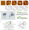
Fig. 1
Fig. 1. Multiple interactions between different serum proteins and graphene-based substrates mediate protein binding and conformation.
a Interactions between serum proteins BSA, Tf, IgG, and BFG, with GO along with the corresponding CD spectra, highlighting structural change with incubation time. CD spectra showed that BSA and Tf after adsorption on GO surface exhibited structural rearrangement from α-helical to enhanced β-sheet characteristics, whereas BFG and Ig showed structural heterogeneity on the GO surface (adapted with permission from ref. . © 2015 American Chemical Society). b Simulation snapshot of BSA molecule after the 20-ns adsorption showing conformational changes with a decrease in α-helical content on a graphite surface (adapted with permission from ref. . © 2011 American Chemical Society). c MD-simulated structural rearrangements of BFG on graphene for 170 ns. Protein aromatic residues Trp, Tyr, and Phe (highlighted in color) oriented and aligned with the graphene surface facilitate π–π stacking (adapted with permission from ref. . © 2015 American Chemical Society). d Different interactions of serum proteins on partial oxidized graphene (adapted with permission from ref. . © 2012 WILEY-VCH Verlag GmbH & Co. KGaA, Weinheim). e RGD is attracted to vacancy-defect graphene surfaces with mono-vacancy showing attraction to COO–. The vacancy is highlighted in ball-and-stick style (adapted with permission from ref. . © 2015 American Chemical Society).
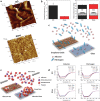
Fig. 2
Fig. 2. Graphene-substrate topography affects protein conformation and cell response.
a AFM 3D topographical images of GO and glass surface. b Difference in surface mean roughness and surface wettability with water contact angles of GO and glass. (The * indicates statistically significant differences for p-values < 0.05. The two-tailed Student's t test was used to make the pairwise comparisons). c Schematic illustrating more adsorption of albumin and fibrinogen on GO than on glass. d Circular dichroism (CD) spectra of albumin and fibrinogen incubated with GO and glass at different concentrations showing changes in protein structure for proteins incubated with GO. e Schematic illustrating the reduced formation of blood clots on albumin–GO surface compared with albumin–glass surface (adapted with permission from ref. . © 2015 Wiley-VCH Verlag GmbH & Co. KGaA, Weinheim).
Fig. 3
Fig. 3. Graphene and GO substrate chemistry affects protein interaction and cell response.
a Cytoplasmic lipid accumulation assessed by Oil Red O staining after 14 days of induction on graphene and GO (scale bar 50 µm). (The asterisk indicates statistically significant differences for p-values < 0.05, using Student’s t test, n = 4 for each group). b Strong adipogenic differentiation of MSCs was observed on GO surfaces, with a significantly higher accumulation of lipid droplets. (The asterisk indicates statistically significant differences for p-values < 0.05, using Student’s t test, n = 4 for each group). c Schematic illustration of insulin adsorption on graphene and GO showing the respective conformations. Note that the schematics of the molecular substrate of protein (insulin), graphene, and GO are not scaling in proportion. d Far UV absorption CD spectra of insulin demonstrate the structural change upon adsorption on graphene and GO (adapted with permission from ref. . © 2011 American Chemical Society).
Fig. 4
Fig. 4. Graphene oxide chemistry affects protein morphology and cell response.
a Fluorescence micrographs illustrating pre-osteoblast proliferation on BSA-adsorbed scaffolds chitosan (CS–BSA) and chitosan–GO (CS–GO–BSA) after 7 and 28 days. Cells were stained with Hoechst to highlight nuclei, but due to autofluorescence of the scaffold nuclei appear as diffuse dots. b Osteogenic differentiation evaluated with energy-dispersive X-ray spectroscopy by mapping Ca and P mineral deposition on scaffolds at day 7. GO scaffolds with BSA adsorbed showed a higher amount of Ca and P mineral corresponding to higher osteogenic differentiation. c Scanning electron micrographs illustrating the structural morphology of pure CS; CS modified with GO (CS–GO) scaffold and morphology of adsorbed BSA protein on the respective scaffold (adapted with permission from ref. . © 2012 Acta Materialia Inc. Published by Elsevier Ltd.).
Fig. 5
Fig. 5. Graphene and RGO substrate chemistry affects protein adsorption and cell response.
a AFM micrograph of GO and rGO substrates and their respective average surface roughness (R). b Immunofluorescence staining for myosin heavy chain (MHC) showing more myotube formation on GO surface. c Change in nitrogen composition on the GO and rGO substrates before and after incubation in serum-containing media due to protein adsorption (adapted with permission from ref. . © 2012 Elsevier Ltd.).
Fig. 6
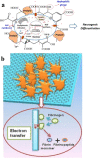
Fig. 6. Graphene-based substrate electrical properties affect protein adsorption and cell response.
a Schematic illustration of protein adsorption from culture media on GO and possible localized electric field mediating calcium-dependent pathway for neurogenic differentiation of ADSCs (adapted with permission from ref. . © 2018 Elsevier B.V.). b Activation of platelet by fibrinogen-adsorbed graphene mediated through electron transfer (adapted with permission from ref. . © 2016 Springer Nature) (Note: schematics of protein, GO, and graphene are not scaling in proportion).
Fig. 7
Fig. 7. Growth factor–graphene-based substrate interaction.
a Schematic illustration for multi-pass caliber-rolled (MPCR) Ti alloy surface coated loaded with RGO adsorbing dexamethasone and promoting osteogenic differentiation of the stem cell for dental application (adapted with permission from ref. . © 2015 American Chemical Society). b Schematic showing the underlying mechanisms of cell interaction with adsorbed fibronectin and response of TGF‐β3 growth factor influencing cell signaling to enhance chondrogenic differentiation (adapted with permission from ref. . © 2014 WILEY-VCH Verlag GmbH & Co. KGaA, Weinheim). c Diagram showing how adsorbed VEGF on GO inhibited angiogenesis due to the structural change in VEFG upon interaction with GO (adapted with permission from ref. . © 2016 Elsevier Ltd.).

Similar articles

Cited by

References

    1. Shin SR, et al. Graphene-based materials for tissue engineering. Adv. Drug Deliv. Rev. 2016;105:255–274. doi: 10.1016/j.addr.2016.03.007. - DOI - PMC - PubMed
    1. Feng L, Liu Z. Graphene in biomedicine: opportunities and challenges. Nanomedicine. 2011;6:317–324. doi: 10.2217/nnm.10.158. - DOI - PubMed
    1. Kumar S, Chatterjee K. Comprehensive review on the use of graphene-based substrates for regenerative medicine and biomedical devices. ACS Appl. Mater. interfaces. 2016;8:26431–26457. doi: 10.1021/acsami.6b09801. - DOI - PubMed
    1. Wei X-Q, et al. Insight into the interaction of graphene oxide with serum proteins and the impact of the degree of reduction and concentration. ACS Appl. Mater. interfaces. 2015;7:13367–13374. doi: 10.1021/acsami.5b01874. - DOI - PubMed
    1. Zhang Y, Wu C, Guo S, Zhang J. Interactions of graphene and graphene oxide with proteins and peptides. Nanotechnol. Rev. 2013;2:27–45. doi: 10.1515/ntrev-2012-0078. - DOI