. 2020 Sep 11;3(1):125.
doi: 10.1038/s42004-020-00372-3.

Quantification of amyloid fibril polymorphism by nano-morphometry reveals the individuality of filament assembly

Affiliations

Quantification of amyloid fibril polymorphism by nano-morphometry reveals the individuality of filament assembly

Liam D Aubrey et al. Commun Chem. .

Abstract

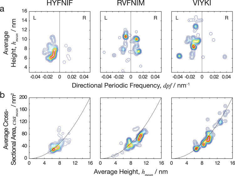
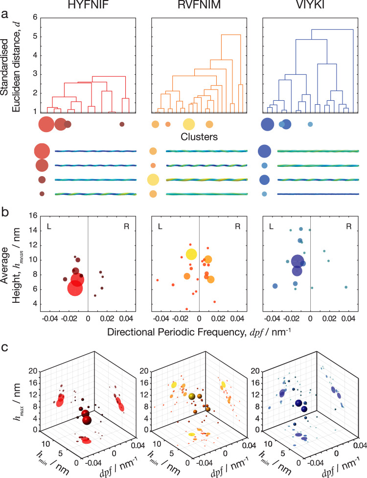
Amyloid fibrils are highly polymorphic structures formed by many different proteins. They provide biological function but also abnormally accumulate in numerous human diseases. The physicochemical principles of amyloid polymorphism are not understood due to lack of structural insights at the single-fibril level. To identify and classify different fibril polymorphs and to quantify the level of heterogeneity is essential to decipher the precise links between amyloid structures and their functional and disease associated properties such as toxicity, strains, propagation and spreading. Employing gentle, force-distance curve-based AFM, we produce detailed images, from which the 3D reconstruction of individual filaments in heterogeneous amyloid samples is achieved. Distinctive fibril polymorphs are then classified by hierarchical clustering, and sample heterogeneity is objectively quantified. These data demonstrate the polymorphic nature of fibril populations, provide important information regarding the energy landscape of amyloid self-assembly, and offer quantitative insights into the structural basis of polymorphism in amyloid populations.


Conflict of interest statement

The authors declare no competing interests.

Figures

Fig. 1
Fig. 1. Amyloid core models of protofilaments formed from the three Waltz peptides.
Predicted models of amyloid protofilaments made from each of the 3 Waltz peptides, HYFNIF (a), RVFNIM (b) and VIYKI (c). These models show possible core packing in the protofilaments, and were generated and validated by comparing simulated and experimental XRFD data as detailed in Morris et al..
Fig. 2
Fig. 2. High resolution AFM imaging of Waltz peptide assemblies.
Fibrils generated by each peptide were deposited onto freshly cleaved mica and imaged using peak-force tapping mode AFM. Each row shows representative image data from each assembly reaction, HYFNIF (a), RVFNIM (b) and VIYKI (c). The original 6 μm by 6 μm images are shown in the left column and each subsequent column shows a 2-fold increase in magnification indicated by the white boxes. Different structural polymorphs are readily visible at high magnification including different twist patterns and height profiles. The colour scale represents the height range from 0 to 12.5 nm and the scale bar represents 1 μm in all images.
Fig. 3
Fig. 3. Comparison of distinct polymorphs from straightened fibril image data.
Around 90 fibrils from each data set were traced and 8 examples from each data set, HYFNIF (a), RVFNIM (b) and VIYKI (c), are displayed. The traced fibrils displayed here were straightened and cropped to 500 nm segments, and no further processing occurred. Qualitatively, each fibril can be distinguished from all of the other fibrils in each data set. Different twist patterns and average heights were readily visible. All of the fibril images analysed (around 90 fibrils for each data set) are displayed in the Supplementary Fig. 1.
Fig. 4
Fig. 4. 3D models of Waltz peptide assemblies.
Reconstructed fibril 3D models from each assembly reaction, HYFNIF (a), RVFNIM (b), and VIYKI (c) are displayed. Each fibril in the three data sets was reconstructed as a 3D models using information and 3D coordinates extracted directly from the AFM data. The average cross-sectional area and the helical symmetry was determined from the generation of each 3D model. All of the models are shown with identical scale and the colour represent the local radius to the screw axis for visualisation. All fibril models displayed here are cropped to 500 nm segments for visualisation if the contour length is longer than 500 nm. The models are shown with their individual index numbers used throughout (see Supplementary Data), are arranged by similarity (see Fig. 7 and Supplementary Fig. 5), and are placed in the same order as Supplementary Fig. 1. A selection of the models matching the images shown in Fig. 3 are displayed in the Supplementary Fig. 2.
Fig. 5
Fig. 5. Nano-morphometry on individual fibrils results in quantitative structural parameters.
a Two example images of straightened fibrils, including a left-hand twisted fibril and a right-hand twisted fibril, are shown together with their corresponding height profiles across the centre line of each straightened fibril. The minimum, maximum and average heights can be determined directly from the height profiles. b FFT of the height profiles are shown, with peaks represent the periodicity describing the repeating units in the height profile. The average length covered by the repeating unit, representing the periodicity of the fibrils, was extracted from this analysis. Because the two example fibrils have different twist handedness, the directional periodic frequency (dpf) was assigned as a negative value for left-hand and a positive value for right-hand twisted fibrils. c A schematic diagram illustrating the quantitative structural parameters obtained from each individual fibril. The solid black line at the bottom representing the mica surface during AFM imaging. The parameters measured include the average height (hmean), the minimum height (hmin), the maximum height (hmax), the directional periodic frequency (dpf), and the cross-sectional area (csa).
Fig. 6
Fig. 6. Comparing the heterogeneity of the polymorphic Waltz peptide assemblies.
a The average height of the fibrils plotted against the number of repeating units per nm, with negative and positive values to distinguish handedness (directional periodic frequency, dpf). b The average cross-sectional area of the fibrils plotted against the average height. The data is represented as a smoothed 2D histogram and visualised as a contour map, where the colouring represents the density of the data-points. The HYFNIF peptide assembly reaction favours one region, whereas the RVFNIM and VIYKI data was distributed across multiple regions. HYFNIF and VIYKI were also predominantly left-handed whereas the RVFNIM fibrils were almost evenly split between left and right-handed. See the Supplementary Fig. 4 for visualisation of the data in other pairs of structural parameters.
Fig. 7
Fig. 7. Natural separations in the clustered data define fibril polymorph classes.
The standardised Euclidean distance was measured in the 5-dimensional space between every possible pair of fibrils in each data set and the data was separated into clusters. a Dendrograms in which the x-axis represents the clusters that were generated, and the y-axis is the standardised Euclidean distance between each cluster. The four data clusters with largest numbers of fibril members are indicated as circles with size corresponding to cluster size, and typical fibril structural models for each of these clusters are shown below the dendrograms. In the dendrograms, the overall distance required to cluster each entire data set reflects the overall heterogeneity of the data. b Scatter plot of clusters shown as spheres for directional periodic frequency vs. average height where the data points are coloured dependent on the clustering at the cut-off level shown by the red line on the dendrograms above (1 standardised Euclidean distance). c 3D scatter plot of clusters, with directional periodic frequency vs. minimum height vs. maximum height with the same colouring of the clusters and projections of the various 2D plots projected onto the back walls of the plot. At this cut-off level, there were 13 clusters in the HYFNIF data set, 22 clusters in RVFNIM and 19 clusters in VIYKI with some clusters containing proportionally more of the data than others, here visualised as sphere size. Further visualisations of the hierarchical clustering analysis are shown in the Supplementary Figs. 5 and 6.

Similar articles

Cited by

References

    1. Sunde M, et al. Common core structure of amyloid fibrils by synchrotron X-ray diffraction. J. Mol. Biol. 1997;11:729–739. doi: 10.1006/jmbi.1997.1348. - DOI - PubMed
    1. Jiménez JL, et al. The protofilament structure of insulin amyloid fibrils. Proc. Natl Acad. Sci. USA. 2002;99:9196–9201. doi: 10.1073/pnas.142459399. - DOI - PMC - PubMed
    1. Serpell LC, et al. The protofilament substructure of amyloid fibrils. J. Mol. Biol. 2000;300:1033–1039. doi: 10.1006/jmbi.2000.3908. - DOI - PubMed
    1. Raleigh D, Zhang X, Hastoy B, Clark A. The β-cell assassin: IAPP cytotoxicity. J. Mol. Endocrinol. 2017;59:R121–R140. doi: 10.1530/JME-17-0105. - DOI - PubMed
    1. Iadanza MG, et al. The structure of a β2 -microglobulin fibril suggests a molecular basis for its amyloid polymorphism. Nat. Commun. 2018;9:1–10. doi: 10.1038/s41467-018-06761-6. - DOI - PMC - PubMed