. 2020 Nov 2;3(1):151.
doi: 10.1038/s42004-020-00394-x.

Converting histidine-induced 3D protein arrays in crystals into their 3D analogues in solution by metal coordination cross-linking

Affiliations

Converting histidine-induced 3D protein arrays in crystals into their 3D analogues in solution by metal coordination cross-linking

Xiaoyi Tan et al. Commun Chem. .

Abstract

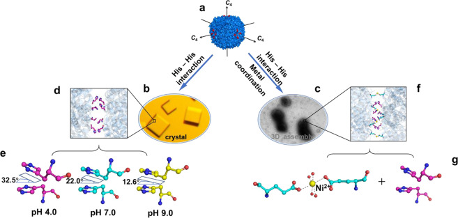
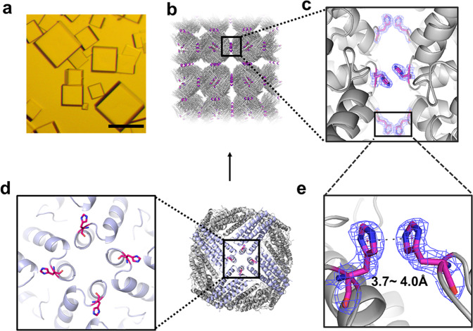
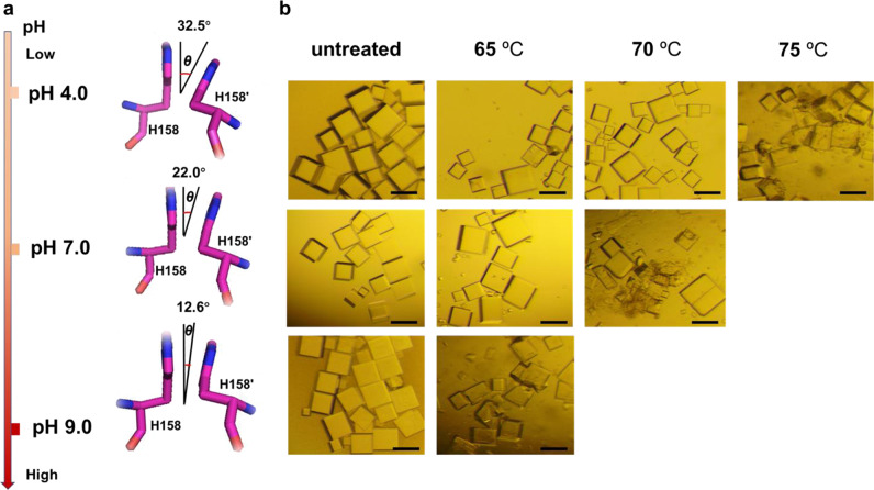
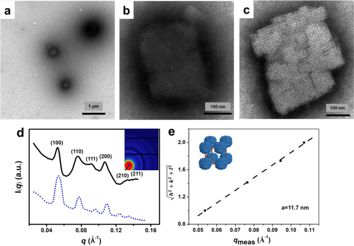
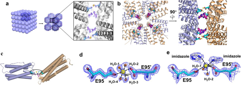
Histidine (His) residues represent versatile motifs for designing protein-protein interactions because the protonation state of the imidazole group of His is the only moiety in protein to be significantly pH dependent under physiological conditions. Here we show that, by the designed His motifs nearby the C4 axes, ferritin nanocages arrange in crystals with a simple cubic stacking pattern. The X-ray crystal structures obtained at pH 4.0, 7.0, and 9.0 in conjunction with thermostability analyses reveal the strength of the π-π interactions between two adjacent protein nanocages can be fine-tuned by pH. By using the crystal structural information as a guide, we constructed 3D protein frameworks in solution by a combination of the relatively weak His-His interaction and Ni2+-participated metal coordination with Glu residues from two adjacent protein nanocages. These findings open up a new way of organizing protein building blocks into 3D protein crystalline frameworks.


Conflict of interest statement

The authors declare no competing interests.

Figures

Fig. 1
Fig. 1. Proposed model of His-induced 3D protein crystals and 3D solid arrays in solution by combining His-His interactions and metal coordination.
a Close-up view from three C4 rotation axes of ferritin which are perpendicular to each other. Single His mutation of each ferritin subunit on the protein outer surface nearby the C4 rotation axes were highlighted in red. b Crystal diagram of ferritin induced by His–His interactions along the C4 rotation axes. c Solid 3D assemblies produced by a combination of His-His interactions and metal coordination. d Crystal structure illustration of four pairs of His–His interactions (colored magenta) between two adjacent ferritin molecules. e The action mode of His–His interactions can be adjusted by pH. f Crystal structure revealed that both His-His (colored magenta) interaction and metal coordination of Ni2+ (colored yellow) with glutamic acid (colored cyan) are responsible for the formation 3D protein arrays. g Close-up views of Ni2+ induced metal coordination and His-His interaction in f.
Fig. 2
Fig. 2. Structural basis of the His-inducible protein crystals.
a Light micrograph of cubic crystals of T158HMjFer. Scale bars represent 100 µm. b Assembly of ferritin into simple cubic (sc) packing in the crystal structure, and closeup views of the four engineered His-induced π–π stacking interactions between two adjacent protein nanocages. c Enlarged view of His-His chemistry between neighboring ferritin molecules. d The orientation of four designed His residues located on the outer surface of T158HMjFer nearby the C4 symmetry axes, which are highlighted in red. e The distance between the centers of imidazole ring of His residues pairs at pH 4.0, pH 7.0, and pH 9.0 is in the range of 3.7–4.0 Å.
Fig. 3
Fig. 3. Structure difference and crystal stability difference of T158HMjFer crystals.
a Effect of pH on the His-induced π–π stacking interactions. The orientation angle about interplanar ring (θ) between H158 and H158′ gradually decreases as pH increases. b Analyses of thermal stability of protein crystals at different pH values. Scale bars represent 200 µm.
Fig. 4
Fig. 4. Characterization of 3D T158HMjFer superlattices.
a TEM images of 3D assembly of T158HMjFer formed by a combination of π–π stacking interactions and Ni2+ coordination. b, c Enlargements of protein superlattices. d SAXS analyses of 3D T158HMjFer superlattices. The (hkl) values in radially averaged 1D SAXS data are labeled above the peaks. The experimental curve (black) matches the simulated pattern (blue dash) well, revealing a simple cubic (sc) structure. The inserted image in d is the 2D SAXS pattern of T158HMjFer assemblies. e Miller indices of assigned reflections for the sc structure versus measured q-vector positions for indexed peaks yield unit cell dimensions of a = 11.7 nm. The inserted image in e is a unit cell of the superlattice composition. TEM conditions: 1.0 µM of T158HMjFer protein in 25 mM Tris-HCl, pH 8.0 containing 500 mM NaCl, 0.7 mM of Ni2+. SAXS samples were prepared by centrifugation of 3D assemblies.
Fig. 5
Fig. 5. The crystal structure of 3D protein nanocage arrays.
a Assembly of T158HMjFer nanocages into sc packing induced by a combination of metal coordination and His-His interactions, and closeup views of π–π stacking and metal coordination. Ni2+ ions are shown as yellow spheres, histidines and glutamic acids are shown as magenta and cyan sticks, respectively. b The overlook view and side view of His-participated π–π stacking interactions and Ni2+-induced coordination between two neighboring nanocages. c Zoomed-in view of a couple of interactional subunits. d A zoomed-in view of one Ni2+ complex where Ni2+ is coordinated to four H2O and glutamic acid residues Glu95 and Glu95′ contributed from two protein nanocages, respectively. The Ni2+ ions and water molecules are shown as yellow sphere and small orange spheres, respectively. The distances of H2O-1, H2O-2, H2O-3 to Ni2+ (2.1 Å) are identical, while the distance of H2O-4 to Ni2+ is 2.4 Å and the carboxylic group of Glu to Ni2+ is 2.5 Å. e A zoomed-in view of one Ni2+ complex where two water molecule ligands are substituted by two imidazole molecules. The distance of water molecules, imidazole molecules and the carboxylic group of Glu to Ni2+ are 2.3 Å, 2.6 Å, and 2.9 Å, respectively. The coordination of Ni2+ with surrounding atoms were marked with black dash lines.

Similar articles

Cited by

References

    1. Hong S, Wang Y, Park SY, Lee H. Progressive fuzzy cation-π assembly of biological catecholamines. Sci. Adv. 2018;4:eaat7457. doi: 10.1126/sciadv.aat7457. - DOI - PMC - PubMed
    1. Nakagawa Y, Kageyama H, Oaki Y, Imai H. Direction control of oriented self-assembly for 1D, 2D, and 3D microarrays of anisotropic rectangular nanoblocks. J. Am. Chem. Soc. 2014;136:3716–3719. doi: 10.1021/ja410183q. - DOI - PubMed
    1. Evers WH, Friedrich H, Filion L, Dijkstra M, Vanmaekelbergh D. Observation of a ternary nanocrystal superlattice and its structural characterization by electron tomography. Angew. Chem. Int. Ed. 2009;48:9655–9657. doi: 10.1002/anie.200904821. - DOI - PubMed
    1. Wang HM, et al. Integrating enzymatic self-assembly and mitochondria targeting for selectively killing cancer cells without acquired drug resistance. J. Am. Chem. Soc. 2016;138:16046–16055. doi: 10.1021/jacs.6b09783. - DOI - PMC - PubMed
    1. Wang HM, Feng ZQQ, Xu B. Intercellular instructed-assembly mimics protein dynamics to Induce cell spheroids. J. Am. Chem. Soc. 2019;141:7271–7274. doi: 10.1021/jacs.9b03346. - DOI - PMC - PubMed