

Efficacy of non-enhanced computer tomography-based radiomics for predicting hematoma expansion: A meta-analysis
- PMID: 36703784
- PMCID: PMC9872032
- DOI: 10.3389/fonc.2022.973104
Efficacy of non-enhanced computer tomography-based radiomics for predicting hematoma expansion: A meta-analysis
Abstract
Background: This meta-analysis aimed to assess the efficacy of radiomics using non-enhanced computed tomography (NCCT) for predicting hematoma expansion in patients with spontaneous intracerebral hemorrhage.
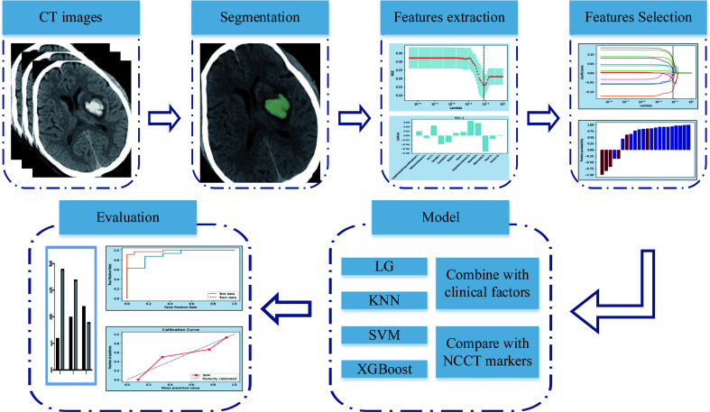
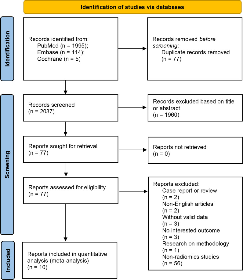
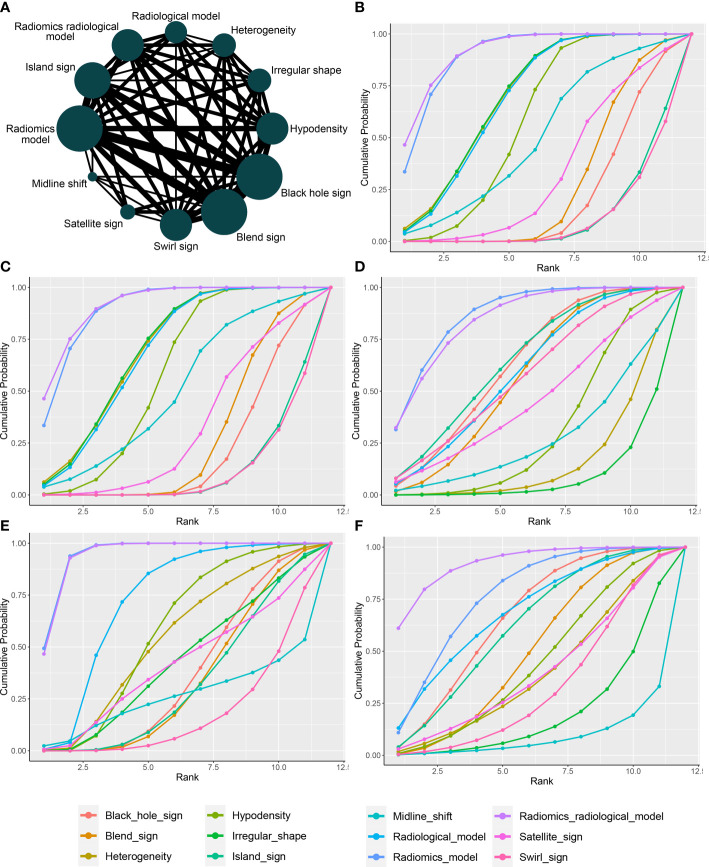
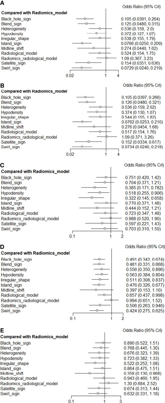
Methods: Throughout the inception of the project to April 11, 2022, a comprehensive search was conducted on PubMed, Embase, and Cochrane Central Register of Controlled Trials. The methodological quality of studies in this analysis was assessed by the radiomics quality scoring system (RQS). A meta-analysis of radiomic studies based on NCCT for predicting hematoma expansion in patients with intracerebral hemorrhage was performed. The efficacy of the radiomics approach and non-contrast CT markers was compared using network meta-analysis (NMA).
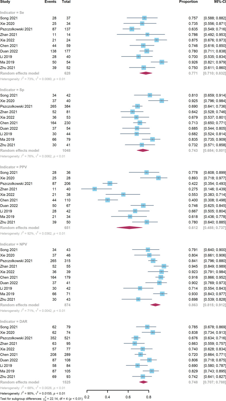
Results: Ten articles comprising a total of 1525 patients were quantitatively analyzed for hematoma expansion after cerebral hemorrhage using radiomics. Based on the included studies, the mean RQS was 14.4. The AUC value (95% confidence interval) of the radiomics model was 0.80 (0.76-0.83). Five articles comprising 846 patients were included in the NMA. The results synthesized according to Bayesian NMA revealed that the predictive ability of the radiomics model outperformed most of the NCCT biomarkers.
Conclusions: The NCCT-based radiomics approach has the potential to predict hematoma expansion. Compared to NCCT biomarkers, we recommend a radiomics approach. Standardization of the radiomics approach is required for further clinical implementation.
Systematic review registration: https://www.crd.york.ac.uk/PROSPERO/display_record.php?RecordID=324034, identifier [CRD42022324034].
Keywords: hematoma expansion; meta-analysis; non-enhanced computer tomography; radiomics; spontaneous intracerebral hemorrhage.
Copyright © 2023 Jiang, Xu, Wang and Chen.
Conflict of interest statement
The authors declare that the research was conducted in the absence of any commercial or financial relationships that could be construed as a potential conflict of interest.
Figures
Similar articles
-
Machine learning for predicting hematoma expansion in spontaneous intracerebral hemorrhage: a systematic review and meta-analysis.Neuroradiology. 2024 Sep;66(9):1603-1616. doi: 10.1007/s00234-024-03399-8. Epub 2024 Jun 12. Neuroradiology. 2024. PMID: 38862772
-
Noncontrast computer tomography-based radiomics model for predicting intracerebral hemorrhage expansion: preliminary findings and comparison with conventional radiological model.Eur Radiol. 2020 Jan;30(1):87-98. doi: 10.1007/s00330-019-06378-3. Epub 2019 Aug 5. Eur Radiol. 2020. PMID: 31385050
-
CT-based radiomics models predict spontaneous intracerebral hemorrhage expansion and are comparable with CT angiography spot sign.Front Neurol. 2024 Feb 26;15:1332509. doi: 10.3389/fneur.2024.1332509. eCollection 2024. Front Neurol. 2024. PMID: 38476195 Free PMC article.
-
Quantitative imaging for predicting hematoma expansion in intracerebral hemorrhage: A multimodel comparison.J Stroke Cerebrovasc Dis. 2024 Jul;33(7):107731. doi: 10.1016/j.jstrokecerebrovasdis.2024.107731. Epub 2024 Apr 23. J Stroke Cerebrovasc Dis. 2024. PMID: 38657831
-
Prognostic value of CT scan-based radiomics in intracerebral hemorrhage patients: A systematic review and meta-analysis.Eur J Radiol. 2024 Sep;178:111652. doi: 10.1016/j.ejrad.2024.111652. Epub 2024 Jul 26. Eur J Radiol. 2024. PMID: 39079323
Cited by
-
Machine learning for predicting hematoma expansion in spontaneous intracerebral hemorrhage: a systematic review and meta-analysis.Neuroradiology. 2024 Sep;66(9):1603-1616. doi: 10.1007/s00234-024-03399-8. Epub 2024 Jun 12. Neuroradiology. 2024. PMID: 38862772
-
The clinical potential of radiomics to predict hematoma expansion in spontaneous intracerebral hemorrhage: a narrative review.Front Neurol. 2024 Jul 19;15:1427555. doi: 10.3389/fneur.2024.1427555. eCollection 2024. Front Neurol. 2024. PMID: 39099779 Free PMC article. Review.
-
The relationship between perihematomal edema and hematoma expansion in acute spontaneous intracerebral hemorrhage: an exploratory radiomics analysis study.Front Neurosci. 2024 Apr 30;18:1394795. doi: 10.3389/fnins.2024.1394795. eCollection 2024. Front Neurosci. 2024. PMID: 38745941 Free PMC article.
References
-
- Pszczolkowski S, Manzano-Patrón JP, Law ZK, Krishnan K, Ali A, Bath PM, et al. . Quantitative CT radiomics-based models for prediction of haematoma expansion and poor functional outcome in primary intracerebral haemorrhage. Eur Radiol (2021) 31:7945–59. doi: 10.1007/s00330-021-07826-9 - DOI - PMC - PubMed
-
- Chen Q, Zhang L, Mo X, You J, Chen L, Fang J, et al. . Current status and quality of radiomic studies for predicting immunotherapy response and outcome in patients with non-small cell lung cancer: A systematic review and meta-analysis. Eur J Nucl Med Mol Imaging (2021) 49:345–60. doi: 10.1007/s00259-021-05509-7 - DOI - PubMed
Publication types
LinkOut - more resources
Full Text Sources
Miscellaneous