

Case report: Streptococcus pneumoniae pneumonia characterized by diffuse centrilobular nodules in both lungs
- PMID: 36703900
- PMCID: PMC9871572
- DOI: 10.3389/fmed.2022.1007160
Case report: Streptococcus pneumoniae pneumonia characterized by diffuse centrilobular nodules in both lungs
Abstract
Background: Streptococcus pneumoniae (S. pneumoniae) is the most common pathogen in community-acquired pneumonia (CAP) and takes the form of lobar pneumonia as typical computed tomography (CT) findings. Various patterns of radiological manifestation have also been reported in patients with S. pneumoniae pneumonia; however, the appearance of diffuse centrilobular nodules in both lungs is rarely reported.
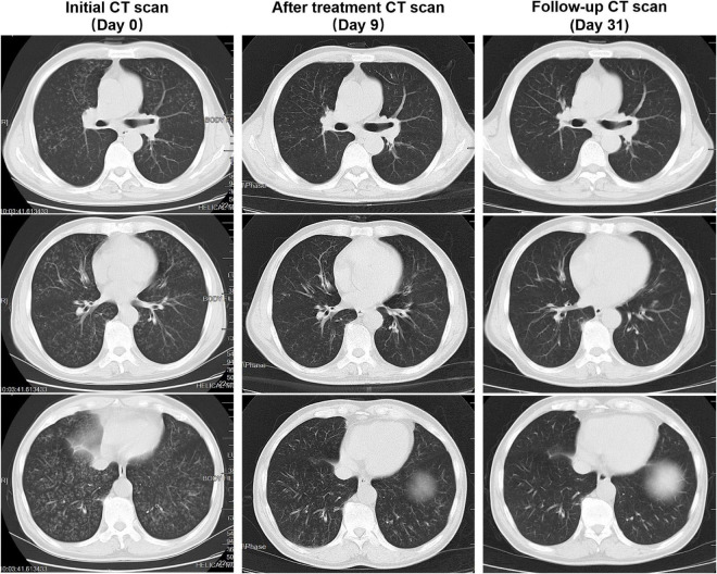
Case presentation: We report the case of a patient with a history of chronic lymphocytic leukemia (CLL) for 9 years who presented with new-onset fever, cough, excess sputum, and shortness of breath for 1 week. He was given intravenous antibacterial (cephalosporin) treatment for 4 days, but his condition did not improve and dyspnea became more serious. The chest CT indicated diffuse centrilobular nodules in both lungs at admission. Patient's bronchoalveolar (BAL) fluid was sent for metagenomic next-generation sequencing, which only supported a diagnosis of S. pneumoniae infection. His condition improved gradually after antimicrobial treatment (moxifloxacin) and a follow-up CT showed that the diffuse centrilobular nodules in both lungs were absorbed completely.
Conclusion: This case highlights a rare CT presentation of S. pneumoniae pneumonia that should alert clinicians, so as to avoid taking unnecessary treatment measures.
Keywords: Streptococcus pneumoniae; case report; centrilobular nodules; chest computed tomography; pneumonia.
Copyright © 2023 Zhang, Qin, Xia, Mao and Li.
Conflict of interest statement
The authors declare that the research was conducted in the absence of any commercial or financial relationships that could be construed as a potential conflict of interest.
Figures
Similar articles
-
Re-evaluation of the etiology and clinical and radiological features of community-acquired lobar pneumonia in adults.J Infect Chemother. 2018 Jun;24(6):463-469. doi: 10.1016/j.jiac.2018.02.001. Epub 2018 Mar 28. J Infect Chemother. 2018. PMID: 29605556
-
Severe community-acquired pneumonia caused by Chlamydia abortus in China: a case report.Front Med (Lausanne). 2024 Jul 22;11:1426577. doi: 10.3389/fmed.2024.1426577. eCollection 2024. Front Med (Lausanne). 2024. PMID: 39104862 Free PMC article.
-
[Clinical analysis of 20 cases with Streptococcus pneumoniae necrotizing pneumonia in China].Zhonghua Er Ke Za Zhi. 2012 Jun;50(6):431-4. Zhonghua Er Ke Za Zhi. 2012. PMID: 22931940 Chinese.
-
Streptococcus salivarius pneumonia-associated pneumomediastinum: a case report and literature review.BMC Infect Dis. 2024 Nov 4;24(1):1238. doi: 10.1186/s12879-024-10138-0. BMC Infect Dis. 2024. PMID: 39497050 Free PMC article. Review.
-
[Immune-related pneumonitis caused by programmed death-1 inhibitor Pembrolizumab: a case report and literature review].Zhonghua Jie He He Hu Xi Za Zhi. 2017 Oct 12;40(10):736-743. doi: 10.3760/cma.j.issn.1001-0939.2017.10.006. Zhonghua Jie He He Hu Xi Za Zhi. 2017. PMID: 29050127 Review. Chinese.
References
Publication types
LinkOut - more resources
Full Text Sources
Miscellaneous