. 2023 Jan 17:2023:7718155.
doi: 10.1155/2023/7718155. eCollection 2023.

Melatonin Repairs Osteoporotic Bone Defects in Iron-Overloaded Rats through PI3K/AKT/GSK-3 β/P70S6k Signaling Pathway

Affiliations

Melatonin Repairs Osteoporotic Bone Defects in Iron-Overloaded Rats through PI3K/AKT/GSK-3 β/P70S6k Signaling Pathway

Maoxian Ren et al. Oxid Med Cell Longev. .

Abstract

It was found recently that iron overload can cause osteoporosis in rats. Through in vitro and in vivo experimentations, the purpose of the present study was to validate and confirm the inhibitory effects of melatonin on iron death of osteoporosis and its role in bone microstructure improvements. Melatonin (100 mol/L) was administered to MC3T3-E1 cells induced by iron overload in vitro for 48 hours. The expression of cleaved caspase-3 and cleaved PARP and the production of ROS (reactive oxygen species) and mitochondrial damage were all exacerbated by iron overload. On the other hand, melatonin restored these impacts in MC3T3-E1 cells produced by iron overload. By evaluating the expression of PI3K/AKT/GSK-3β/P70S6k signaling pathway-related proteins (RUNX2, BMP2, ALP, and OCN) using RT-PCR and Western blot, osteogenic-related proteins were identified. Alizarin red S and alkaline phosphatase were utilized to evaluate the osteogenic potential of MC3T3-E1 cells. Melatonin significantly improved the osteogenic ability and phosphorylation rates of PI3K, AKT, GSK-3β, and P70S6k in iron overload-induced MC3T3-E1 cells. In vivo, melatonin treated iron overload-induced osteoporotic bone defect in rats. Rat skeletal microstructure was observed using micro-CT and bone tissue pathological section staining. ELISA was utilized to identify OCN, PINP, CTX-I, and SI in the serum of rats. We discovered that melatonin increased bone trabecular regeneration and repair in osteoporotic bone defects caused by iron overload. In conclusion, melatonin enhanced the osteogenic ability of iron overload-induced MC3T3-E1 cells by activating the PI3K/AKT/GSK-3β/P70S6k signaling pathway and promoting the healing of iron overload-induced osteoporotic bone defects in rats.


Conflict of interest statement

The authors declare that they have no conflicts of interest.

Figures

Figure 1
Figure 1
FAC induced osteoporosis in SD rats. (a) Analysis of femoral bone mineral density of SD rats in each group by micro-CT examination. (b) Comparison of BMD of SD rats in each group. (c) Detection of trabecular bone density in SD rats in each group by HE staining. (d) Adipocyte density was compared between the two groups using bone marrow samples from SD rats. N.e.Lc/B.Ar stands for number of empty lacunae per bone area; CON stands for control group; HE stands for hematoxylin-eosin staining; FAC stands for ferric ammonium citrate group; BMD stands for bone density; P < 0.05 and ∗∗∗P < 0.001.
Figure 2
Figure 2
MC3T3-E1 cells that were iron-overloaded. MT decreased ROS levels. (a) The intracellular ROS concentration of MC3T3-E1 cells in each group was discovered using an inverted fluorescent phase contrast microscope (magnification: 100 and scale bar: 250 m). (b) Using flow cytometry to measure the ROS levels in each group's MC3T3-E1 cells. Ferric ammonium citrate plus melatonin is referred to as FAC+MT; P < 0.05 and ∗∗∗P < 0.001.
Figure 3
Figure 3
Iron-overloaded MC3T3-E1 cells are more capable of osteogenic mineralization when treated with MT. (a) Alkaline phosphatase expression in MC3T3-E1 cells in each group was determined using an ALP staining. Images obtained from a gross scanner are shown on the left (scale bar: 1 mm), enlarged images are displayed in the middle (magnification: 100 and scale bar: 250 μm), and the quantitative results of the gross scanner images are displayed on the right. (b) ALP, Runx2, BMP2, and OCN mRNA expressions in MC3T3-E1 cells in each group were determined by RT-PCR. (c) Alizarin red S staining was used to identify each group's osteogenic mineralization of MC3T3-E1 cells. Images obtained from a gross scanner are shown on the left (scale bar: 1 mm), magnified images are displayed in the middle (magnification: 100 and scale bar: 250 μm), and a quantitative analysis of the left gross scanning images is displayed on the right. (d) The protein expression of OCN, ALP, Runx2, and BMP2 in MC3T3-E1 cells was determined by using Western blotting. ALP stands for alkaline phosphatase, RUNX2 for runt-related transcription factor 2, OCN for osteocalcin, and BMP2 for bone morphogenetic protein 2; P < 0.05 and ∗∗∗P < 0.001.
Figure 4
Figure 4
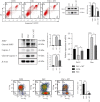
MT decreased the rate of apoptosis in MC3T3-E1 cells that were iron-overloaded. (a) The Annexin V-FITC/PI double labelling technique was used to determine the MC3T3-E1 cells' apoptosis rate in each group. (b) Bax and Bcl2 protein expressions were found using a Western blot analysis. (c) Caspase-3, cleaved PARP, PARP, cleaved caspase-3, and β-actin marker expressions were found using a Western blotting in MC3T3-E1 cells between groups. (d) We compared the levels of Bcl-2 and Bax mRNA expressions in MC3T3-E1 cells across treatments using RT-PCR. (e) Each group's MC3T3-E1 cells had their mitochondrial membrane potential measured using flow cytometry. Bcl-2 stands for B-cell lymphoma-2, PARP stands for poly ADP-ribose polymerase, and cleaved PARP stands for cleaved poly ADP-ribose polymerase; P < 0.05 and ∗∗∗P < 0.001.
Figure 5
Figure 5
Showing how MT uses the PI3K/AKT/GSK-3β/P70S6k signaling pathway to treat iron-overloaded MC3T3-E1 cells. (a) All mRNA expression levels in MC3T3-E1 cells were determined by using RT-PCR for p-PI3K, p-GSK-3β, p-AKT, and p-P70S6k. (b) Western blotting was used to analyze the expression of PI3K, AKT, GSK-3β, P70S6k, p-PI3K, p-AKT, p-GSK-3β, p-P70S6k, and β-actin proteins in MC3T3-E1 cells across treatment groups. MT stands for melatonin group; FAC+MT+LY294002 stands for the ferric ammonium citrate+melatonin+LY294002 group; PI3K stands for phosphatidylinositol kinase; p-PI3K stands for phosphorylated phosphatidylinositol kinase; AKT stands for protein kinase B; p-AKT stands for phosphorylated protein kinase B; p-GSK-3β stands for phosphorylated glycogen synthase kinase-3β; GSK-3β stands for glycogen synthase kinase-3β; P70S6 stands for ribosomal S6 protein kinase; p-P70S6k stands for phosphorylated ribosomal S6 protein kinase; P < 0.05 and ∗∗∗P < 0.001.
Figure 6
Figure 6
In iron-overloaded SD mice with osteoporotic bone abnormalities, melatonin stimulates trabecular bone repair while decreasing bone marrow adipocyte density. (a) Chemical labeling of nascent bone trabeculae and adipocytes in the bone marrow of SD rats. (b) Rats were divided into two groups, and the pace at which new bone formed in the defect location was compared using the standard deviation. (c) Dissecting the differences between normal and SD rats' bone marrow adipocyte densities in relation to the abnormality's location. Masson staining; N.e.Lc/B.d.Ar, the ratio of the number of empty lacunae to the total bone defect area; significance level for the amount of new bone trabeculae (P < 0.05 and ∗∗∗P < 0.001). TB = trabecular bone; BM = bone marrow cavity.
Figure 7
Figure 7
In rats with iron overload and osteoporotic bone abnormalities, melatonin promotes bone healing. (a) By using micro-CT imaging, the bone defect area of SD rats in each group was examined. (b) Comparison of BV/TV, BMD, Tb.Th, Tb.N, and Tb.Sp in each group in the location of the femoral defect in SD rats. (c) Comparison of each group's SD rat's serum levels of SI, PINP, OCN, and CTX-I. 3DR stands for three-dimensional reconstruction; 2D stands for two dimensions; BV/TV stands for bone volume/total mass; Tb.Th stands for trabecular thickness, Tb.N for trabeculae number, and Tb.Sp for trabecular space. SI stands for serum iron; PINP stands for type I procollagen N-terminal propeptide; CTX-I stands for type I collagen C-terminal peptide and OCN for osteocalcin; P < 0.05 and ∗∗∗P < 0.001.

Similar articles

Cited by

References

    1. Consensus development conference: diagnosis, prophylaxis, and treatment of osteoporosis. The American Journal of Medicine . 1993;94(6):646–650. doi: 10.1016/0002-9343(93)90218-E. - DOI - PubMed
    1. NIH Consensus Development Panel on Osteoporosis Prevention, Diagnosis, and Therapy. Osteoporosis prevention, diagnosis, and therapy. JAMA: The Journal of the American Medical Association . 2001;285(6):785–795. doi: 10.1001/jama.285.6.785. - DOI - PubMed
    1. Bliuc D., Nguyen N. D., Alarkawi D., Nguyen T. V., Eisman J. A., Center J. R. Accelerated bone loss and increased post-fracture mortality in elderly women and men. Osteoporosis International . 2015;26(4):1331–1339. doi: 10.1007/s00198-014-3014-9. - DOI - PubMed
    1. Cauley J. A. Public health impact of osteoporosis. The Journals of Gerontology Series A: Biological Sciences and Medical Sciences . 2013;68(10):1243–1251. doi: 10.1093/gerona/glt093. - DOI - PMC - PubMed
    1. Adachi J. D., Ioannidis G., Pickard L., et al. The association between osteoporotic fractures and health-related quality of life as measured by the health utilities index in the Canadian multicentre osteoporosis study (CaMos) Osteoporosis International . 2003;14(11):895–904. doi: 10.1007/s00198-003-1483-3. - DOI - PubMed

MeSH terms