. 2023 Jan 10:13:752900.
doi: 10.3389/fphys.2022.752900. eCollection 2022.

Habituation of the stress response multiplex to repeated cold pressor exposure

Affiliations

Habituation of the stress response multiplex to repeated cold pressor exposure

Tom Bullock et al. Front Physiol. .

Abstract

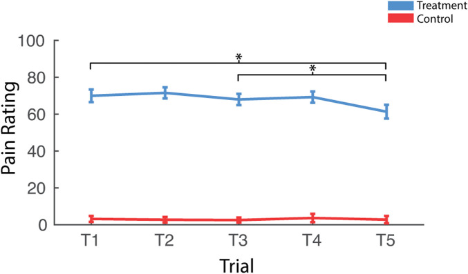
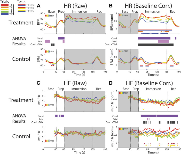
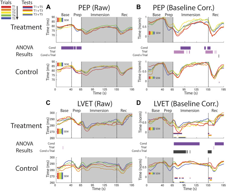
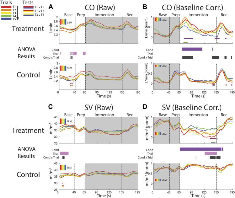
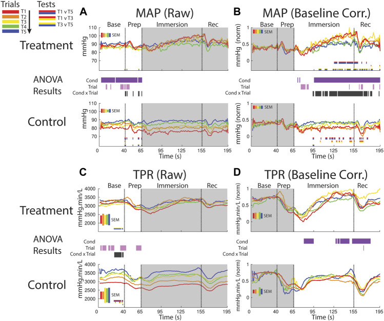
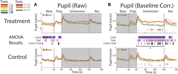
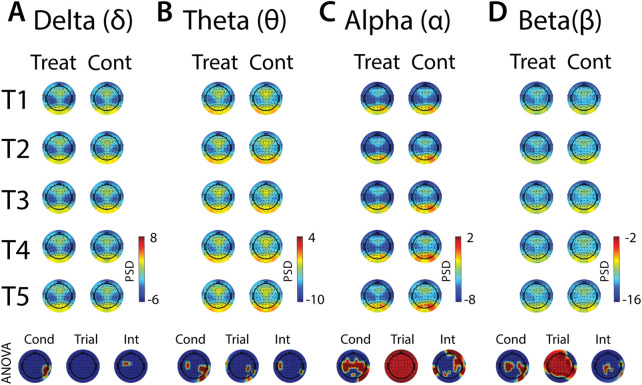
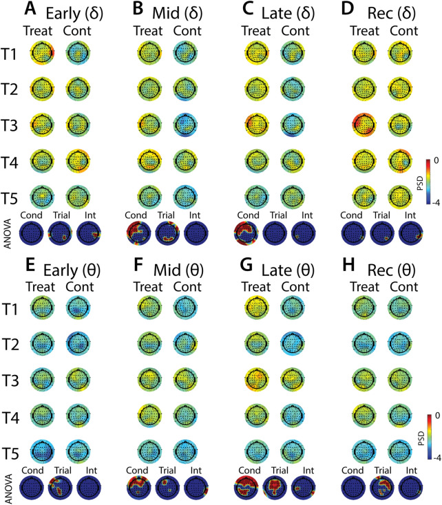
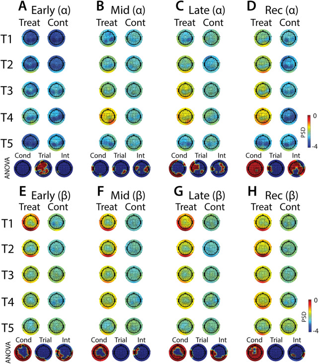
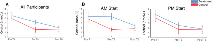
Humans show remarkable habituation to aversive events as reflected by changes of both subjective report and objective measures of stress. Although much experimental human research focuses on the effects of stress, relatively little is known about the cascade of physiological and neural responses that contribute to stress habituation. The cold pressor test (CPT) is a common method for inducing acute stress in human participants in the laboratory; however, there are gaps in our understanding of the global state changes resulting from this stress-induction technique and how these responses change over multiple exposures. Here, we measure the stress response to repeated CPT exposures using an extensive suite of physiologic measures and state-of-the-art analysis techniques. In two separate sessions on different days, participants underwent five 90 s CPT exposures of both feet and five warm water control exposures, while electrocardiography (ECG), impedance cardiography, continuous blood pressure, pupillometry, scalp electroencephalography (EEG), salivary cortisol and self-reported pain assessments were recorded. A diverse array of adaptive responses are reported that vary in their temporal dynamics within each exposure as well as habituation across repeated exposures. During cold-water exposure there was a cascade of changes across several cardiovascular measures (elevated heart rate (HR), cardiac output (CO) and Mean Arterial Pressure (MAP) and reduced left ventricular ejection time (LVET), stroke volume (SV) and high-frequency heart rate variability (HF)). Increased pupil dilation was observed, as was increased power in low-frequency bands (delta and theta) across frontal EEG electrode sites. Several cardiovascular measures also habituated over repeated cold-water exposures (HR, MAP, CO, SV, LVET) as did pupil dilation and alpha frequency activity across the scalp. Anticipation of cold water induced stress effects in the time-period immediately prior to exposure, indexed by increased pupil size and cortical disinhibition in the alpha and beta frequency bands across central scalp sites. These results provide comprehensive insight into the evolution of a diverse array of stress responses to an acute noxious stressor, and how these responses adaptively contribute to stress habituation.

Keywords: EEG; adaptation; cardiac physiology; cold pressor; habituation; pupillometry; repeated exposure; stress.


Conflict of interest statement

The authors declare that the research was conducted in the absence of any commercial or financial relationships that could be construed as a potential conflict of interest.

Figures

FIGURE 1
FIGURE 1
Pressor protocol. (A) An example of a fully instrumented participant in the treatment condition. (B) The standard protocol for each CPT or WPT trial. Each protocol consisted of a pre-immersion baseline recording (40 s) followed by a preparatory period (25 s) where the participant was required to position their feet on the edge of the water bucket in preparation for immersion. The participant then lowered their feet into the water (90 s) and removed them from the water for the recovery period (40 s). They then provided a pain rating. (C) Schematic of the complete experiment protocol. Participants completed five CPT exposures on the Treatment Day or five WPT exposures on the Control Day (T1—T5). The time between the onset of each exposure was ∼25 min. Saliva samples were collected prior to T1 and T3 and immediately after T5.
FIGURE 2
FIGURE 2
PEP and LVET measures extracted from ICG and ECG waveforms. The ICG waveform (green line) is shown overlapping the ECG waveform (blue line). The ICG B and X points represent the open and closing of the aortic valve, respectively. The Q point of the ECG represents the initial ventricular depolarization. The time interval between Q and B represents PEP, the sum of electromechanical delay and isovolumic contraction of the ventricle. LVET is the time interval between B and X.
FIGURE 3
FIGURE 3
Effects of treatment and repeated trial exposure on self-reported pain. Participants were asked to provide pain ratings at the termination of each treatment or control trial. *p null<0.05. Error bars = ± SEM.
FIGURE 4
FIGURE 4
Effects of treatment and repeated trial exposure on heartbeat rate [HR; (A,B)] and high frequency HR variability [HF; (C,D)]. Pre-exposure and evoked stress effects are reflected in raw (left column) and baseline corrected/normalized (right column) plots, respectively. Each panel contains a separate plot for the treatment and control condition, with the mean response across participants in each repetition trial (T1-T5) represented in each plot by a different color (ROYGB). Horizontal lines positioned between treatment and control plots indicate statistically significant ANOVA main effects of condition (dark purple), trial (light purple) and the condition x trial interaction (dark gray) at designated timepoints (p null<0.05). Horizontal red/blue, red/yellow and yellow/blue lines at the base of treatment and control plots respectively indicate statistically significant pairwise comparison results for T1 v T5, T1 v T3 and T3 v T5 at the designated timepoints (p null<0.05). Post-immersion raw data and pre-immersion baseline corrected normalized data are greyed out as these periods are not relevant to the pre-exposure/evoked stress analyses. The set of five vertical error bars labelled SEM on each plot represent ±SEM of the data averaged over the time-course of interest.
FIGURE 5
FIGURE 5
Effects of treatment and repeated trial exposure on sympathetic drive [PEP; (A,B)] and cardiac timing [LVET; (C,D)]. Pre-exposure and evoked stress effects are reflected in raw (left column) and baseline corrected/normalized (right column) plots, respectively. Each panel contains a separate plot for the treatment and control condition, with the mean response across participants in each repetition trial (T1-T5) represented in each plot by a different color (ROYGB). Horizontal lines positioned between treatment and control plots indicate statistically significant ANOVA main effects of condition (dark purple), trial (light purple) and the condition x trial interaction (dark gray) at designated timepoints (p null<0.05). Horizontal red/blue, red/yellow and yellow/blue lines at the base of treatment and control plots respectively indicate statistically significant pairwise comparison results for T1 v T5, T1 v T3 and T3 v T5 at the designated timepoints (p null<0.05). Post-immersion raw data and pre-immersion baseline corrected normalized data are greyed out as these periods are not relevant to the pre-exposure/evoked stress analyses. The set of five vertical error bars labelled SEM on each plot represent ±SEM of the data averaged over the time-course of interest.
FIGURE 6
FIGURE 6
Effects of treatment and repeated trial exposure on cardiac output [CO; (A,B)] and stroke volume [SV; (C,D)]. Pre-exposure and evoked stress effects are reflected in raw (left column) and baseline corrected/normalized (right column) plots, respectively. Each panel contains a separate plot for the treatment and control condition, with the mean response across participants in each repetition trial (T1-T5) represented in each plot by a different color (ROYGB). Horizontal lines positioned between treatment and control plots indicate statistically significant ANOVA main effects of condition (dark purple), trial (light purple) and the condition x trial interaction (dark gray) at designated timepoints (p null<0.05). Horizontal red/blue, red/yellow and yellow/blue lines at the base of treatment and control plots respectively indicate statistically significant pairwise comparison results for T1 v T5, T1 v T3 and T3 v T5 at the designated timepoints (p null<0.05). Post-immersion raw data and pre-immersion baseline corrected normalized data are greyed out as these periods are not relevant to the pre-exposure/evoked stress analyses. The set of five vertical error bars labelled SEM on each plot represent ±SEM of the data averaged over the time-course of interest.
FIGURE 7
FIGURE 7
Effects of treatment and repeated trial exposure on mean arterial pressure [MAP; (A,B)] and total peripheral resistance [TPR; (C,D)]. Pre-exposure and evoked stress effects are reflected in raw (left column) and baseline corrected/normalized (right column) plots, respectively. Each panel contains a separate plot for the treatment and control condition, with the mean response across participants in each repetition trial (T1-T5) represented in each plot by a different color (ROYGB). Horizontal lines positioned between treatment and control plots indicate statistically significant ANOVA main effects of condition (dark purple), trial (light purple) and the condition x trial interaction (dark gray) at designated timepoints (p null<0.05). Horizontal red/blue, red/yellow and yellow/blue lines at the base of treatment and control plots respectively indicate statistically significant pairwise comparison results for T1 v T5, T1 v T3 and T3 v T5 at the designated timepoints (p null<0.05). Post-immersion raw data and pre-immersion baseline corrected normalized data are greyed out as these periods are not relevant to the pre-exposure/evoked stress analyses. The set of five vertical error bars labelled SEM on each plot represent ±SEM of the data averaged over the time-course of interest.
FIGURE 8
FIGURE 8
Effects of treatment and repeated trial exposure on pupil size. Pre-exposure and evoked stress effects are reflected in (A) raw and (B) baseline corrected/normalized plots, respectively. Each panel contains a separate plot for the treatment and control condition, with the mean response across participants in each repetition trial (T1-T5) represented in each plot by a different color (ROYGB). Horizontal lines positioned between treatment and control plots indicate statistically significant ANOVA main effects of condition (dark purple), trial (light purple) and the condition x trial interaction (dark gray) at designated timepoints (p null<0.05). Horizontal red/blue, red/yellow and yellow/blue lines at the base of treatment and control plots respectively indicate statistically significant pairwise comparison results for T1 v T5, T1 v T3 and T3 v T5 at the designated timepoints (p null<0.05). Post-immersion raw data and pre-immersion baseline corrected normalized data are greyed out as these periods are not relevant to the pre-exposure/evoked stress analyses. The set of five vertical error bars labelled SEM on each plot represent ±SEM of the data averaged over the time-course of interest. * Due to technical issues with eye-tracking, this pupillometry analysis was underpowered relative to analyses of the other measures, so the results of the timepoint-by-timepoint analysis are not definitive. Accordingly, the data were averaged across the immersion time period and submitted to an additional ANOVA, revealing a robust trial x condition interaction, driven by early-session habituation.
FIGURE 9
FIGURE 9
Effects of treatment and repeated trial exposure on anticipatory neural activity. Topographic plots in each panel (AD) depict pre-exposure oscillatory neural activity in the delta, theta, alpha and beta frequency bands, respectively. Each panel contains separate columns of plots for the treatment and control conditions, with each repetition trial (T1-T5) represented by each row. Each plot depicts brain activity averaged across the baseline period. Condition (treatment, control) by trial (T1-T5) ANOVA results at each electrode are plotted at the base of each panel, with condition, trial and interaction effects depicted in separate topographic plots. Scalp regions overlaid in red indicate significant effects (p null<0.05).
FIGURE 10
FIGURE 10
Effects of treatment and repeated trial exposure on stress-evoked oscillatory neural activity in delta and theta frequency bands. Topographic plots in each panel depict evoked stress effects on oscillatory neural activity in the delta (AD) and theta (EH) frequency bands. Topographic plots in panels AD and EH respectively represent brain activity averaged across the early (65–94 s), mid (95–124 s), late (125–154 s) and recovery (155–190 s) periods of the immersion period. Each panel contains separate columns of plots for the treatment and control conditions, with each repetition trial (T1-T5) represented by each row. Condition (treatment, control) by trial (T1-T5) ANOVA results at each electrode are plotted at the base of each panel, with condition, trial and interaction effects depicted in separate topographic plots. Scalp regions overlaid in red indicate significant effects (p null<0.05).
FIGURE 11
FIGURE 11
Effects of treatment and repeated trial exposure on stress-evoked oscillatory neural activity in alpha and beta frequency bands. Topographic plots in each panel depict evoked stress effects on oscillatory neural activity in the alpha (AD) and beta (EH) frequency bands. Topographic plots in panels AD and EH respectively represent brain activity averaged across the early (65–94 s), mid (95–124 s), late (125–154 s) and recovery (155–190 s) periods of the immersion period. Each panel contains separate columns of plots for the treatment and control conditions, with each repetition trial (T1-T5) represented by each row. Condition (treatment, control) by trial (T1-T5) ANOVA results at each electrode are plotted at the base of each panel, with condition, trial and interaction effects depicted in separate topographic plots. Scalp regions overlaid in red indicate significant effects (p null<0.05).
FIGURE 12
FIGURE 12
Cortisol. Saliva samples were acquired prior to the first and third pressor tests and at the end of the session. Plots depict cortisol levels averaged across all participants (A) and split by participant start time (B). Error bars represent ±SEM.

Similar articles

Cited by

References

    1. Al Maskati H. A., Zbrozyna A. W. (1989). Stimulation in prefrontal cortex area inhibits cardiovascular and motor components of the defence reaction in rats. J. Auton. Nerv. Syst. 28, 117–125. 10.1016/0165-1838(89)90084-2 - DOI - PubMed
    1. Al’Absi M., Petersen K. L., Wittmers L. E. (2002). Adrenocortical and hemodynamic predictors of pain perception in men and women. Pain 96, 197–204. 10.1016/S0304-3959(01)00447-X - DOI - PubMed
    1. Alvarez R. P., Chen G., Bodurka J., Kaplan R., Grillon C. (2011). Phasic and sustained fear in humans elicits distinct patterns of brain activity. Neuroimage 55, 389–400. 10.1016/j.neuroimage.2010.11.057 - DOI - PMC - PubMed
    1. Bachmann P., Zhang X., Larra M. F., Rebeck D., Schönbein K., Koch K. P., et al. (2018). Validation of an automated bilateral feet cold pressor test. Int. J. Psychophysiol. 124, 62–70. 10.1016/j.ijpsycho.2017.10.013 - DOI - PubMed
    1. Backonja M., Howland E. W., Wang J., Smith J., Salinsky M., Cleeland C. S. (1991). Tonic changes in alpha power during immersion of the hand in cold water. Electroencephalogr. Clin. Neurophysiol. 79, 192–203. 10.1016/0013-4694(91)90137-S - DOI - PubMed