. 2023 Jan 10:13:1023553.
doi: 10.3389/fimmu.2022.1023553. eCollection 2022.

Alpha-1 antitrypsin limits neutrophil extracellular trap disruption of airway epithelial barrier function

Affiliations

Alpha-1 antitrypsin limits neutrophil extracellular trap disruption of airway epithelial barrier function

K M Hudock et al. Front Immunol. .

Abstract

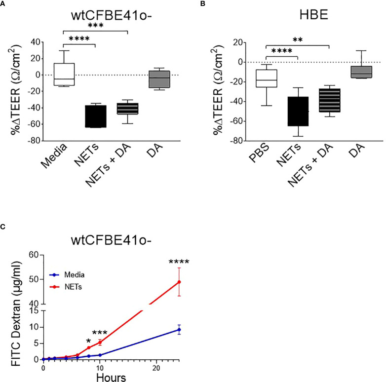
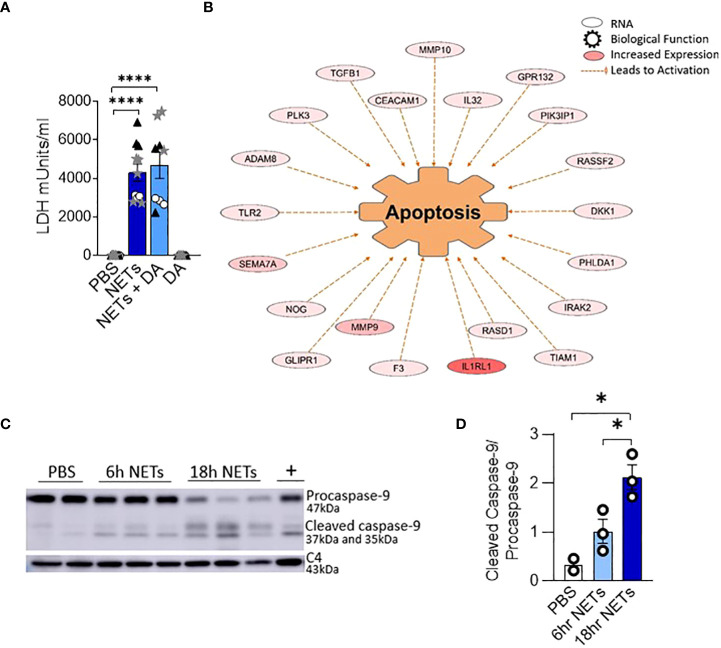
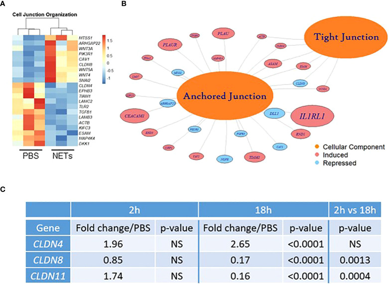
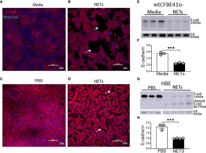
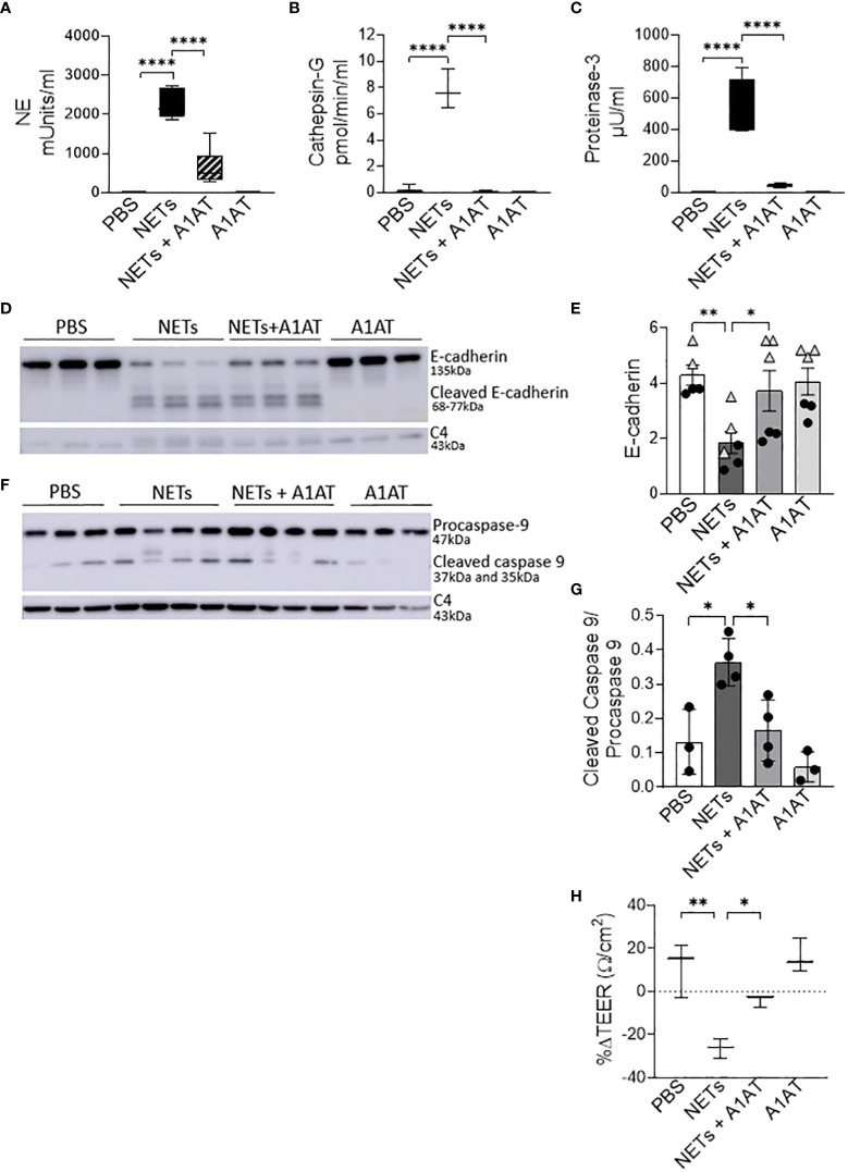
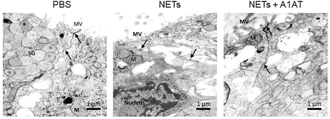
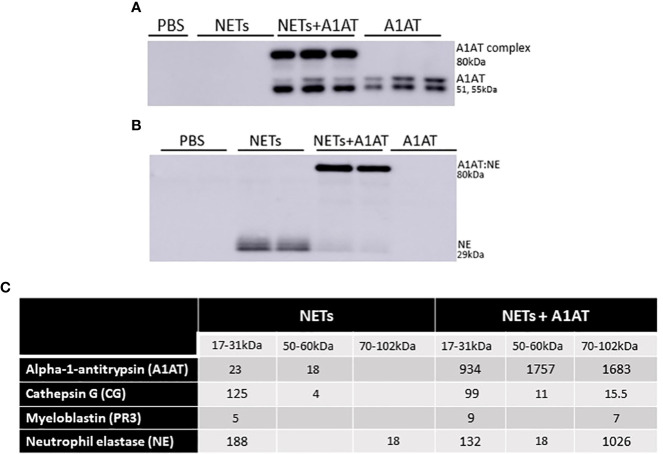
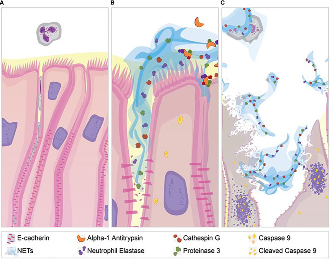
Neutrophil extracellular traps contribute to lung injury in cystic fibrosis and asthma, but the mechanisms are poorly understood. We sought to understand the impact of human NETs on barrier function in primary human bronchial epithelial and a human airway epithelial cell line. We demonstrate that NETs disrupt airway epithelial barrier function by decreasing transepithelial electrical resistance and increasing paracellular flux, partially by NET-induced airway cell apoptosis. NETs selectively impact the expression of tight junction genes claudins 4, 8 and 11. Bronchial epithelia exposed to NETs demonstrate visible gaps in E-cadherin staining, a decrease in full-length E-cadherin protein and the appearance of cleaved E-cadherin peptides. Pretreatment of NETs with alpha-1 antitrypsin (A1AT) inhibits NET serine protease activity, limits E-cadherin cleavage, decreases bronchial cell apoptosis and preserves epithelial integrity. In conclusion, NETs disrupt human airway epithelial barrier function through bronchial cell death and degradation of E-cadherin, which are limited by exogenous A1AT.

Keywords: E-cadherin (CDH1); NETs (neutrophil extracellular traps); alpha-1 antitrypsin (A1AT); barrier function; bronchial epithelia.


Conflict of interest statement

The authors declare that the research was conducted in the absence of any commercial or financial relationships that could be construed as a potential conflict of interest.

Figures

Figure 1
Figure 1
Neutrophil extracellular traps (NETs) disrupt epithelial barrier function. (A) wtCFBE41o- grown to confluence and (B) primary normal human epithelia (HBE) grown at air-liquid interface (ALI) were exposed to media or PBS control (respectively), 5µg/ml NETs, NETs+0.5µg/ml dornase alpha (DA), or DA in the apical compartment for 18h in triplicate wells. Transepithelial electrical resistance (TEER) was measured pre and post exposure in duplicate per well and expressed as a percent change. Data analyzed by one-way ANOVA followed by Bonferroni’s multiple comparisons test. (experiments=3, HBE donors=3, NET donors=4). (C) Apical to basolateral flux of FITC labeled dextran across wtCFBE41o- exposed to media control or NETs for 0-24h. Data analyzed by Least Squares Means, F-test=264.58, ****p<0.0001 from 8-24 hours (experiments=2, NET donors=2). *p<0.05, **p<0.01, ***p<0.001, ****p<0.0001.
Figure 2
Figure 2
NETs activate apoptosis in normal human bronchial epithelia. HBE were grown at ALI and exposed to PBS control, 5µg/ml NETs, NETs+0.5µg/ml dornase alpha (DA), or DA in the apical compartment for 18h in triplicate wells. (A) LDH concentration measured in apical supernatant. Data analyzed by one-way ANOVA followed by Bonferroni’s multiple comparisons test (experiments=2, HBE donors=2, NET donors=2). (B) Differential gene expression assessed by RNA sequencing in HBE after exposure to 5µg/ml NETs compared to PBS. Network representation of impacted genes predicted to activate apoptosis. Color intensity indicates increased expression. Data analyzed using Ingenuity Pathway Analysis (IPA) (p=1.07e-13, experiments=3, HBE donors=3, NET donors=3). (C, D) Representative western blot and analysis demonstrate decreased procaspase-9 and increased cleaved caspase-9, indicating apoptosis activation in HBE exposed to NETs at 6h and 18h compared to PBS. Positive control (+) 16HBE cells treated with staurosporine (5µM, 2h). Data analyzed by one-way ANOVA followed by Bonferroni’s multiple comparisons test (experiments=3, HBE donors=3, NET donors=4). *p < 0.05, ****p < 0.0001.
Figure 3
Figure 3
NETs alter bronchial epithelial cell junction genes. HBE grown at ALI exposed to PBS control or 5µg/ml NETs in the apical compartment in triplicate wells. (A) Differentially expressed genes involved in cell junction organization visualized in a z-scored normalized heat map of fragments per kilobase of transcript per million mapped reads (FPKM) demonstrate significant changes in HBE exposed to NETs for 18h. (B) Functionally enriched RNAs for cellular components of anchored junctions and tight junctions in HBE impacted by NET exposure for 18h, analyzed using ToppGene (experiments=3, HBE donors=3, NET donors=3). (C) Changes in junctional RNAs after 2 or 18h exposure to NETs were confirmed by RT-qPCR and normalized to 18S. Data analyzed by unpaired t-test (NS, not significant; experiments=3, HBE donors=4, NET donors=3).
Figure 4
Figure 4
NETs disrupt epithelia monolayers and decrease E-cadherin. Confocal microscopy images (20x) of human epithelia stained for E-cadherin/AF647 and DNA/Hoechst. (A) Confluent wtCFBE41o- exposed to media or (B) 15µg/ml NETs for 18h, the latter with visible holes (arrows) in the monolayer. (C) HBE grown at ALI exposed to PBS or (D) 5μg/ml NETs for 18h, the latter with disruptions (arrows) in the monolayer. (E) Representative western blot and (F) analysis of E-cadherin protein from lysates of wtCFBE41o- exposed to PBS or 15µg/ml NETs for 18h. (G) Representative western blot and (H) analysis of E-cadherin protein from lysates of HBE exposed to PBS or 5µg/ml NETs for 18h. E-cadherin was normalized to C4-actin. Data analyzed by unpaired t-test (experiments=3, HBE donors=3, NET donors=3). ***p<0.001.
Figure 5
Figure 5
Alpha-1 antitrypsin reduces NET-driven barrier dysfunction. HBE were exposed to PBS, 5µg/ml NETs, NETs+A1AT or A1AT for 18h in triplicate wells. (A–C) Activity of neutrophil elastase (NE), cathepsin G (CG), and proteinase 3 (PR3) were reduced in the apical supernatants of HBE exposed to NETs+A1AT. Data analyzed by one-way ANOVA followed by Bonferroni’s multiple comparisons test (experiments=3, HBE donors=4, NET donor=2). (D, E) Representative western blot and analysis of E-cadherin protein from lysates of HBE demonstrates that NETs decrease full-length E-cadherin and increases cleaved E-cadherin peptides compared to PBS control. A1AT limits NET-driven reductions in full-length E-cadherin protein. (F, G) Representative western blot and analysis demonstrate decreased cleaved caspase 9 (less activation of apoptosis) in HBE exposed to NETs with A1AT. Western blots were normalized to C4-actin and analyzed by one-way ANOVA followed by Bonferroni’s multiple comparisons test (experiments=2, HBE donors=2, NET donors=3). (H) A1AT maintained HBE TEER with exposure to NETs (representative graph). Data analyzed by one-way ANOVA followed by Bonferroni’s multiple comparisons test (experiments=3, HBE donors=3, NETs=3). *p < 0.05, **p < 0.01, ****p < 0.0001.
Figure 6
Figure 6
Alpha-1 antitrypsin preserves epithelial cell junction integrity disrupted by NETs. Representative images from transmission electron microscopy of HBE exposed to PBS, 5µg/ml NETs, NETs + A1AT or A1AT for 18 h. NET exposure resulted in partial disruption of epithelial tight junctions. A1AT helped maintain tight junctions in HBE exposed to NETs. (M=mitochondria, MV=microvilli, SG=secretory granules in a goblet cell, (→) =cell junction, experiments=1, HBE donors=1, NET donors=1).
Figure 7
Figure 7
Alpha-1 antitrypsin binds serine proteases in NET exposed HBE. HBE grown at ALI were exposed to PBS, 5µg/ml NETs, NETs + A1AT or A1AT for 18h in triplicate wells. (A) Western blot of HBE apical supernatants probed with A1AT antibody demonstrate the colocalization of A1AT (51-55kDa) with a 29kDa protein in NETs. (B) Western blot of HBE apical supernatants probed with NE antibody demonstrate the colocalization of A1AT with NE (29kDa) in NETs forming an 80kDa complex. (C) Representative mass spectrometry results from bands shown on western blot in 7A & B confirm that HBE exposed to NETs have peptide spectral matches (PSMs) that correspond to CG, PR3, and NE at the expected 17-31kDa sizes. HBE exposed to NETs + A1AT demonstrate PSMs corresponding to NE, CG, PR3 and A1AT at 70-102kDa indicating formation of a A1AT:NE, A1AT:CG and A1AT:PR3 complexes (mass spec run=3, HBE donors=2, NET donors=3).
Figure 8
Figure 8
NET effect on bronchial epithelial barrier function. (A) Pseudostratified bronchial epithelia form a monolayer with cell-cell contact via junctional proteins. This epithelial barrier tightly controls the paracellular flux of molecules and cells. (B) Neutrophils expel NETs, which contain biologically active proteases that degrade adherens junction protein E-cadherin creating gaps in the monolayer. A1AT limits NET degradation of E-cadherin to help restore barrier function. (C) NETs also disrupt barrier function by causing epithelial cell death via apoptosis, which is limited by A1AT.

Similar articles

Cited by

References

    1. Young RL, Malcolm KC, Kret JE, Caceres SM, Poch KR, Nichols DP, et al. . Neutrophil extracellular trap (NET)-mediated killing of pseudomonas aeruginosa: evidence of acquired resistance within the CF airway, independent of CFTR. PloS One (2011) 6(9):e23637. doi: 10.1371/journal.pone.0023637 - DOI - PMC - PubMed
    1. Veras FP, Pontelli MC, Silva CM, Toller-Kawahisa JE, de Lima M, Nascimento DC, et al. . SARS-CoV-2-triggered neutrophil extracellular traps mediate COVID-19 pathology. J Exp Med (2020) 217(12):e20201129. doi: 10.1084/jem.20201129 - DOI - PMC - PubMed
    1. Keshari RS, Jyoti A, Kumar S, Dubey M, Verma A, Srinag BS, et al. . Neutrophil extracellular traps contain mitochondrial as well as nuclear DNA and exhibit inflammatory potential. Cytom Part A J Int Soc Anal Cytol (2012) 81(3):238–47. doi: 10.1002/cyto.a.21178 - DOI - PubMed
    1. Kenny EF, Herzig A, Kruger R, Muth A, Mondal S, Thompson PR, et al. . Diverse stimuli engage different neutrophil extracellular trap pathways. eLife (2017) 6:2–21. doi: 10.7554/eLife.24437 - DOI - PMC - PubMed
    1. McDonald B, Urrutia R, Yipp BG, Jenne CN, Kubes P. Intravascular neutrophil extracellular traps capture bacteria from the bloodstream during sepsis. Cell Host Microbe (2012) 12(3):324–33. doi: 10.1016/j.chom.2012.06.011 - DOI - PubMed

Publication types