

Applications of spatially resolved omics in the field of endocrine tumors
- PMID: 36704039
- PMCID: PMC9873308
- DOI: 10.3389/fendo.2022.993081
Applications of spatially resolved omics in the field of endocrine tumors
Abstract
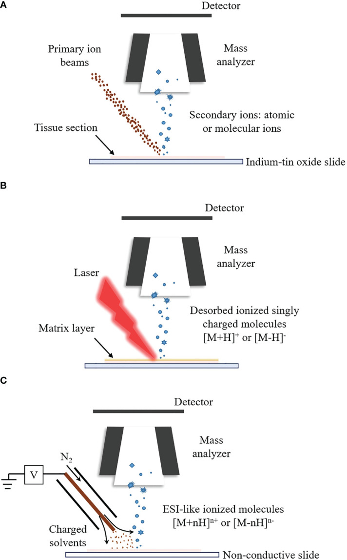
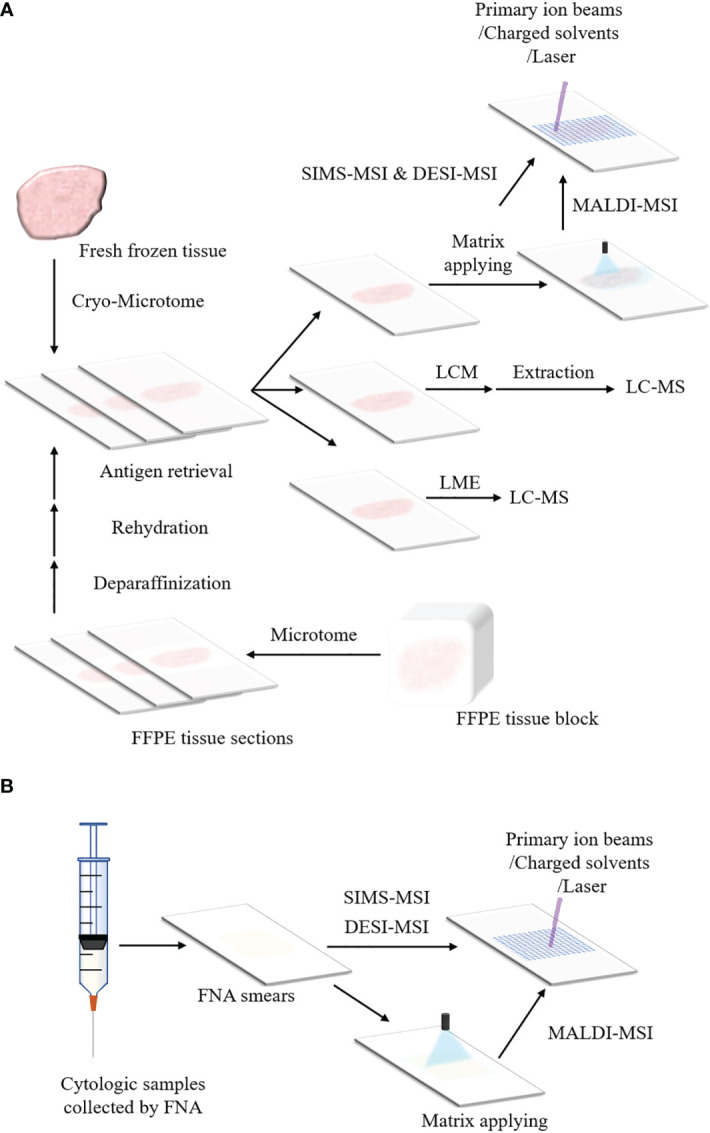
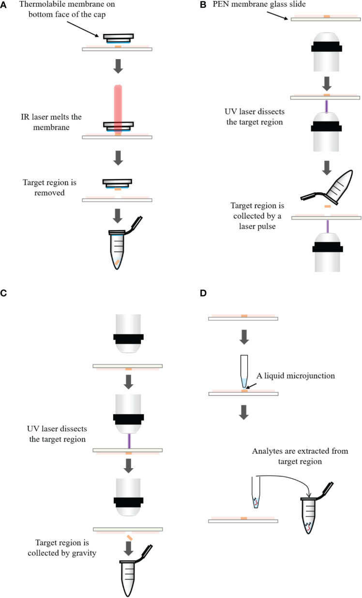
Endocrine tumors derive from endocrine cells with high heterogeneity in function, structure and embryology, and are characteristic of a marked diversity and tissue heterogeneity. There are still challenges in analyzing the molecular alternations within the heterogeneous microenvironment for endocrine tumors. Recently, several proteomic, lipidomic and metabolomic platforms have been applied to the analysis of endocrine tumors to explore the cellular and molecular mechanisms of tumor genesis, progression and metastasis. In this review, we provide a comprehensive overview of spatially resolved proteomics, lipidomics and metabolomics guided by mass spectrometry imaging and spatially resolved microproteomics directed by microextraction and tandem mass spectrometry. In this regard, we will discuss different mass spectrometry imaging techniques, including secondary ion mass spectrometry, matrix-assisted laser desorption/ionization and desorption electrospray ionization. Additionally, we will highlight microextraction approaches such as laser capture microdissection and liquid microjunction extraction. With these methods, proteins can be extracted precisely from specific regions of the endocrine tumor. Finally, we compare applications of proteomic, lipidomic and metabolomic platforms in the field of endocrine tumors and outline their potentials in elucidating cellular and molecular processes involved in endocrine tumors.
Keywords: endocrine tumors; liquid chromatography-mass spectrometry; mass spectrometry imaging; microextraction; multi-omics; spatially resolved microproteomics.
Copyright © 2023 Hou, Gao, Guo, Zhang, Chen and Zhang.
Conflict of interest statement
The authors declare that the research was conducted in the absence of any commercial or financial relationships that could be construed as a potential conflict of interest.
Figures
Similar articles
-
Droplet-Based Liquid Extraction for Spatially-Resolved Microproteomics Analysis of Tissue Sections.Methods Mol Biol. 2017;1618:49-63. doi: 10.1007/978-1-4939-7051-3_6. Methods Mol Biol. 2017. PMID: 28523499
-
Neurotrauma investigation through spatial omics guided by mass spectrometry imaging: Target identification and clinical applications.Mass Spectrom Rev. 2023 Jan;42(1):189-205. doi: 10.1002/mas.21719. Epub 2021 Jul 29. Mass Spectrom Rev. 2023. PMID: 34323300 Review.
-
MS Imaging-Guided Microproteomics for Spatial Omics on a Single Instrument.Proteomics. 2020 Dec;20(23):e1900369. doi: 10.1002/pmic.201900369. Epub 2020 Aug 19. Proteomics. 2020. PMID: 32767647
-
De novo discovery of metabolic heterogeneity with immunophenotype-guided imaging mass spectrometry.Mol Metab. 2020 Jun;36:100953. doi: 10.1016/j.molmet.2020.01.017. Epub 2020 Feb 14. Mol Metab. 2020. PMID: 32278304 Free PMC article.
-
Recent Advances in Mass Spectrometry-Based Spatially Resolved Molecular Imaging of Drug Disposition and Metabolomics.Drug Metab Dispos. 2023 Oct;51(10):1273-1283. doi: 10.1124/dmd.122.001069. Epub 2023 Jun 9. Drug Metab Dispos. 2023. PMID: 37295949 Review.
Cited by
-
Spatial proteomics: unveiling the multidimensional landscape of protein localization in human diseases.Proteome Sci. 2024 Sep 20;22(1):7. doi: 10.1186/s12953-024-00231-2. Proteome Sci. 2024. PMID: 39304896 Free PMC article. Review.
-
Application of multi-omics in the study of traditional Chinese medicine.Front Pharmacol. 2024 Sep 6;15:1431862. doi: 10.3389/fphar.2024.1431862. eCollection 2024. Front Pharmacol. 2024. PMID: 39309011 Free PMC article. Review.
References
-
- Pang ALY, Chan W-Y. Molecular basis of diseases of the endocrine system. In: Coleman WB, editor. Molecular pathology, 2nd ed. (2018) London, United Kingdom: Academic Press. p. 477–505.
-
- Lloyd RV, Osamura RY, Klöppel G, Rosai J. eds. World health organization classification of endocrine tumours. In: World health organization classification of tumours, 4, vol. 10 . Lyon: IARC Press.
Publication types
MeSH terms
LinkOut - more resources
Full Text Sources
Medical