Macrophage elastase derived from adventitial macrophages modulates aortic remodeling
- PMID: 36704203
- PMCID: PMC9871815
- DOI: 10.3389/fcell.2022.1097137
Macrophage elastase derived from adventitial macrophages modulates aortic remodeling
Abstract
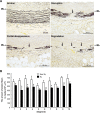
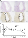
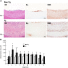
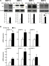
Abdominal aortic aneurysm (AAA) is pathologically characterized by intimal atherosclerosis, disruption and attenuation of the elastic media, and adventitial inflammatory infiltrates. Although all these pathological events are possibly involved in the pathogenesis of AAA, the functional roles contributed by adventitial inflammatory macrophages have not been fully documented. Recent studies have revealed that increased expression of matrix metalloproteinase-12 (MMP-12) derived from macrophages may be particularly important in the pathogenesis of both atherosclerosis and AAA. In the current study, we developed a carrageenan-induced abdominal aortic adventitial inflammatory model in hypercholesterolemic rabbits and evaluated the effect of adventitial macrophage accumulation on the aortic remodeling with special reference to the influence of increased expression of MMP-12. To accomplish this, we compared the carrageenan-induced aortic lesions of transgenic (Tg) rabbits that expressed high levels of MMP-12 in the macrophage lineage to those of non-Tg rabbits. We found that the aortic medial and adventitial lesions of Tg rabbits were greater in degree than those of non-Tg rabbits, with the increased infiltration of macrophages and prominent destruction of elastic lamellae accompanied by the frequent appearance of dilated lesions, while the intimal lesions were slightly increased. Enhanced aortic lesions in Tg rabbits were focally associated with increased dilation of the aortic lumens. RT-PCR and Western blotting revealed high levels of MMP-12 in the lesions of Tg rabbits that were accompanied by elevated levels of MMP-2 and -3, which was caused by increased number of macrophages. Our results suggest that adventitial inflammation constitutes a major stimulus to aortic remodeling and increased expression of MMP-12 secreted from adventitial macrophages plays an important role in the pathogenesis of vascular diseases such as AAA.
Keywords: MMP-12; abdominal aortic aneurysm; atherosclerosis; elastin; macrophage; transgenic rabbits.
Copyright © 2023 Chen, Yang, Kitajima, Quan, Wang, Zhu, Liu, Lai, Yan and Fan.
Conflict of interest statement
The authors declare that the research was conducted in the absence of any commercial or financial relationships that could be construed as a potential conflict of interest.
Figures


Similar articles
-
Macrophage metalloelastase accelerates the progression of atherosclerosis in transgenic rabbits.Circulation. 2006 Apr 25;113(16):1993-2001. doi: 10.1161/CIRCULATIONAHA.105.596031. Circulation. 2006. PMID: 16636188
-
Influence of hypercholesterolemia and adventitial inflammation on the development of aortic aneurysm in rabbits.Arterioscler Thromb Vasc Biol. 1997 Jan;17(1):10-7. doi: 10.1161/01.atv.17.1.10. Arterioscler Thromb Vasc Biol. 1997. PMID: 9012631
-
Macrophage-derived MMP-9 enhances the progression of atherosclerotic lesions and vascular calcification in transgenic rabbits.J Cell Mol Med. 2020 Apr;24(7):4261-4274. doi: 10.1111/jcmm.15087. Epub 2020 Mar 3. J Cell Mol Med. 2020. PMID: 32126159 Free PMC article.
-
Adventitial matrix metalloproteinase production and distribution of immunoglobulin G4-related abdominal aortic aneurysms.JVS Vasc Sci. 2020 Jul 16;1:151-165. doi: 10.1016/j.jvssci.2020.06.001. eCollection 2020. JVS Vasc Sci. 2020. PMID: 34617043 Free PMC article.
-
Immune cells and molecular mediators in the pathogenesis of the abdominal aortic aneurysm.Cardiol Rev. 2009 Sep-Oct;17(5):201-10. doi: 10.1097/CRD.0b013e3181b04698. Cardiol Rev. 2009. PMID: 19690470 Review.
Cited by
-
The functional role of cellular senescence during vascular calcification in chronic kidney disease.Front Endocrinol (Lausanne). 2024 Jan 22;15:1330942. doi: 10.3389/fendo.2024.1330942. eCollection 2024. Front Endocrinol (Lausanne). 2024. PMID: 38318291 Free PMC article. Review.
-
Unraveling Elastic Fiber-Derived Signaling in Arterial Aging and Related Arterial Diseases.Biomolecules. 2025 Jan 21;15(2):153. doi: 10.3390/biom15020153. Biomolecules. 2025. PMID: 40001457 Free PMC article. Review.
-
Pharmacological Inhibition of MMP-12 Exerts Protective Effects on Angiotensin II-Induced Abdominal Aortic Aneurysms in Apolipoprotein E-Deficient Mice.Int J Mol Sci. 2024 May 27;25(11):5809. doi: 10.3390/ijms25115809. Int J Mol Sci. 2024. PMID: 38891996 Free PMC article.
-
The contribution of matrix metalloproteinases and their inhibitors to the development, progression, and rupture of abdominal aortic aneurysms.Front Cardiovasc Med. 2023 Sep 19;10:1248561. doi: 10.3389/fcvm.2023.1248561. eCollection 2023. Front Cardiovasc Med. 2023. PMID: 37799778 Free PMC article. Review.
-
PCDH11X mutation as a potential biomarker for immune checkpoint therapies in lung adenocarcinoma.J Mol Med (Berl). 2024 Jul;102(7):899-912. doi: 10.1007/s00109-024-02450-8. Epub 2024 May 13. J Mol Med (Berl). 2024. PMID: 38739269
References
LinkOut - more resources
Full Text Sources
Miscellaneous

