

Incidence of Dentinal Microcracks during Root Canal Preparation with Self Adjusting File, Reciproc Blue and ProTaper Next
- PMID: 36704321
- PMCID: PMC9723216
- DOI: 10.22037/iej.v15i1.26667
Incidence of Dentinal Microcracks during Root Canal Preparation with Self Adjusting File, Reciproc Blue and ProTaper Next
Abstract
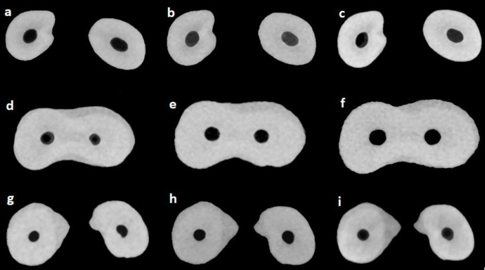
Introduction: Forces formed during root canal instrumentation could cause the crack formation in dentinal walls. Their propagation may result in vertical root fracture and eventually tooth loss. The aim of the study was to explore microcrack formation after root canal preparation with Self-adjusting File (SAF), Reciproc Blue (RB), and ProTaper Next (PTN) instruments on young premolars by means of micro-computed tomography (micro CT).
Methods and materials: Forty-five upper premolars with two canals, were extracted due to orthodontic reasons from patients aged 16 to 20 years and stored for up to two months. The teeth were scanned with a micro-CT (Nikon XT H 225, Tring, UK) at structural resolution of 20.2 µm and randomly divided into three groups: SAF, RB, and PTN. Specimens were instrumented and irrigation was performed with 12 mL of 2.5% sodium hypochlorite (NaOCl) and 4 mL of 17% ethylenediaminetetraacetic acid (EDTA) per root canal. Subsequently, the specimens were scanned under the same conditions as before, in wet condition and 24 h after drying. The presence of microcracks in dentinal walls was evaluated using the image-processing software Volume Graphics VGStudio Max 3.
Results: No dentinal defect was found in any evaluated specimen, neither in pre-nor post-operative scans in wet and dry condition.
Conclusion: Under the circumstances of this in vitro study instruments with improved design and metallurgy do not cause dentinal microcracks in young premolar teeth.
Keywords: Dentin; Micro-Computed Tomography; Nickel-Titanium; Root Canal Preparation.
Conflict of interest statement
‘None declared’.
Figures
Similar articles
-
Effect of ProTaper Gold, Self-Adjusting File, and XP-endo Shaper Instruments on Dentinal Microcrack Formation: A Micro-computed Tomographic Study.J Endod. 2017 Jul;43(7):1166-1169. doi: 10.1016/j.joen.2017.02.005. Epub 2017 May 2. J Endod. 2017. PMID: 28476466
-
Formation of dentinal microcracks after root canal preparation with four kinds of mechanical nickel-titanium files.Hua Xi Kou Qiang Yi Xue Za Zhi. 2024 Feb 1;42(1):75-81. doi: 10.7518/hxkq.2023.2023257. Hua Xi Kou Qiang Yi Xue Za Zhi. 2024. PMID: 38475954 Free PMC article. Chinese, English.
-
Micro-computed tomography evaluation of dentinal cracks after root canal preparation with different endodontic rotary files: An ex vivo study.Dent Med Probl. 2025 Jan-Feb;62(1):89-98. doi: 10.17219/dmp/149733. Dent Med Probl. 2025. PMID: 40019238
-
Micro-computed Tomographic Evaluation of Dentinal Microcracks after Preparation of Curved Root Canals with ProTaper Gold, WaveOne Gold, and ProTaper Next Instruments.J Endod. 2021 Feb;47(2):309-314. doi: 10.1016/j.joen.2020.10.014. Epub 2020 Oct 21. J Endod. 2021. PMID: 33096193
-
A critical analysis of research methods and experimental models to study dentinal microcracks.Int Endod J. 2022 Mar;55 Suppl 1:178-226. doi: 10.1111/iej.13660. Epub 2021 Nov 27. Int Endod J. 2022. PMID: 34743355 Review.
Cited by
-
Effect of Root Canal Preparation on Propagation of Dentinal Microcracks.Iran Endod J. 2021;16(2):90-96. doi: 10.22037/iej.v16i2.26744. Iran Endod J. 2021. PMID: 36704217 Free PMC article.
-
Root Fracture Resistance of Maxillary Premolars Obturated with Three Root Canal Sealers after Passive Utrasonic Irrigation: An in Vitro study.Iran Endod J. 2020 Summer;15(3):166-172. doi: 10.22037/iej.v15i3.26426. Iran Endod J. 2020. PMID: 36703808 Free PMC article.
-
Dentinal microcracks induced by endodontic procedures: A scientometric and bibliometric analysis.J Conserv Dent. 2022 Jan-Feb;25(1):78-87. doi: 10.4103/jcd.jcd_469_21. Epub 2022 May 2. J Conserv Dent. 2022. PMID: 35722065 Free PMC article.
References
-
- Burklein S, Tsotsis P, Schafer E. Incidence of dentinal defects after root canal preparation: reciprocating versus rotary instrumentation. J Endod. 2013;39(4):501–4. - PubMed
-
- Aydin U, Aksoy F, Karataslioglu E, Yildirim C. Effect of ethylenediaminetetraacetic acid gel on the incidence of dentinal cracks caused by three novel nickel-titanium systems. Aust Endod J. 2015;41(3):104–10. - PubMed
-
- Shemesh H, Bier CA, Wu MK, Tanomaru-Filho M, Wesselink PR. The effects of canal preparation and filling on the incidence of dentinal defects. Int Endod J. 2009;42(3):208–13. - PubMed
LinkOut - more resources
Full Text Sources