

Case Reports
doi: 10.1016/j.jhsg.2022.10.011.
eCollection 2023 Jan.
Calcific Tendinitis of the Proximal Flexor Carpi Radialis in a Throwing Athlete
Affiliations
- PMID: 36704383
- PMCID: PMC9870810
- DOI: 10.1016/j.jhsg.2022.10.011
Item in Clipboard
Case Reports
Calcific Tendinitis of the Proximal Flexor Carpi Radialis in a Throwing Athlete
J Hand Surg Glob Online.
.
Abstract
Flexor carpi radialis tendinitis is a condition that almost exclusively affects the distal aspect of the tendon where it lies in a tight fibro-osseous tunnel. Tendinitis of the proximal aspect of the tendon at its myotendinous junction is extremely rare. Herein, we present a single case of calcific tendinitis of the flexor carpi radialis tendon at its myotendinous junction in a throwing athlete. Diagnostic challenges and treatments are discussed.
Keywords: Athlete; Calcific tendinitis; FCR; Flexor carpi radialis; Injury.
© 2022 The Authors.
Figures
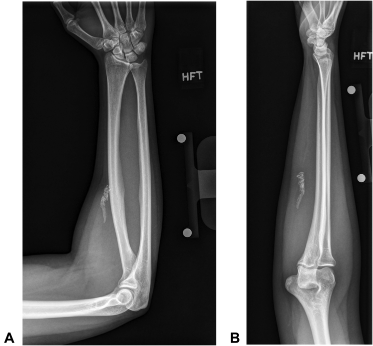
A Anteroposterior and B lateral plain forearm radiographs showing the details of the calcified tendon.

A Point-of-care ultrasound imaging taken in the short axis revealed intact muscle belly proximally. B and C Partial tear and calcification at the myotendinous junction. D Intact tendon distally. Dashed line outlines the FCR myotendinous junction.

Noncontrast T2 weighted magnetic resonance imaging showing calcification about the proximal aspect of the FCR tendon (arrows). A Axial view, B Sagittal view, C Coronal view.

Longitudinal incision of the mass revealed calcified tendon at the FCR myotendinous junction.

Dissected tendon from the FCR myotendinous junction. Remaining slip of the tendon is visible in the wound bed.

Macroscopic picture of the excised calcified tendon. The units of the reference scale are in centimeters.
References
-
- Bishop A.T., Gabel G., Carmichael S.W. Flexor carpi radialis tendinitis. Part I: operative anatomy. J Bone Joint Surg Am. 1994;76(7):1009–1014. - PubMed
-
- Uhthoff H.K., Loehr J.W. Calcific tendinopathy of the rotator cuff: pathogenesis, diagnosis, and management. J Am Acad Orthop Surg. 1997;5(4):183–191. - PubMed
-
- Wo S., Mulcahy H., Richardson M.L., et al. Pathologies of the shoulder and elbow affecting the overhead throwing athlete. Skeletal Radiol. 2017;46(7):873–888. - PubMed
-
- Kanevsky J., Zammit D., Brutus J.P. Rupture of the flexor carpi radialis tendon secondary to trauma: case report and literature review. Plast Aesthet Res. 2015;2:138–139.
Publication types
LinkOut - more resources
Full Text Sources
