. 2023 Jan 10:16:1097467.
doi: 10.3389/fnana.2022.1097467. eCollection 2022.

The olfactory limbus of the red fox (Vulpes vulpes). New insights regarding a noncanonical olfactory bulb pathway

Affiliations

The olfactory limbus of the red fox (Vulpes vulpes). New insights regarding a noncanonical olfactory bulb pathway

Irene Ortiz-Leal et al. Front Neuroanat. .

Abstract

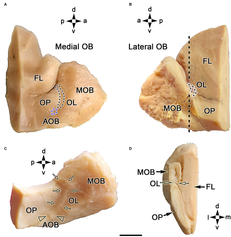
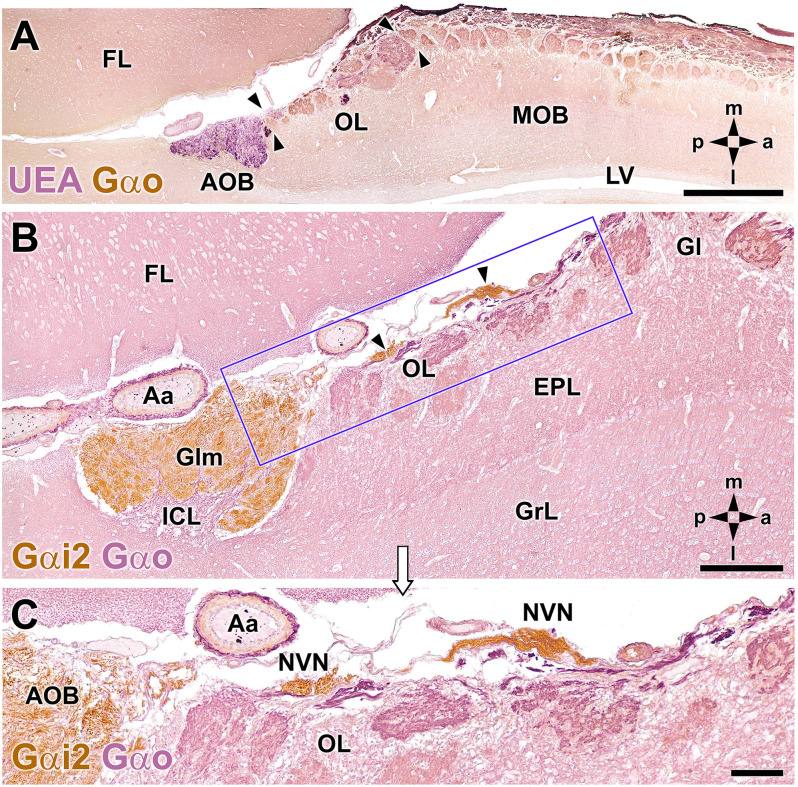
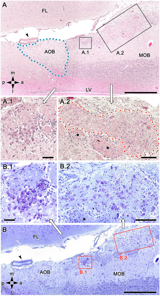
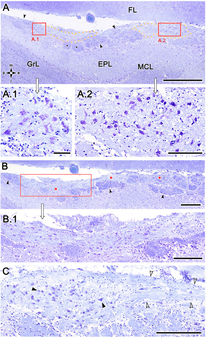
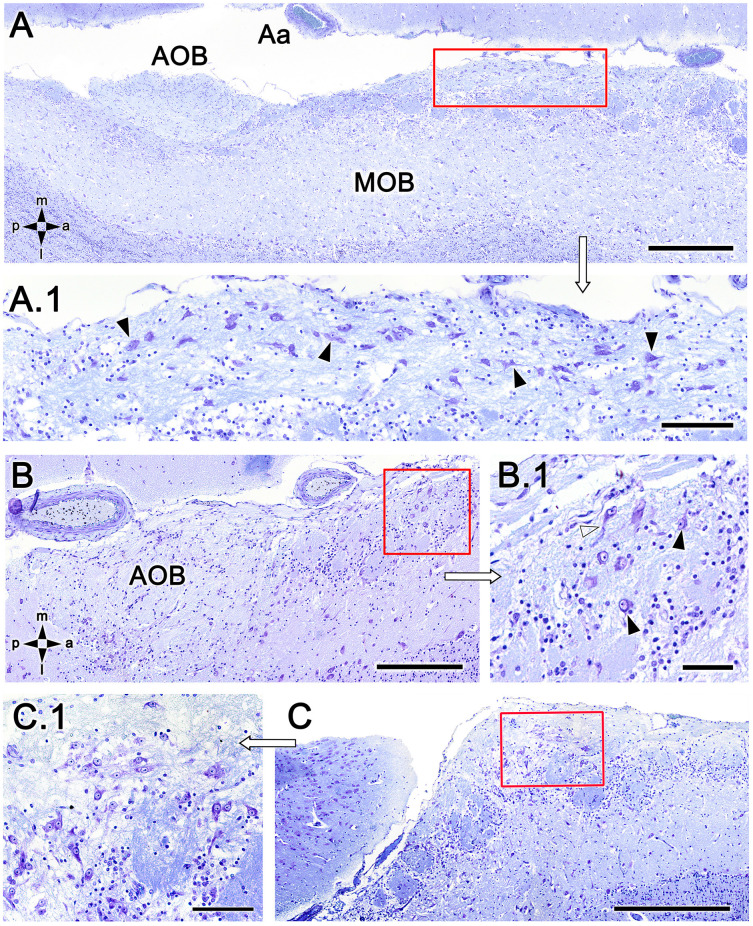
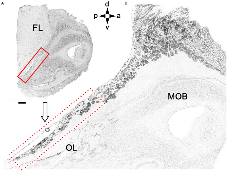
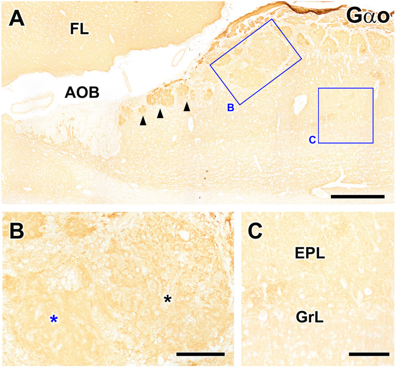
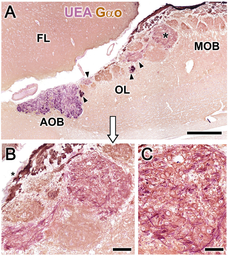
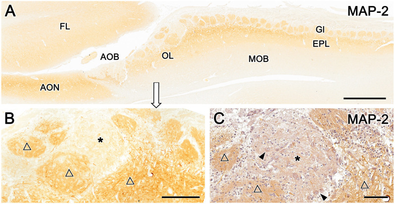
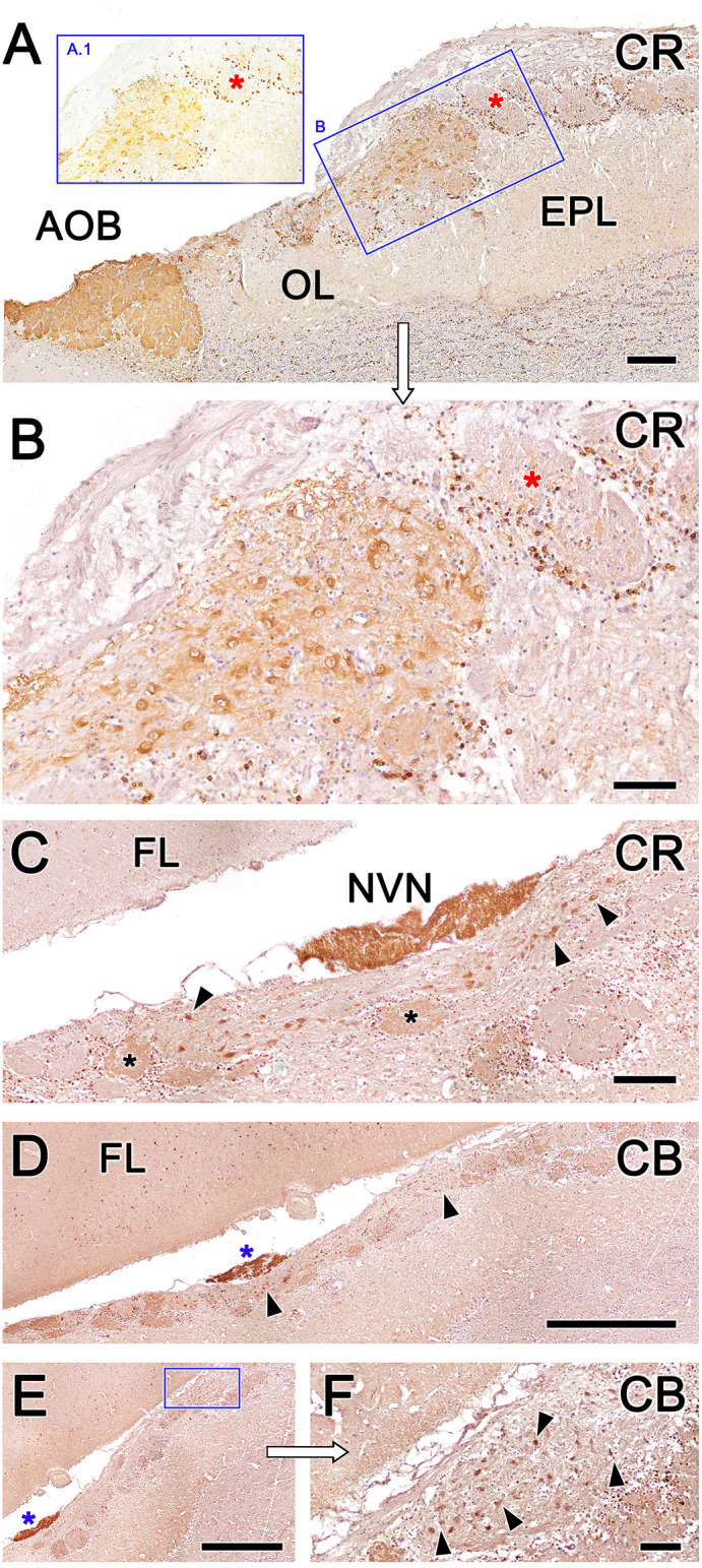
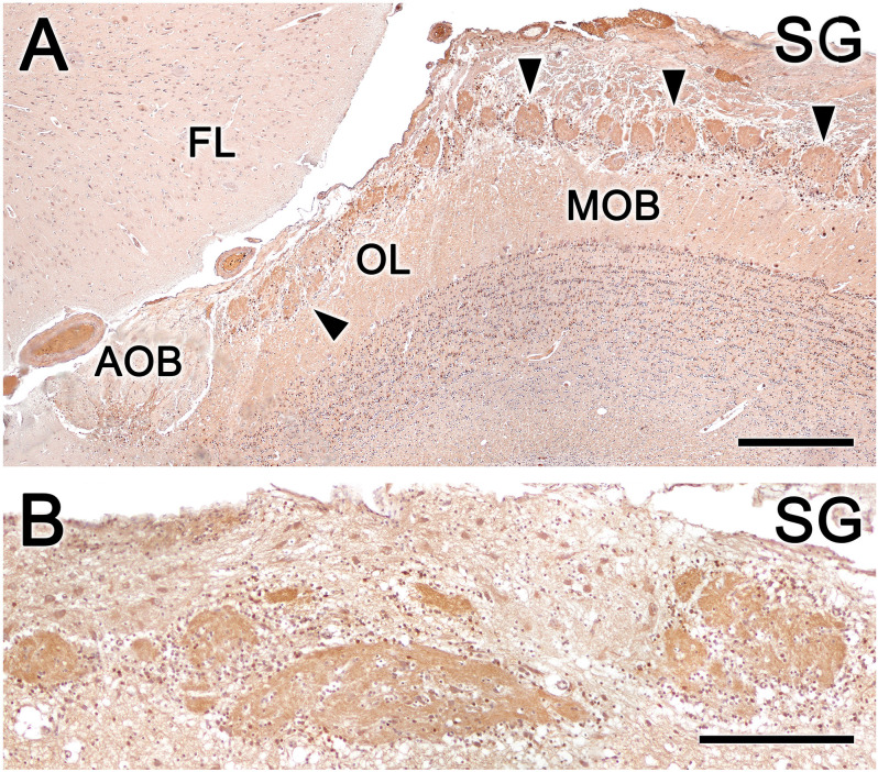
Introduction: The olfactory system in most mammals is divided into several subsystems based on the anatomical locations of the neuroreceptor cells involved and the receptor families that are expressed. In addition to the main olfactory system and the vomeronasal system, a range of olfactory subsystems converge onto the transition zone located between the main olfactory bulb (MOB) and the accessory olfactory bulb (AOB), which has been termed the olfactory limbus (OL). The OL contains specialized glomeruli that receive noncanonical sensory afferences and which interact with the MOB and AOB. Little is known regarding the olfactory subsystems of mammals other than laboratory rodents. Methods: We have focused on characterizing the OL in the red fox by performing general and specific histological stainings on serial sections, using both single and double immunohistochemical and lectin-histochemical labeling techniques. Results: As a result, we have been able to determine that the OL of the red fox (Vulpes vulpes) displays an uncommonly high degree of development and complexity. Discussion: This makes this species a novel mammalian model, the study of which could improve our understanding of the noncanonical pathways involved in the processing of chemosensory cues.

Keywords: Canidae; fox; immunohistochemistry; lectins; olfaction; olfactory limbus.


Conflict of interest statement

The authors declare that the research was conducted in the absence of any commercial or financial relationships that could be construed as a potential conflict of interest.

Figures

Figure 1
Figure 1
Macroscopic topographical anatomy of the fox olfactory limbus (OL). (A) Medial view of the left main olfactory bulb (MOB). The olfactory peduncle (OP) and the caudomedial margin of the olfactory bulb are visualized. The accessory olfactory bulb (AOB, blue dots) and the olfactory limbus (OL, black dots) are located along the MOB’s caudal surface. (B) Lateral view of the left main olfactory bulb. The lateral most portion of the OL is located in the caudal margin of the MOB. (C) Dorsolateral view of the right MOB after removal of the frontal lobe (FL). The entire length of the OL (arrows) and the AOB (arrowheads) can be visualized. (D) Caudal view of a transverse section of the left MOB and OP at the level depicted by a dashed line in (B). The OL is located in the caudoventral portion of the MOB (open arrows). a, anterior; d, dorsal; l, lateral; m, medial; p, posterior; v, ventral. Scale bar: 500 μm.
Figure 2
Figure 2
Double-immunohistochemical labeling of the fox olfactory limbus (OL). (A) Double immunostaining with Ulex europaeus agglutinin (UEA) lectin (magenta) and Gαo antibody (brown). The vomeronasal nerve and glomerular layers of the accessory olfactory bulb (AOB) are strongly labeled with UEA. Anti-Gαo immunolabeling shows a widespread immunopositive pattern, more intensely in both the olfactory nerve and glomerular layers of the MOB. The OL, delimited by arrowheads, comprises irregularly shaped glomeruli, without a homogeneous immunostaining pattern. (B,C) Double immunostaining for Gαi2 (brown) and Gαo (magenta). Anti-Gαi2 stains the superficial AOB and the nervus vomeronasalis (NVN, arrowheads). Anti-Gαo stains the internal cellular layer (ICL) of the AOB and the neuropil of the olfactory bulb. The box in (B) is enlarged in (C), showing that the OL comprises irregularly shaped glomeruli without a homogeneous immunostaining pattern. The branches of the NVN, which are Gαi2-immunopositive, contrast with the Gαo-immunopositive nerve endings that project to the OL glomeruli. Aa, artery; EPL, external plexiform layer; FL, frontal lobe; Gl, MOB glomerular layer; Glm, AOB glomerular layer; GrL, MOB granular cell layer; LV, lateral ventricle. The compass indicates the orientation, as follows: m, medial; l, lateral; p, posterior; a, anterior. Scale bars: (A): 1 cm. (B,C): 250 μm.
Figure 3
Figure 3
Histological study of the fox olfactory limbus (OL). (A) General view of the OL, in the horizontal plane. The accessory olfactory bulb (AOB), close to a large artery (arrowhead), is framed by dots. The two most strikingly discernible features of the OL are framed and shown at higher magnification in (A.1) and (A.2): the dense neuronal cluster (A.1) and the macroglomerular complex (MGC, delimited by red dots in A.2), a broad nervous formation consisting of neuronal somata distributed within a neuropil with clearly distinct boundaries. Small, irregularly shaped glomeruli were observed deep to both structures (asterisks). (B) Consecutive, Nissl-stained serial sections showing the morphology of the neuronal somata. Both the denser aggregates (B.1) and the MGC (B.2), possess polygonal, ellipsoidal, and rounded somata. FL, frontal lobe; LV, lateral ventricle; MOB, main olfactory bulb. Orientation: m, medial; l, lateral; p, posterior; a, anterior. Scale bars: (A,B): 500 μm. (A.1,B.1): 100 μm. (A.2,B.2): 250 μm.
Figure 4
Figure 4
Nissl-stained sections of the fox olfactory limbus (OL) at different horizontal levels. Serial Nissl-stained sections from a single specimen reveal the development of the OL in this species. (A) A neuronal cluster (box enlarged in A.1) and the superficial macroglomerular complex (MGC, encircled by yellow dots and partially enlarged in A.2) were observed in a section at the ventral level. Most glomeruli proximal to the MGC had irregular and atypical shapes (arrowheads), but some appeared spherical (black asterisk). (B) A more dorsal section shows the extent of the MGC (red asterisks). The red box is shown at a higher magnification in (B.1). Atypical glomeruli were also observed (arrowheads). (C) An even more dorsal section shows the morphologies of MGC neurons (black arrowheads) and their association with a prominent fascicle of nerve fibers (open arrowheads). FL, frontal lobe of the telencephalon; GrL, granular layer; EPL, external plexiform layer; MCL, mitral cell layer. Orientation: m, medial; l, lateral; p, posterior; a, anterior. Scale bars: (A): 1 mm. (B): 500 μm. (A.2,B.1,C): 250 μm. (A.1): 100 μm.
Figure 5
Figure 5
Nissl-stained sections of the fox olfactory limbus (OL). (A) A ventral section shows the topography of the nervous formation, located on the bulbar surface, superficial to the glomeruli. (A.1) A higher magnification of the red box in (A) shows numerous neuronal somata (arrowheads). (B) Horizontal section at the level of the AOB. Anterior to the AOB, a superficial neuronal cluster surrounded by atypical glomeruli can be observed (red box, magnified in B.1). (B.1) The neuronal somata have oval shapes (arrowheads), and the origin of thick processes is visible (open arrowhead). (C) In this specimen, the neuronal cluster is located in a more anterior position, very close to the pial surface. (C.1) At a higher magnification, the neurons show similar morphology to that of the specimen shown in (B). Aa, artery; AOB, accessory olfactory bulb; MOB, main olfactory bulb. Orientation: m, medial; l, lateral; p, posterior; a, anterior. Scale bars: (A,C): 500 μm. (B): 250 μm. (A.1,C.1): 100 μm. (B.1): 50 μm.
Figure 6
Figure 6
Schematic drawing of a sagittal histological section of the olfactory bulb. (A) Magnification of the area corresponding to the OL (box in B). Orientation: m, medial; l, lateral; p, posterior; a, anterior. Scale bar: 500 μm.
Figure 7
Figure 7
Immunohistochemical labeling of the fox olfactory limbus (OL) using anti-Gαo. (A) Immunolabeling for the G-protein subunit Gαo revealed uniform expression of Gαo throughout the frontal lobe of the telencephalon (FL) and the olfactory bulb, but no immunostaining in the nerve or glomerular layers of the accessory olfactory bulb (AOB). Atypical glomeruli proximal to AOB (arrowheads) are more intensely immunolabeled with anti-Gαo than typical glomeruli in the main olfactory bulb (MOB). The macroglomerular complex (MGC, box B) shows a labeling intensity similar to the typical MOB glomeruli. (B) Higher magnification of box (B), showing that the MGC possesses two clearly differentiated areas: one containing numerous neuronal somata (black asterisk) and one devoid of somata (blue asterisk). Somata in the MGC are Gαo-immunopositive, unlike the mitral cell somata in the MOB, which are immunonegative (box C, C). EPL, external plexiform layer; GrL, granular layer. Scale bars: (A): 500 μm. (B,C): 125 μm.
Figure 8
Figure 8
Double-immunohistochemical labeling of the fox olfactory limbus (OL). Double immunostaining with the lectin Ulex europaeus agglutinin (UEA, magenta) and Gαo antibody (brown). (A) The nerve and glomerular layers of the accessory olfactory bulb (AOB) are intensely labeled by UEA, whereas the main olfactory bulb (MOB) is not. In the OL, some glomeruli are UEA-negative, whereas smaller glomeruli are strongly UEA-positive (arrowheads). The macroglomerular complex (MGC, asterisk) is also UEA-positive. (B) Higher magnification of the MGC area, showing Gαo-positive somata embedded in a dense network of UEA-positive fibers. The vomeronasal nerve fibers segregate into UEA-positive (black asterisk) and Gαo-positive (white asterisk components). (C) The MGC somata are oval in shape and Gαo-positive. FL, Frontal lobe of the telencephalon. Scale bars: (A): 500 μm. (B): 50 μm.
Figure 9
Figure 9
Immunohistochemical labeling of the fox olfactory limbus (OL) with anti-microtubule-associated protein 2 (MAP-2). (A) Immunostaining with anti-MAP-2 results in the strong and uniform immunolabeling of glomeruli (Gl) and the external plexiform layer (EPL) of the main olfactory bulb (MOB). (B) A higher magnification view of the rostral part of the OL shows the staining of atypical Gl formations (open triangles), in addition to a patch of the nervous formation that remains unstained (*). (C) Anti-MAP-2 immunostained serial sections were counterstained with hematoxylin, confirming that the unlabeled area corresponds to an area of the macroglomerular complex that is rich in neuronal bodies (arrowheads) and receives the innervation from Ulex europaeus agglutinin-positive fibers. AOB, accessory olfactory bulb; AON, anterior olfactory nucleus; EPL, external plexiform layer; FL, front lobe of the telencephalon; Gl, glomeruli; MOB, main olfactory bulb; OL, olfactory limbus. Scale bars: (A): 1 mm. (B): 250 μm. (C): 100 μm.
Figure 10
Figure 10
Fox olfactory limbus (OL) sections immunolabeled with the calcium-binding proteins calretinin (CR) and calbindin (CB) and counterstained with hematoxylin. (A) Anti-CR immunostaining produces intense labeling in the macroglomerular complex (MGC) of the OL (box B) and in the accessory olfactory bulb (AOB). The glomeruli of the main olfactory bulb (MOB) do not show any immunolabeling in their neuropil (red asterisks), although their periglomerular cells are clearly labeled, which is particularly evident in non-counterstained sections (box A.1). (B) A higher magnification image of box (B) from panel (A). In the MGC, both neuronal somata and the neuropil (asterisk) are immunolabeled with anti-CR. (C) Section at the level of the vomeronasal nerve (NVN) shows intense anti-CR immunopositivity. The atypical nervous formation beneath the NVN is elongated and contains scattered neuronal somata (arrowheads). A subpopulation of glomeruli is CR-positive (asterisks). (D) Anti-CB immunostaining produces a similar pattern to anti-CR immunostaining. The NVN is intensely immunopositive to anti-CB (blue asterisk). The MGC is delimited by arrowheads. (E) Atypical glomeruli are variably labeled with anti-CB. (F) Higher magnification of the inset in (E). Somata belonging to the MGC are intensely stained (arrowheads). AOB, accessory olfactory bulb; OL, olfactory limbus; EPL, external plexiform layer; FL, the frontal lobe of the telencephalon; NVN, vomeronasal nerve Scale bars: (D,E): 1 mm. (A,C): 250 μm. (B,F): 100 μm.
Figure 11
Figure 11
Fox olfactory limbus (OL) sections immunolabeled for the calcium-binding protein secretagogin (SG) and counterstained with hematoxylin. (A) Anti-SG immunostaining results in immunolabeling of the entire glomeruli population (arrowheads) in both the OL and the main olfactory bulb (MOB) but not the glomerular layer of the accessory olfactory bulb (AOB). Periglomerular cells are intensely labeled. (B) In the macroglomerular complex, both the neuropil and neurons are SG-immunopositive, with variable labeling intensity. FL, the frontal lobe of the telencephalon. Scale bars: (A): 500 μm. (B): 250 μm.

Similar articles

Cited by

References

    1. Adams D. R. (1972). Olfactory and non-olfactory epithelia in the nasal cavity of the mouse, Peromyscus. Am. J. Anat. 133, 37–49. 10.1002/aja.1001330104 - DOI - PubMed
    1. Adrio F., Rodríguez-Moldes I., Anadón R. (2011). Distribution of glycine immunoreactivity in the brain of the siberian sturgeon (Acipenser baeri): comparison with γ-aminobutyric acid. J. Comp. Neurol. 519, 1115–1142. 10.1002/cne.22556 - DOI - PubMed
    1. Alonso J. R., Porteros A., Crespo C., Arevalo R., Briñon J. G., Weruaga E., et al. . (1998). Chemical anatomy of the macaque monkey olfactory bulb: NADPH-diaphorase/nitric oxide synthase activity. J. Comp. Neurol. 402, 419–434. 10.1002/(SICI)1096-9861(19981221)402:3<419::AID-CNE9>3.0.CO;2-C - DOI - PubMed
    1. Alpár A., Attems J., Mulder J., Hökfelt T., Harkany T. (2012). The renaissance of Ca2+-binding proteins in the nervous system: secretagogin takes center stage. Cell. Signal. 24, 378–387. 10.1016/j.cellsig.2011.09.028 - DOI - PMC - PubMed
    1. Baimbridge K. G., Celio M. R., Rogers J. H. (1992). Calcium-binding proteins in the nervous system. Trends Neurosci. 15, 303–308. 10.1016/0166-2236(92)90081-i - DOI - PubMed