

Encephalo-Arterio-Synangiosis with Cranioplasty after Treatment of Acute Subdural Hematoma Associated with Subcortical Hemorrhage Due to Unilateral Moyamoya Disease
- PMID: 36704418
- PMCID: PMC9873458
- DOI: 10.1155/2023/1787738
Encephalo-Arterio-Synangiosis with Cranioplasty after Treatment of Acute Subdural Hematoma Associated with Subcortical Hemorrhage Due to Unilateral Moyamoya Disease
Abstract
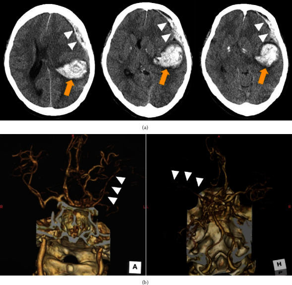
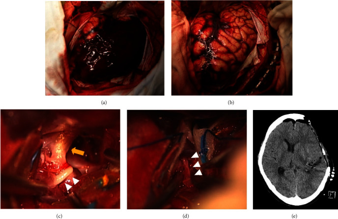
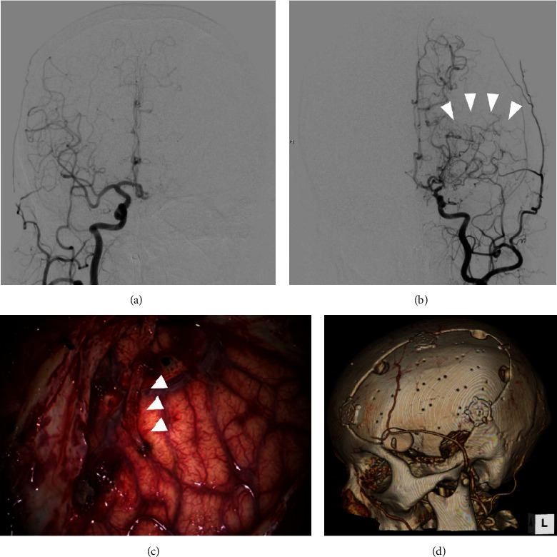
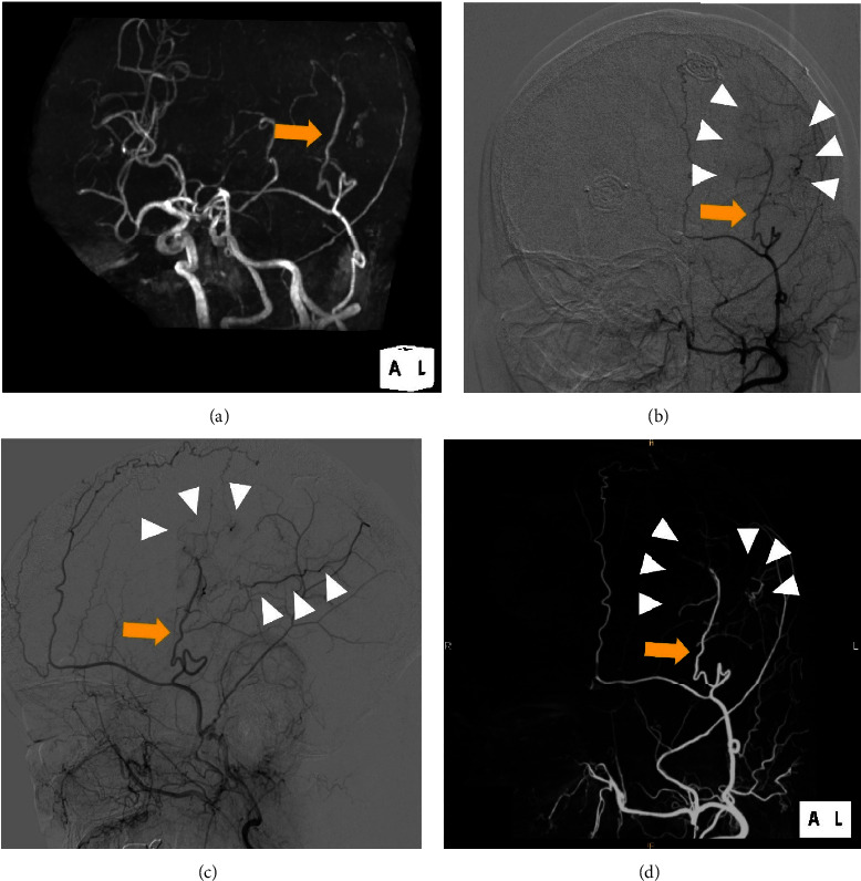
Moyamoya disease is often diagnosed after intracranial hemorrhage in adult patients. Here, we report a case of unilateral moyamoya disease treated with indirect revascularization combined with cranioplasty after treatment for acute subdural hematoma and subcortical hemorrhage. A middle-aged woman with disturbed consciousness was transferred to our hospital. Computed tomography (CT) revealed an acute subdural hematoma with left temporoparietal subcortical hemorrhage. Three-dimensional CT angiography indicated a scarcely enhanced left middle cerebral artery (MCA) that was suspected to be delayed or nonfilling due to increased intracranial pressure. Subsequently, hematoma evacuation and external decompression were performed. Postoperative digital subtraction angiography (DSA) revealed stenosis of the left MCA and moyamoya vessels, indicating unilateral moyamoya disease. Forty-five days after the initial procedure, we performed encephalo-arterio-synangiosis (EAS) using the superficial temporal artery simultaneously with cranioplasty for the skull defect. The modified Rankin Scale score of the patient one year after discharge was 1, and the repeat DSA showed good patency of the EAS. Revascularization using EAS in the second step can be an option for revascularization for hemorrhagic moyamoya disease if the patient required cranioplasty for postoperative skull defect after decompressive craniotomy.
Copyright © 2023 Naoki Kato et al.
Conflict of interest statement
The authors declare that there are no conflicts of interest regarding the publication of this article.
Figures
Similar articles
-
A Preliminary Report of One-Session Treatment with Cranioplasty and Superficial Temporal Artery-Middle Cerebral Artery Bypass for Hemorrhagic Moyamoya Disease Patients with Skull Defect.World Neurosurg. 2022 Aug;164:276-280. doi: 10.1016/j.wneu.2022.05.071. Epub 2022 May 23. World Neurosurg. 2022. PMID: 35618236
-
The importance of encephalo-myo-synangiosis in surgical revascularization strategies for moyamoya disease in children and adults.World Neurosurg. 2015 May;83(5):691-9. doi: 10.1016/j.wneu.2015.01.016. Epub 2015 Feb 3. World Neurosurg. 2015. PMID: 25655688
-
"STA-MCA bypass with encephalo-duro-myo-synangiosis combined with bifrontal encephalo-duro-periosteal-synangiosis" as a one-staged revascularization strategy for pediatric moyamoya vasculopathy.Childs Nerv Syst. 2015 May;31(5):765-72. doi: 10.1007/s00381-015-2665-y. Epub 2015 Feb 27. Childs Nerv Syst. 2015. PMID: 25722049
-
Successful Surgical Management of Traumatic Intracranial Hemorrhaging After Revascularization Surgery for Moyamoya Vasculopathy: A Case Report and Review of Literature.World Neurosurg. 2020 May;137:24-28. doi: 10.1016/j.wneu.2020.01.184. Epub 2020 Jan 31. World Neurosurg. 2020. PMID: 32014547 Review.
-
Anastomosis of the superficial temporal artery to the middle cerebral artery with the interposed occipital artery graft in moyamoya disease: case report.Surg Neurol. 1997 Dec;48(6):615-9. doi: 10.1016/s0090-3019(97)00015-3. Surg Neurol. 1997. PMID: 9400645 Review.
Cited by
-
Case Report: A rare presentation of rapidly progressive moyamoya disease refractory to unilateral surgical revascularization.Front Surg. 2024 Aug 16;11:1409692. doi: 10.3389/fsurg.2024.1409692. eCollection 2024. Front Surg. 2024. PMID: 39220621 Free PMC article.
-
Rare Presentation of Moyamoya Disease with an Acute Subdural Hemorrhage from a Rare Location of Aneurysm-Related Moyamoya Disease.Asian J Neurosurg. 2025 Jan 17;20(2):367-372. doi: 10.1055/s-0044-1801374. eCollection 2025 Jun. Asian J Neurosurg. 2025. PMID: 40485810 Free PMC article.
References
-
- Takahashi J. C., Funaki T., Houkin K., et al. Impact of cortical hemodynamic failure on both subsequent hemorrhagic stroke and effect of bypass surgery in hemorrhagic moyamoya disease: a supplementary analysis of the Japan Adult Moyamoya Trial. Journal of Neurosurgery . 2020;134(3):940–945. doi: 10.3171/2020.1.JNS192392. - DOI - PubMed
-
- Matsushima T., Inoue T., Suzuki S. O., Fujii K., Fukui M., Hasuo K. Surgical treatment of moyamoya disease in pediatric patients--comparison between the results of indirect and direct revascularization procedures. Neurosurgery . 1992;31(3):401–405. doi: 10.1097/00006123-199209000-00003. - DOI - PubMed
-
- Shen W. C., Lee W. Y. Moyamoya disease causes acute subdural hematomas and sudden death: a case report. Zhonghua yi xue za zhi = Chinese medical journal; Free China ed . 1998;61(10):619–623. - PubMed
Publication types
LinkOut - more resources
Full Text Sources
