

3D echocardiography in mitral valve prolapse
- PMID: 36704460
- PMCID: PMC9871497
- DOI: 10.3389/fcvm.2022.1050476
3D echocardiography in mitral valve prolapse
Abstract
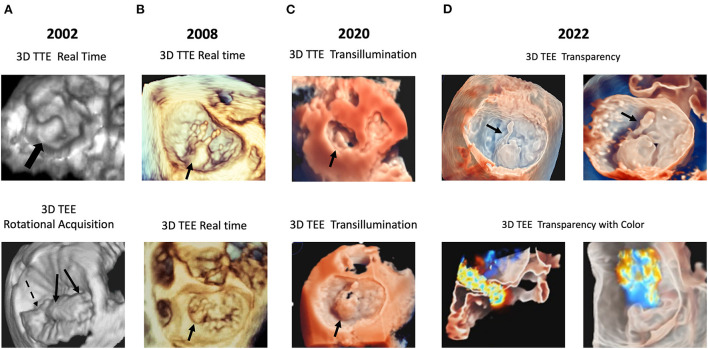
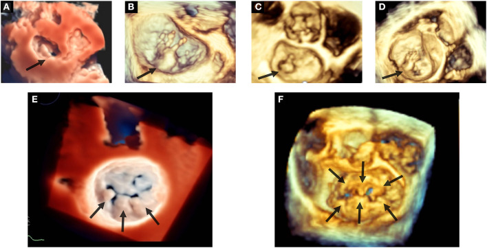
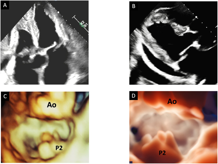
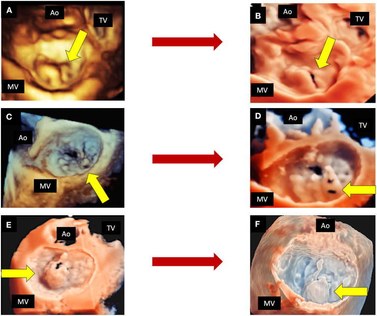
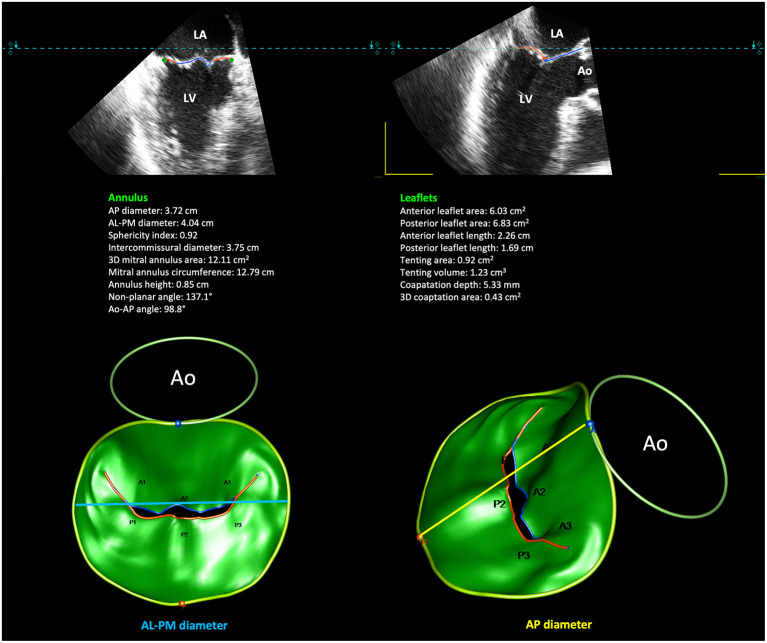
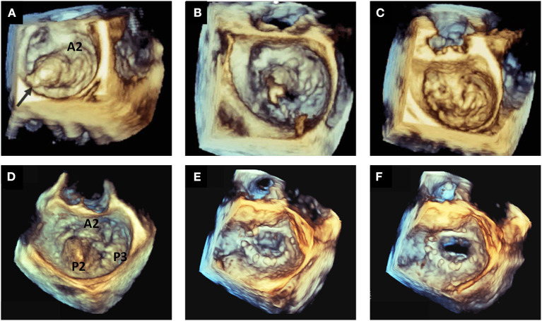
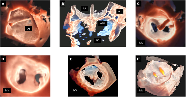
Mitral valve prolapse (MVP) is the leading cause of mitral valve surgery. Echocardiography is the principal imaging modality used to diagnose MVP, assess the mitral valve morphology and mitral annulus dynamics, and quantify mitral regurgitation. Three-dimensional (3D) echocardiographic (3DE) imaging represents a consistent innovation in cardiovascular ultrasound in the last decades, and it has been implemented in routine clinical practice for the evaluation of mitral valve diseases. The focus of this review is the role and the advantages of 3DE in the comprehensive evaluation of MVP, intraoperative and intraprocedural monitoring.
Keywords: mitral annulus (MA); mitral regurgitation (MR); mitral valve prolapse (MVP); mitral valve surgery; percutaneous mitral valve repair; three-dimensional echocardiography (3DE).
Copyright © 2023 Mantegazza, Gripari, Tamborini, Muratori, Fusini, Ghulam Ali, Garlaschè and Pepi.
Conflict of interest statement
The authors declare that the research was conducted in the absence of any commercial or financial relationships that could be construed as a potential conflict of interest.
Figures
Similar articles
-
Usefulness of three-dimensional echocardiography for the evaluation of mitral valve prolapse: an intraoperative study.J Heart Valve Dis. 2000 May;9(3):341-9. J Heart Valve Dis. 2000. PMID: 10888088
-
Valve Strain Quantitation in Normal Mitral Valves and Mitral Prolapse With Variable Degrees of Regurgitation.JACC Cardiovasc Imaging. 2021 Jun;14(6):1099-1109. doi: 10.1016/j.jcmg.2021.01.006. Epub 2021 Mar 17. JACC Cardiovasc Imaging. 2021. PMID: 33744129
-
Comparison of mitral valve dimensions and motion in mitral valve prolapse with severe mitral regurgitation to uncomplicated mitral valve prolapse and to mitral regurgitation without mitral valve prolapse.Am J Cardiol. 1988 Aug 1;62(4):257-63. doi: 10.1016/0002-9149(88)90222-6. Am J Cardiol. 1988. PMID: 3400603
-
Three-dimensional echocardiography in the assessment of congenital mitral valve disease.J Am Soc Echocardiogr. 2014 Feb;27(2):142-54. doi: 10.1016/j.echo.2013.11.011. Epub 2013 Dec 18. J Am Soc Echocardiogr. 2014. PMID: 24360740 Review.
-
Three-Dimensional Echocardiography: Advancements in Qualitative and Quantitative Analyses of Mitral Valve Morphology in Mitral Valve Prolapse.J Cardiovasc Echogr. 2014 Jan-Mar;24(1):1-9. doi: 10.4103/2211-4122.131985. J Cardiovasc Echogr. 2014. PMID: 28465897 Free PMC article. Review.
Cited by
-
Arrhythmic Mitral Valve Prolapse: A Comprehensive Review.Diagnostics (Basel). 2023 Sep 6;13(18):2868. doi: 10.3390/diagnostics13182868. Diagnostics (Basel). 2023. PMID: 37761235 Free PMC article. Review.
-
Comprehensive Analysis of Cardiovascular Diseases: Symptoms, Diagnosis, and AI Innovations.Bioengineering (Basel). 2024 Dec 7;11(12):1239. doi: 10.3390/bioengineering11121239. Bioengineering (Basel). 2024. PMID: 39768057 Free PMC article. Review.
-
Three-Dimensional Transesophageal Echocardiography in Percutaneous Catheter-Based Cardiac Interventions.J Clin Med. 2023 Aug 31;12(17):5664. doi: 10.3390/jcm12175664. J Clin Med. 2023. PMID: 37685731 Free PMC article. Review.
-
Intraoperative Transesophageal Echocardiography Quantification of Mitral Regurgitation by Regurgitation Volume and Fraction Using 2D and 3D Techniques.Ann Card Anaesth. 2025 Apr 1;28(2):161-169. doi: 10.4103/aca.aca_218_24. Epub 2025 Apr 16. Ann Card Anaesth. 2025. PMID: 40237663 Free PMC article.
-
Delineating thrombus versus myxoma: Perioperative 3D transesophageal echocardiography to the rescue!J Clin Imaging Sci. 2024 Feb 29;14:6. doi: 10.25259/JCIS_136_2023. eCollection 2024. J Clin Imaging Sci. 2024. PMID: 38469175 Free PMC article.
References
Publication types
LinkOut - more resources
Full Text Sources
Miscellaneous