

Mycochemistry, antioxidant content, and antioxidant potentiality of the ethanolic extract of Pleurotus florida and its anti-cancerous effect on HeLa cancer cell line, and antitumor effect on HeLa-implanted mice
- PMID: 36704494
- PMCID: PMC9832905
Mycochemistry, antioxidant content, and antioxidant potentiality of the ethanolic extract of Pleurotus florida and its anti-cancerous effect on HeLa cancer cell line, and antitumor effect on HeLa-implanted mice
Abstract
Objectives: Cervical cancer is increasing worldwide and is becoming resistant to the existing drugs in clinical practice. Here, ethanolic extract of fruit body of Pleurotus florida was evaluated as antioxidant, anticancer agent against HeLa cell lines and anti-tumor against cervical cancer in mice model.
Methods: Fruit bodies of P. florida in 90% ethanol, and the P. florida ethanolic extract (PFEE) was subsequently investigated for its antioxidant content and activity, anticancer properties against the cervical cancer cell line, HeLa, and antitumor activity against HeLa implanted mice.
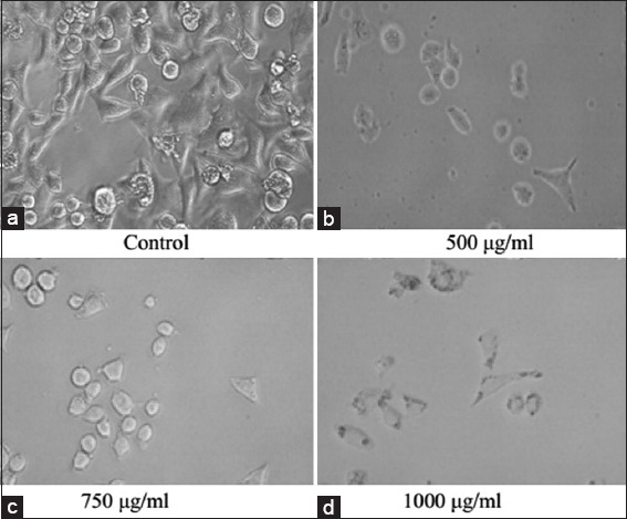
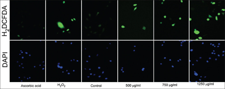
Results: The antioxidant activity bioassay showed that the IC50 of PFEE was 41.17 ± 1.42a μg/ml. The cytotoxicity assay revealed that PFEE caused inhibition of cell proliferation. At the highest dose (1,250 μg/ml) after 24 h, 48 h, or 72 h of treatment, the percentages of cell growth inhibition were 75.22%, 77.77%, and 84.65%, respectively. It revealed that PFEE-treated cells became rounded and the nuclei became fragmented. PFEE induced intracellular generation of reactive oxygen species and reduced the mitochondrial membrane potential. PFEE also led to an up regulation of the apoptotic genes for caspases-3, -9, and Bax, whereas Bcl-2 gene was down regulated, and it also promoted the expression of p53. Cell cycle analysis revealed that cell cycle was arrested at the G0/G1 checkpoint. PFEE suppressed metastasis and colonization. At a dosage of PFEE of 50 mg/kg of body weight, a 66.72% reduction in the size of tumors and an 87.44% reduction in the tumor weight were observed in mice.
Conclusions: It has demonstrated that PFEE is a highly potent anti-cervical cancer agent in vitro and in vivo.
Keywords: Apoptosis; Cytotoxicity; colonization; metastasis; migration.
Copyright: © International Journal of Health Sciences.
Figures


Similar articles
-
Cultivated and wild Pleurotus ferulae ethanol extracts inhibit hepatocellular carcinoma cell growth via inducing endoplasmic reticulum stress- and mitochondria-dependent apoptosis.Sci Rep. 2018 Sep 18;8(1):13984. doi: 10.1038/s41598-018-32225-4. Sci Rep. 2018. PMID: 30228276 Free PMC article.
-
Chemical composition, antioxidant and antitumor activities of sub-fractions of wild and cultivated Pleurotus ferulae ethanol extracts.PeerJ. 2018 Dec 20;6:e6097. doi: 10.7717/peerj.6097. eCollection 2018. PeerJ. 2018. PMID: 30595979 Free PMC article.
-
Evaluation of antioxidant, anti-inflammatory and anticancer activities of diosgenin enriched Paris polyphylla rhizome extract of Indian Himalayan landraces.J Ethnopharmacol. 2021 Apr 24;270:113842. doi: 10.1016/j.jep.2021.113842. Epub 2021 Jan 16. J Ethnopharmacol. 2021. PMID: 33460752
-
Dendrobium chrysanthum ethanolic extract induces apoptosis via p53 up-regulation in HeLa cells and inhibits tumor progression in mice.J Complement Integr Med. 2017 Jun 1;14(2):/j/jcim.2017.14.issue-2/jcim-2016-0070/jcim-2016-0070.xml. doi: 10.1515/jcim-2016-0070. J Complement Integr Med. 2017. PMID: 28195549
-
Potent anti-cancer activity of Sphaerocoryne affinis fruit against cervical cancer HeLa cells via inhibition of cell proliferation and induction of apoptosis.BMC Complement Med Ther. 2023 Aug 19;23(1):290. doi: 10.1186/s12906-023-04127-0. BMC Complement Med Ther. 2023. PMID: 37598145 Free PMC article.
Cited by
-
Therapeutic effect of bromelain and papain on intestinal injury induced by indomethacin in male rats.Int J Health Sci (Qassim). 2023 Sep-Oct;17(5):23-30. Int J Health Sci (Qassim). 2023. PMID: 37692988 Free PMC article.
-
Antimicrobial and cytotoxic activities of natural (Z)-13-docosenamide derived from Penicillium chrysogenum.Front Cell Infect Microbiol. 2025 Feb 27;15:1529104. doi: 10.3389/fcimb.2025.1529104. eCollection 2025. Front Cell Infect Microbiol. 2025. PMID: 40083907 Free PMC article.
-
Mushroom Bioactive Molecules as Anticancerous Agents: An Overview.Food Sci Nutr. 2025 Jul 14;13(7):e70580. doi: 10.1002/fsn3.70580. eCollection 2025 Jul. Food Sci Nutr. 2025. PMID: 40661813 Free PMC article. Review.
References
-
- Sadler M, Saltmarsh M. Functional Foods:The Consumer, the Products and the Evidence. United Kingdom: Royal Society of Chemistry; 1998. p. 215.
-
- Zhanga M, Cuia SW, Cheungb PC, Wanga Q. Antitumor polysaccharides from mushrooms:A review on their isolation process, structural characteristics and antitumor activity. Trends Food Sci Technol. 2007;18:4–19.
-
- Zaidman BZ, Yassin M, Mahajna J, Wasser SP. Medicinal mushroom modulators of molecular targets as cancer therapeutics. Appl Microbiol Biotechnol. 2005;67:453–68. - PubMed
LinkOut - more resources
Full Text Sources
Research Materials
Miscellaneous