. 2023 Mar;32(3):e4578.
doi: 10.1002/pro.4578.

Design of a protease-activated PD-L1 inhibitor

Affiliations

Design of a protease-activated PD-L1 inhibitor

Odessa J Goudy et al. Protein Sci. 2023 Mar.

Abstract

Immune checkpoint inhibitors that bind to the cell surface receptor PD-L1 are effective anti-cancer agents but suffer from immune-related adverse events as PD-L1 is expressed on both healthy and cancer cells. To mitigate toxicity, researchers are testing prodrugs that have low affinity for checkpoint targets until activated with proteases enriched in the tumor microenvironment. Here, we engineer a prodrug form of a PD-L1 inhibitor. The inhibitor is a soluble PD-1 mimetic that was previously engineered to have high affinity for PD-L1. In the basal state, the binding surface of the PD-1 mimetic is masked by fusing it to a soluble variant of its natural ligand, PD-L1. Proteolytic cleavage of the linker that connects the mask to the inhibitor activates the molecule. To optimize the mask so that it effectively blocks binding to PD-L1 but releases upon cleavage, we tested a set of mutants with varied affinity for the inhibitor. The top-performing mask reduces the affinity of the prodrug for PD-L1 120-fold, and binding is nearly fully recovered upon cleavage. In a cell-based assay measuring inhibition of the PD-1:PD-L1 interaction on the surface of cells, the IC50s of the masked inhibitors were up to 40-fold higher than their protease-treated counterparts. The changes in activity we observe upon protease treatment are comparable to systems currently tested in the clinic and provide evidence that natural binding partners are an excellent starting point for creating a prodrug.

Keywords: PD-1; PD-L1; immune checkpoint inhibitor; prodrug; protein engineering.


Conflict of interest statement

The authors declare no conflict of interest.

Figures

FIGURE 1
FIGURE 1
Structure and mutations of masked complexes. (a) AlphaFold prediction of the wild‐type masked complex illustrating HA‐PD1 (gray) followed by a 28‐linker (yellow) containing a protease recognition site (orange) and a PD‐L1 masking domain (pink). (b) Zoomed in view of interface interactions highlighting the Y56‐mediated intramolecular bond within PD‐L1 (pink) and an intermolecular bond to HA‐PD1 (gray) via Y123. Some residues were hidden for clarity. (c) Cartoons illustrating the alanine mutations (white circles) in the masking domain (pink) of each masked complex. Note, although NoMC lacks a masking domain, it retains a cleavable linker. NoMC, unmasked complex, or no masked complex.
FIGURE 2
FIGURE 2
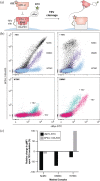
Masked complex yeast surface expression and binding to PD‐L1. (a) Masked complexes were displayed on the surface of yeast (blue cell) through the Aga2p system. Using flow cytometry, the overall presence of the mask was monitored with an anti‐cMyc‐FITC antibody (green star). Binding of biotinylated PD‐L1 (light pink) to HA‐PD1 of the complex was measured through streptavidin‐633 (light pink star). (b) Bivariate expression and binding profiles of NoMC, DMMC, and WTMC without (top left) and with (top right) TEV incubation. To directly compare a single complex with (teal) and without (pink) protease treatment, the expression and binding profiles for WTMC and DMMC are also shown (bottom left and right, respectively). The default number (8000) of dots is shown for each condition. (c) The change in the gMFI of FITC and 633 upon protease treatment are shown for NoMC, DMMC, and WTMC, calculated using the following equation: (gMFI+TEV − gMFI−TEV)/(gMFI−TEV). Additional gating details found in the Supporting Information. DMMC, double mutant masked complex, or Y56A and 123A PD‐L1 masked complex; NoMC, unmasked complex, or no masked complex; TEV, tobacco etch virus; WTMC, wild‐type PD‐L1 masked complex.
FIGURE 3
FIGURE 3
Masks reduce affinity to PD‐L1. Biotinylated PD‐L1 was immobilized on a NeutrAvidin chip. (a) On–off rate map indicating the mean binding kinetics parameters of the various masked complexes without protease treatment. Reported error represents SD. The map was constructed using analyte concentrations from 12 nM to 1 μM for WTMC, 56MC, and 123MC; 1.2 to 100 nM for DMMC; and 0.12 to 10 nM for NoMC. (b) Representative surface plasmon resonance (SPR) sensorgrams of the masked complexes in the absence (pink) and presence (teal) of overnight TEV proteolysis. Corresponding responses were simultaneously collected with analyte concentrations from 1.2 to 100 nM. For direct comparison, across the panels are the maximum response of WTMC in the absence and presence of TEV (dotted lines). (c) Table of mean kinetic parameters for the various masked complexes before and after TEV treatment, including the fold change in KD over that of the NoMC control, and the change before and after TEV proteolysis for each complex. In the absence of TEV, experimental conditions as mentioned in (a); in the presence of TEV, conditions as mentioned in (b). Error represents SD. “n” represents the number of runs, which includes experiments collected from at least two different sensor chips for each condition; individual data included in the Supporting Information. DMMC, double mutant masked complex, or Y56A and 123A PD‐L1 masked complex; NoMC, unmasked complex, or no masked complex; SD, standard deviation; TEV, tobacco etch virus; WTMC, wild‐type PD‐L1 masked complex.
FIGURE 4
FIGURE 4
Masked complexes conditionally block the PD‐1:PD‐L1 endogenous interaction on the surface of cells. In the assay, when the endogenous PD‐1:PD‐L1 interaction is disrupted, TCR activation induces luminescence. (a) The luminescence signal at various concentrations of the masked complexes with (teal) and without (pink) TEV protease treatment. Two technical replicates from a single experiment, presented as mean ± SEM are shown. (b) To directly compare the activity of the various complexes, the luminescence responses without (left) and with (right) TEV incubation are shown. (c) Data from two separate experiments were analyzed in Prism and the mean LogIC50 windows with and without TEV incubation for each masked complex are shown. The fold change of the IC50 upon protease treatment is labeled for each complex. Individual data from the two separate experiments are included in the Supporting Information. SEM, standard error of the mean; TCR, T‐cell receptor; TEV, tobacco etch virus.

References

    1. Assi HH, Wong C, Tipton KA, Mei L, Wong K, Razo J, et al. Conditional PD‐1/PD‐L1 probody therapeutics induce comparable antitumor immunity but reduced systemic toxicity compared with traditional anti‐PD‐1/PD‐L1 agents. Cancer Immunol Res. 2021;9:1451–64. - PMC - PubMed
    1. Baek M, DiMaio F, Anishchenko I, Dauparas J, Ovchinnikov S, Lee GR, et al. Accurate prediction of protein structures and interactions using a three‐track neural network. Science. 2021;1979:373–876. - PMC - PubMed
    1. Bagchi S, Yuan R, Engleman EG. Immune checkpoint inhibitors for the treatment of cancer: clinical impact and mechanisms of response and resistance. Annu Rev Pathol. 2021;16:223–49. - PubMed
    1. Benatuil L, Perez JM, Belk J, Hsieh CM. An improved yeast transformation method for the generation of very large human antibody libraries. Protein Eng Des Sel. 2010;23:155–9. - PubMed
    1. Bleuez C, Koch WF, Urbach C, Hollfelder F, Jermutus L. Exploiting protease activation for therapy. Drug Discov Today. 2022;27:1743–54. - PMC - PubMed

Publication types

MeSH terms