

Increased Great Saphenous Vein Diameter at the Level of Knee among Patients with Varicose Veins in a Tertiary Care Centre: A Descriptive Cross-sectional Study
- PMID: 36705219
- PMCID: PMC9446489
- DOI: 10.31729/jnma.7543
Increased Great Saphenous Vein Diameter at the Level of Knee among Patients with Varicose Veins in a Tertiary Care Centre: A Descriptive Cross-sectional Study
Abstract
Introduction: Colour Doppler ultrasonography plays an important role in determining the morphological and hemodynamic information of the venous system. This study aimed to find out the prevalence of increased great saphenous vein diameter at the level of the knee among patients with varicose veins in a tertiary care centre.
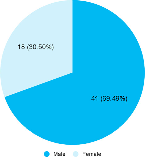
Methods: A descriptive cross-sectional study was carried out in the Department of Radiology at a tertiary care centre from 30 October 2021 to 31 March 2022 after taking ethical approval from the Institutional Review Committee (Reference number: 028-077/078). A convenience sampling technique was used for the study. The study group consisted of patients over 18 years, coming for ultrasonography examination of the lower limb with the clinical symptoms and signs of varicose veins. The great saphenous vein diameter was measured at the level of the medial femoral condyle of the knee using the software in the ultrasonography unit. B mode, colour Doppler and spectral analysis were done. A cut-off value of 5 mm for the diameter of the great saphenous vein was taken to indicate the presence or absence of varicosity and saphenofemoral reflux. Point estimate and 90% Confidence Interval were calculated.
Results: Among 72 patients with varicose veins, the diameter of the great saphenous vein was increased in 59 (81.94%) (74.50-89.38, 90% Confidence Interval) patients.
Conclusions: The mean diameter of the great saphenous vein in our study was similar when compared to other studies conducted in similar settings.
Keywords: saphenous vein; ultrasonography; varicose veins.
Conflict of interest statement
Figures
Similar articles
-
Association between the saphenous vein diameter and venous reflux on computed tomography venography in patients with varicose veins.PLoS One. 2022 Feb 15;17(2):e0263513. doi: 10.1371/journal.pone.0263513. eCollection 2022. PLoS One. 2022. PMID: 35167584 Free PMC article.
-
The Cut-off Value of Great Saphenous Vein Diameter at the Level of Femoral Condyle to Predict the Sapheno-Femoral Junction Incompetence.Kathmandu Univ Med J (KUMJ). 2022 Jul-Sep;20(79):280-283. Kathmandu Univ Med J (KUMJ). 2022. PMID: 37042366
-
Ultrasound-based topographic analysis of tributary vein connection with the saphenous vein during ambulatory conservative hemodynamic correction of chronic venous insufficiency.J Vasc Surg Venous Lymphat Disord. 2019 May;7(3):356-363. doi: 10.1016/j.jvsv.2018.09.011. Epub 2019 Feb 15. J Vasc Surg Venous Lymphat Disord. 2019. PMID: 30777672
-
[Postoperative recurrence of varicosities at the level of the popliteal fossa. Anatomic data guiding the ultrasonographic exploration and surgical sequelae].J Mal Vasc. 1998 Feb;23(1):54-60. J Mal Vasc. 1998. PMID: 9551354 Review. French.
-
A systematic review supporting the Society for Vascular Surgery, the American Venous Forum, and the American Vein and Lymphatic Society guidelines on the management of varicose veins.J Vasc Surg Venous Lymphat Disord. 2022 Sep;10(5):1155-1171. doi: 10.1016/j.jvsv.2021.08.011. Epub 2021 Aug 24. J Vasc Surg Venous Lymphat Disord. 2022. PMID: 34450355
References
-
- Azhar MA. Role of colour flow duplex sonography in the evaluation of chronic venous insufficiency in lower limbs. International Journal of Contemporary Medicine Surgery and Radiology. 2017;2(3):80–4.
MeSH terms
LinkOut - more resources
Full Text Sources
Medical