. 2023 Feb 7;62(3):782-796.
doi: 10.1021/acs.biochem.2c00635. Epub 2023 Jan 27.

What Drives Chorismate Mutase to Top Performance? Insights from a Combined In Silico and In Vitro Study

Affiliations

What Drives Chorismate Mutase to Top Performance? Insights from a Combined In Silico and In Vitro Study

Helen V Thorbjørnsrud et al. Biochemistry. .

Abstract

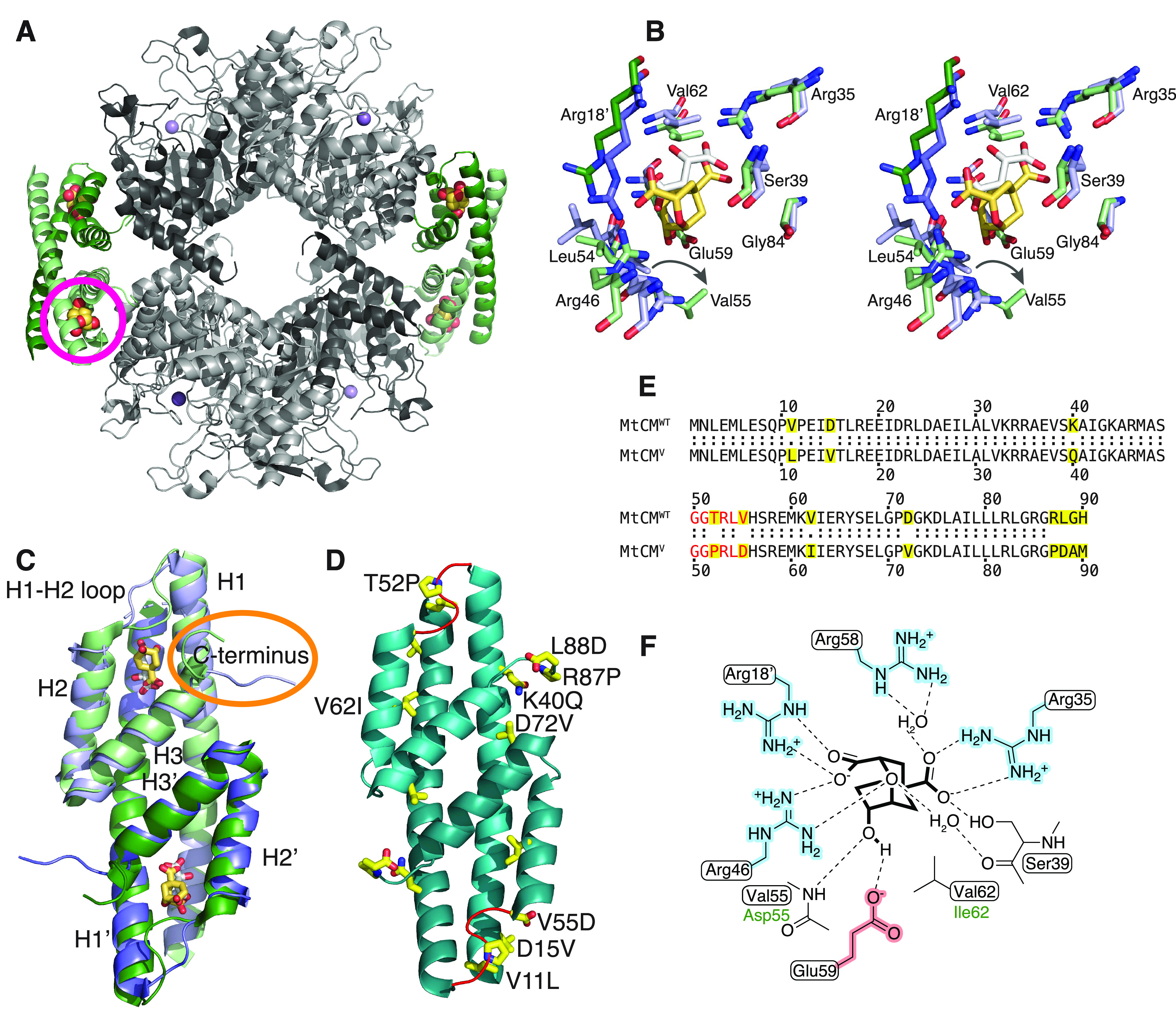
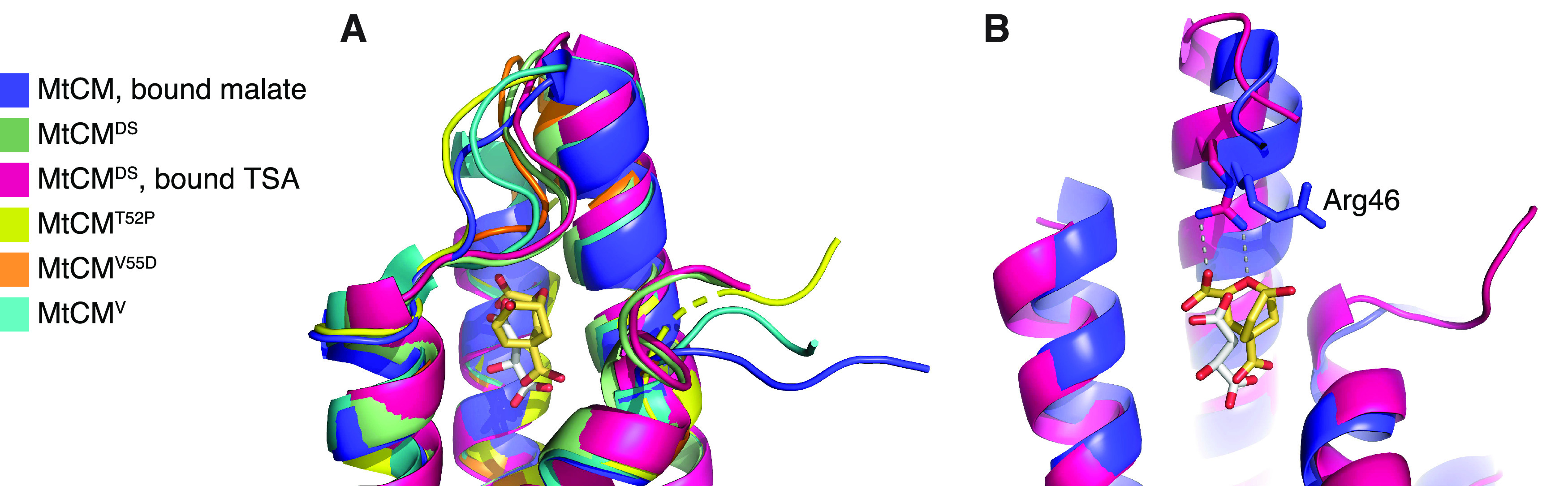
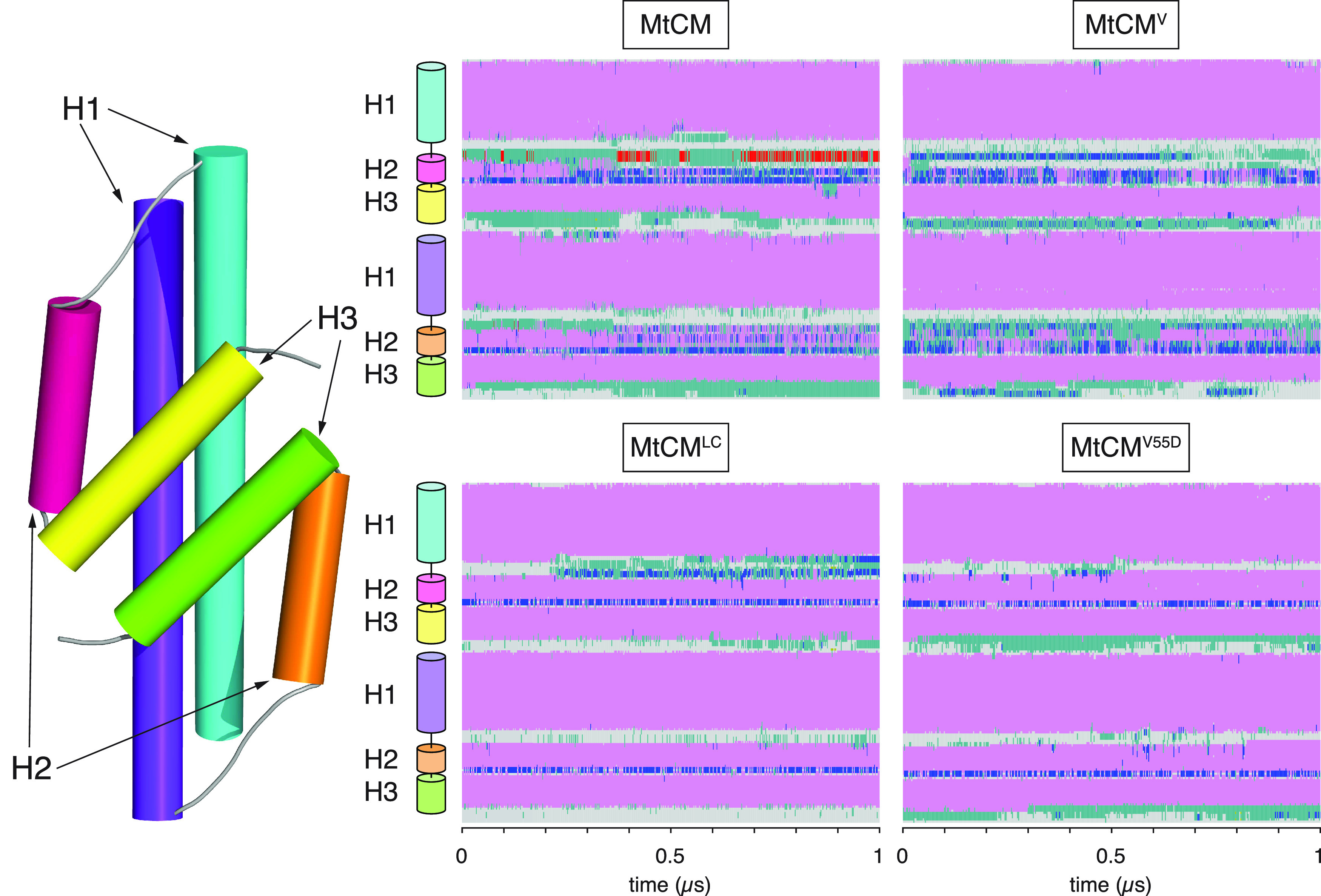
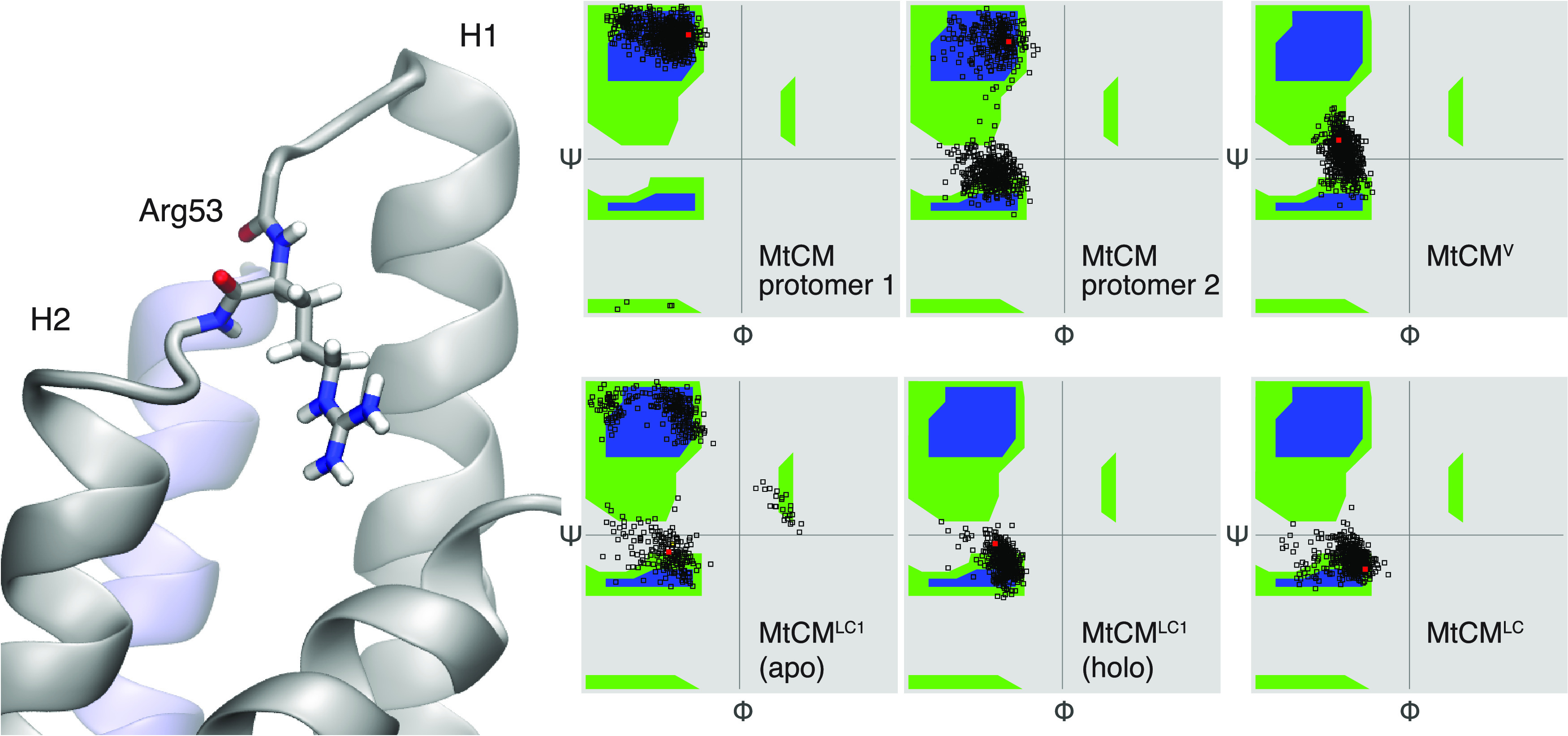
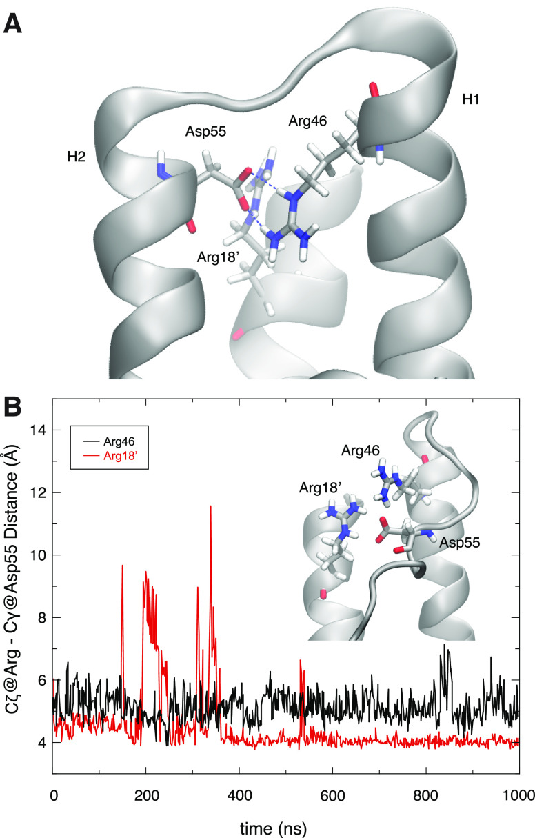
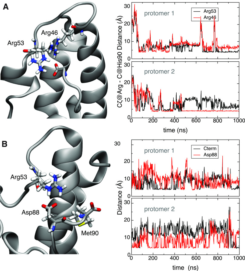
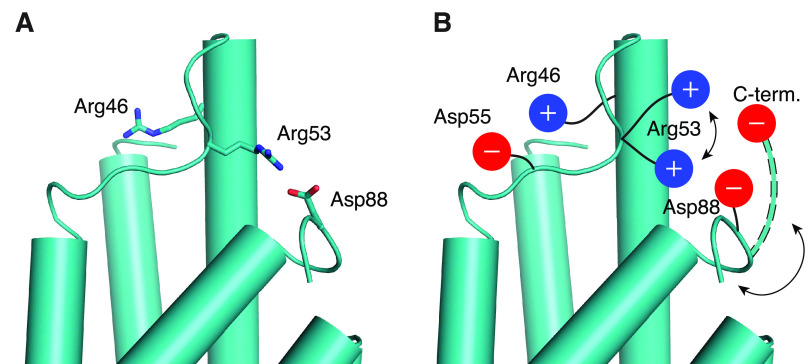
Unlike typical chorismate mutases, the enzyme from Mycobacterium tuberculosis (MtCM) has only low activity on its own. Remarkably, its catalytic efficiency kcat/Km can be boosted more than 100-fold by complex formation with a partner enzyme. Recently, an autonomously fully active MtCM variant was generated using directed evolution, and its structure was solved by X-ray crystallography. However, key residues were involved in crystal contacts, challenging the functional interpretation of the structural changes. Here, we address these challenges by microsecond molecular dynamics simulations, followed up by additional kinetic and structural analyses of selected sets of specifically engineered enzyme variants. A comparison of wild-type MtCM with naturally and artificially activated MtCMs revealed the overall dynamic profiles of these enzymes as well as key interactions between the C-terminus and the active site loop. In the artificially evolved variant of this model enzyme, this loop is preorganized and stabilized by Pro52 and Asp55, two highly conserved residues in typical, highly active chorismate mutases. Asp55 stretches across the active site and helps to appropriately position active site residues Arg18 and Arg46 for catalysis. The role of Asp55 can be taken over by another acidic residue, if introduced at position 88 close to the C-terminus of MtCM, as suggested by molecular dynamics simulations and confirmed by kinetic investigations of engineered variants.


Conflict of interest statement

The authors declare no competing financial interest.

Figures

Scheme 1
Scheme 1. Chorismate Mutase Reaction
The Claisen rearrangement catalyzed by chorismate mutase converts chorismate (1) to prephenate (2) and proceeds via a highly polarized chair-like transition state carrying partial charges at the C–O bond that is broken during the reaction.
Figure 1
Figure 1
Structural information on M. tuberculosis chorismate mutase. (A) Cartoon illustration of the heterooctameric complex of MtCM with DAHP synthase (MtDS) (Protein Data Bank (PDB) ID: 2W1A). MtCM is colored in shades of green and MtDS in shades of gray to emphasize individual subunits; Bartlett’s transition state analogue (TSA) is shown with golden spheres. The location of one of the four active sites of MtCM is marked with a circle. (B) Stereo superimposition of CM active sites of MtCM (shades of violet, with malate bound, white sticks) (PDB ID: 2VKL) and of the MtCM–MtDS complex (with TSA bound, golden sticks) (PDB ID: 2W1A), showing several active site residues as sticks. Shades of violet/green (and prime notation for Arg18′) illustrate separate protomers of MtCM/MtCM–MtDS structures, respectively. An arrow shows the shift in the position of Val55 upon MtDS binding, allowing H-bond formation of its backbone to TSA. (C) Cartoon superimposition of MtCM (PDB ID: 2VKL; violet, with white sticks for malate ligand) and activated MtCM (MtCMDS) from the MtCM–MtDS complex (PDB ID: 2W1A; green, with golden sticks for TSA). The biggest structural changes upon activation are a kink observed in the H1–H2 loop and interaction of the C-terminus (circled in orange) with the active site of MtCM. (D) Cartoon representation of the artificially evolved MtDS-independent super-active MtCM variant N-s4.15 (PDB ID: 5MPV; cyan), dubbed MtCMV in this work, having a kcat/Km typical for the most efficient CMs known to date. Amino acid replacements accumulated after four cycles of directed evolution are emphasized as yellow side-chain sticks (A89 and M90 are not resolved) and labeled for one of the protomers. The H1–H2 loop (shown in red) adopts a kinked conformation similar to that observed for the MtDS-activated MtCMDS shown in (C). (E) Sequence alignment of wild-type MtCM (MtCMWT) and the highly active variant N-s4.15 (MtCMV). Substituted residues are highlighted in yellow, and the H1–H2 loop is colored red. (F) Schematic representation of the active site of MtCM with bound TSA. Boxed residues refer to the wild-type enzyme, and green font color (Asp55, Ile62) refers to those substituted in MtCMV. Charged residues are highlighted in red and blue.
Figure 2
Figure 2
Comparison of MtCM crystal structures, with focus on Arg46 and H1–H2 loop. (A) Superimposition of the active site of MtCM (PDB ID: 2VKL; violet), MtCMDS (PDB ID: 2W19; green), MtCMDS–TSA complex (PDB ID: 2W1A; pink), MtCMT52P (PDB ID: 6YGT, this work; yellow), MtCMV55D (this work; orange), and top-evolved MtCMV (PDB ID: 5MPV; cyan); cartoon representation featuring the H1–H2 loop, with the ligands depicted as sticks. (B) Superimposition of MtCM (PDB ID: 2VKL; violet, with bound malate in gray sticks) and MtCM in the MtCM–MtDS complex (PDB ID: 2W1A; pink, with TSA in golden sticks, corresponding to MtCMLC) in cartoon representation, with the catalytically important Arg46 depicted as sticks. MtDS binding promotes the catalytically competent conformation of Arg46. Helix H2 was removed for clarity.
Figure 3
Figure 3
Secondary structure changes of MtCM during MD simulations. Estimated secondary structure of MtCM over 1 μs of MD simulation. Color code: α-helical structure (magenta), 310 helix (blue), π-helix (red), turn (green), coil (gray). The top panels report data for apo MtCM and MtCMV, showing clear instability of H2. The bottom panels present the data for the holo-MtCMLC system and for the single V55D variant (apo structure), which in contrast retained all secondary structure elements within the simulation time.
Figure 4
Figure 4
Conformation of Arg53 in the H1–H2 loop. Ramachandran plot showing backbone dihedral angles Φ and ψ for Arg53. Red dots mark starting conformations. For MtCM, the H1–H2 loops from the two protomers (left and middle plots in the top row) assume different ensembles of conformations; overall, the catalytically favored conformation (Ψ ∼ 0) is observed less frequently than the nonproductive one. In contrast, in MtCMV, both loops retained the active conformation during the entire course of the simulation, similarly to that observed for MtCMLC. When only one ligand was bound (MtCMLC1), the TSA-loaded site (holo) retained the active conformation, while the loop in the other protomer (apo) remained flexible.
Figure 5
Figure 5
Role of MtCMV residue Asp55 in positioning active site residues. (A) Extension of H2 and stabilization of the H1–H2 loop by residue Asp55. Substitution of Val55 by Asp stabilizes helix H2 through interactions with Arg18′ and Arg46 across the active site (the image shows the structure of V55D after 1 μs of MD simulations). Note that Arg46 is a catalytically essential residue for MtCM and its correct orientation is critical for catalytic proficiency. (B) Distance plotted between MtCMV Arg46 (black, chain A) or Arg18′ (red, from chain B), and Asp55 (chain A) observed during the simulation. In both cases, the distance measured is between Asp Cγ and Arg Cζ, using PDB nomenclature.
Figure 6
Figure 6
Interaction between C-terminal carboxylate of MtCM and H1–H2 loop. (A) Right panels show the distance between the Cζ carbon of arginine residues 53 or 46 and the carboxylate carbon of the C-terminus of MtCM in the two protomers (top and bottom panels). Formation of a steady contact (<5 Å) with Arg46 (bottom panel) corresponds to stabilization of the catalytically productive H1–H2 loop conformation, which allows for stabilization of the transition state of the chorismate to prephenate rearrangement (Figure 1B,F). (B) Interaction between C-terminal residues and the H1–H2 loop in MtCMV. Salt bridge contacts between Arg53 and the carboxyl groups of Asp88 (red line) and Met90 (C-terminus; black line) in MtCMV in the two protomers. The top and bottom panels on the right show the evolution of the distances over time between Arg53′s Cζ and the corresponding carboxylate carbons for each of the two protomers of MtCMV.
Figure 7
Figure 7
Shielding interaction mediated by the H1–H2 loop. (A) Conformation of important Arg residues in chain A of MtCMV (cyan) after 31.7 ns of MD simulations. The key active site residue Arg46 is positioned on the opposite side of the H1–H2 loop, which in turn is bolted to the C-terminus by a salt bridge between Arg53 and Asp88 (cartoon representation, with side chains shown as sticks). (B) Cartoon summarizing the important stabilizing interactions in the top-evolved variant MtCMV depicted in (A) that properly position Arg46 for catalysis. Asp55 stabilizes the stretched-out conformation of Arg46, whereas alternating salt bridges accessible for Arg53 with the negatively charged groups present in the C-terminal region hinder Arg46 from adopting an unfavorable interaction with the C-terminal carboxylate. One example of an alternative backbone conformation that allows for interactions between the C-terminal carboxylate and Arg53 is depicted with dashed outlines.

Similar articles

Cited by

References

    1. Ohashi M.; Liu F.; Hai Y.; Chen M.; Tang M.-C.; Yang Z.; Sato M.; Watanabe K.; Houk K. N.; Tang Y. SAM-dependent enzyme-catalysed pericyclic reactions in natural product biosynthesis. Nature 2017, 549, 502–506. 10.1038/nature23882. - DOI - PMC - PubMed
    1. Minami A.; Oikawa H. Recent advances of Diels-Alderases involved in natural product biosynthesis. J. Antibiot. 2016, 69, 500–506. 10.1038/ja.2016.67. - DOI - PubMed
    1. Tang M.-C.; Zou Y.; Watanabe K.; Walsh C. T.; Tang Y. Oxidative cyclization in natural product biosynthesis. Chem. Rev. 2017, 117, 5226–5333. 10.1021/acs.chemrev.6b00478. - DOI - PMC - PubMed
    1. Lin C.-I.; McCarty R. M.; Liu H.-W. The enzymology of organic transformations: A survey of name reactions in biological systems. Angew. Chem., Int. Ed. 2017, 56, 3446–3489. 10.1002/anie.201603291. - DOI - PMC - PubMed
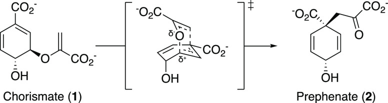
    1. Sogo S. G.; Widlanski T. S.; Hoare J. H.; Grimshaw C. E.; Berchtold G. A.; Knowles J. R. Stereochemistry of the rearrangement of chorismate to prephenate: Chorismate mutase involves a chair transition state. J. Am. Chem. Soc. 1984, 106, 2701–2703. 10.1021/ja00321a039. - DOI

Publication types

MeSH terms

Substances