

Significant difference in cardiac ventricular dimensions when measured using two different standard methods
- PMID: 36705885
- PMCID: PMC10752913
- DOI: 10.1007/s12024-023-00579-5
Significant difference in cardiac ventricular dimensions when measured using two different standard methods
Abstract
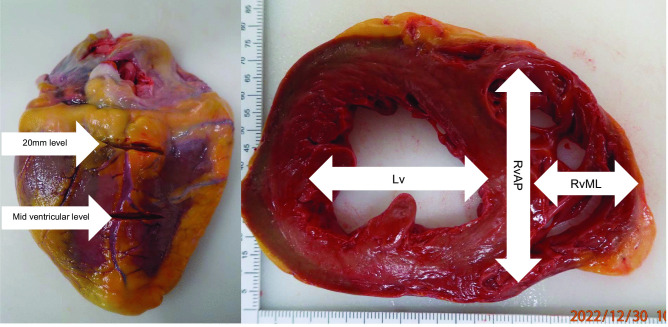
Cardiac ventricular dimensions measured at postmortem examination are used to assess whether there is hypertrophy of the heart chambers. However, there is no clear consensus on where these measurements should be taken. Some have proposed this should be measured at the mid-ventricular level, but others advocate it should be measured at a set distance (e.g. 20 mm) from the base of the heart. Twenty consecutive adult hearts were examined and showed the ventricular dimensions were significantly higher (mean: 5-15 mm, p < 0.01) when measured at a level 20 mm from the base of the heart compared to the mid-ventricular level. Of clinical significance is that in slightly less than half the cases, normal ventricular dimensions at mid ventricle level fell within the criteria considered pathological (> 40 mm) when measured at 20 mm from the base of the heart. In terms of actual ventricular dimensions, only the left ventricle diameter measured at 20 mm from the base of the heart correlated significantly (albeit moderately) with heart weight, suggesting it can be a predictor for cardiac hypertrophy.
Keywords: Autopsy; Dimensions; Heart; Hypertrophy; Postmortem; Ventricle.
© 2023. The Author(s).
Conflict of interest statement
The authors declare no competing interests.
Figures
Similar articles
-
Comparison of ante- and postmortem ventricular wall thickness using echocardiography and autopsy findings.Virchows Arch. 2025 Apr;486(4):833-842. doi: 10.1007/s00428-024-03960-z. Epub 2024 Nov 8. Virchows Arch. 2025. PMID: 39511013 Free PMC article.
-
Gross Heart Dimensions From Postmortem Computed Tomography and Postmortem Examination Measurements: Heart Weight and Cardiac Hypertrophy.Am J Forensic Med Pathol. 2023 Sep 1;44(3):176-182. doi: 10.1097/PAF.0000000000000846. Epub 2023 May 26. Am J Forensic Med Pathol. 2023. PMID: 37249480
-
Intravital and postmortem diagnostics of myocardial hypertrophy of the left ventricle:identity or convention?Ter Arkh. 2018 Sep 20;90(9):73-80. doi: 10.26442/terarkh201890973-80. Ter Arkh. 2018. PMID: 30701739
-
Structural features of the athlete heart as defined by echocardiography.J Am Coll Cardiol. 1986 Jan;7(1):190-203. doi: 10.1016/s0735-1097(86)80282-0. J Am Coll Cardiol. 1986. PMID: 2934463 Review.
-
Borderline left ventricle.Eur J Cardiothorac Surg. 2005 Jan;27(1):67-73. doi: 10.1016/j.ejcts.2004.10.034. Eur J Cardiothorac Surg. 2005. PMID: 15621473 Review.
References
-
- Dolinak D, Matshes E, Lew E. Forensic Pathology. Murlington MA: Elsevier; 2005.
-
- Saukko P, Knight B. Knight's Forensic Pathology. 4. Boca Raton: CRC Press, Taylor & Francis Group; 2016.
MeSH terms
LinkOut - more resources
Full Text Sources
