

A Spike-destructing human antibody effectively neutralizes Omicron-included SARS-CoV-2 variants with therapeutic efficacy
- PMID: 36706160
- PMCID: PMC9907810
- DOI: 10.1371/journal.ppat.1011085
A Spike-destructing human antibody effectively neutralizes Omicron-included SARS-CoV-2 variants with therapeutic efficacy
Abstract
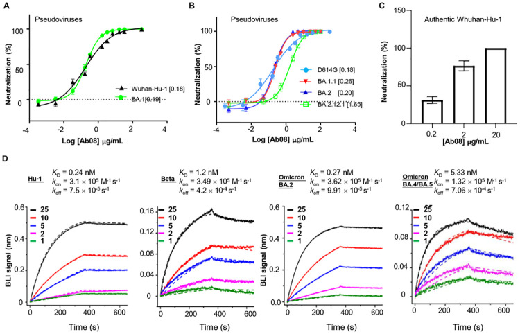
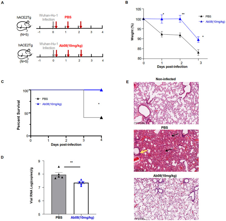
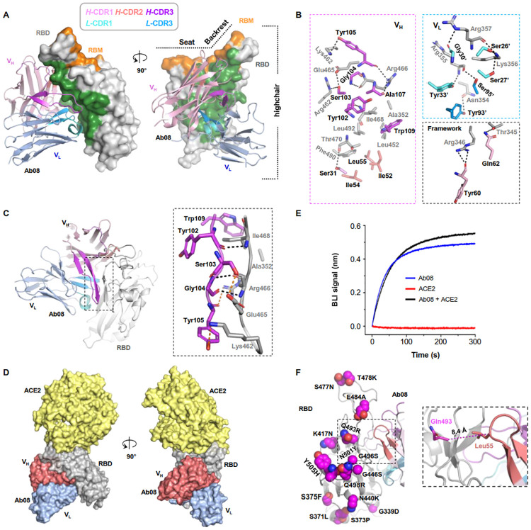
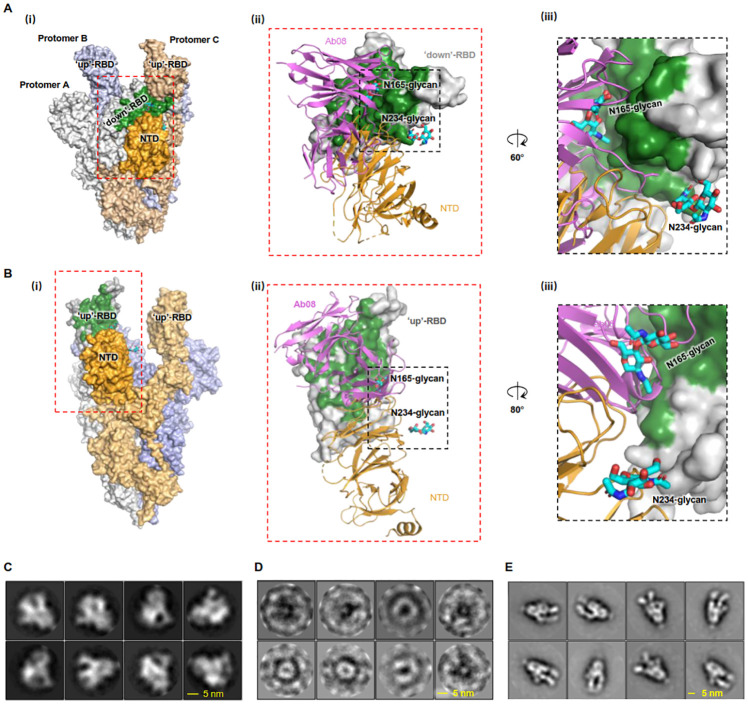
Neutralizing antibodies (nAbs) are important assets to fight COVID-19, but most existing nAbs lose the activities against Omicron subvariants. Here, we report a human monoclonal antibody (Ab08) isolated from a convalescent patient infected with the prototype strain (Wuhan-Hu-1). Ab08 binds to the receptor-binding domain (RBD) with pico-molar affinity (230 pM), effectively neutralizes SARS-CoV-2 and variants of concern (VOCs) including Alpha, Beta, Gamma, Mu, Omicron BA.1 and BA.2, and to a lesser extent for Delta and Omicron BA.4/BA.5 which bear the L452R mutation. Of medical importance, Ab08 shows therapeutic efficacy in SARS-CoV-2-infected hACE2 mice. X-ray crystallography of the Ab08-RBD complex reveals an antibody footprint largely in the β-strand core and away from the ACE2-binding motif. Negative staining electron-microscopy suggests a neutralizing mechanism through which Ab08 destructs the Spike trimer. Together, our work identifies a nAb with therapeutic potential for COVID-19.
Copyright: © 2023 Meng et al. This is an open access article distributed under the terms of the Creative Commons Attribution License, which permits unrestricted use, distribution, and reproduction in any medium, provided the original author and source are credited.
Conflict of interest statement
I have read the journal’s policy and the authors of this manuscript have the following competing interests: A patent application for monoclonal antibody therapy for the treatment of COVID-19 has been filed for Ab08 with application No. 202110125913.6 (China National Intellectual Property Administration). Congli Jiang is an employee of the Shenzhen Kangtai Biological Product Company.
Figures
Similar articles
-
A human monoclonal antibody neutralizing SARS-CoV-2 Omicron variants containing the L452R mutation.J Virol. 2024 Dec 17;98(12):e0122324. doi: 10.1128/jvi.01223-24. Epub 2024 Nov 4. J Virol. 2024. PMID: 39494911 Free PMC article.
-
Structure-Based Optimization of One Neutralizing Antibody against SARS-CoV-2 Variants Bearing the L452R Mutation.Viruses. 2024 Apr 5;16(4):566. doi: 10.3390/v16040566. Viruses. 2024. PMID: 38675908 Free PMC article.
-
Structural Study of SARS-CoV-2 Antibodies Identifies a Broad-Spectrum Antibody That Neutralizes the Omicron Variant by Disassembling the Spike Trimer.J Virol. 2022 Aug 24;96(16):e0048022. doi: 10.1128/jvi.00480-22. Epub 2022 Aug 4. J Virol. 2022. PMID: 35924918 Free PMC article.
-
Current Status and Perspectives of Therapeutic Antibodies Targeting the Spike Protein S2 Subunit against SARS-CoV-2.Biol Pharm Bull. 2024;47(5):917-923. doi: 10.1248/bpb.b23-00639. Biol Pharm Bull. 2024. PMID: 38692869 Review.
-
Status and Developing Strategies for Neutralizing Monoclonal Antibody Therapy in the Omicron Era of COVID-19.Viruses. 2023 May 31;15(6):1297. doi: 10.3390/v15061297. Viruses. 2023. PMID: 37376597 Free PMC article. Review.
Cited by
-
Is there any revival of the use of plasma therapy or neutralizing convalescent antibody therapy to treat SARS-CoV-2 variants and are we rethinking preparedness plans?Transfus Apher Sci. 2023 Jun;62(3):103726. doi: 10.1016/j.transci.2023.103726. Epub 2023 May 8. Transfus Apher Sci. 2023. PMID: 37169698 Free PMC article. Review. No abstract available.
-
A monoclonal antibody targeting a large surface of the receptor binding motif shows pan-neutralizing SARS-CoV-2 activity.Nat Commun. 2024 Feb 5;15(1):1051. doi: 10.1038/s41467-024-45171-9. Nat Commun. 2024. PMID: 38316751 Free PMC article.
-
A human monoclonal antibody neutralizing SARS-CoV-2 Omicron variants containing the L452R mutation.J Virol. 2024 Dec 17;98(12):e0122324. doi: 10.1128/jvi.01223-24. Epub 2024 Nov 4. J Virol. 2024. PMID: 39494911 Free PMC article.
-
Structure-Based Optimization of One Neutralizing Antibody against SARS-CoV-2 Variants Bearing the L452R Mutation.Viruses. 2024 Apr 5;16(4):566. doi: 10.3390/v16040566. Viruses. 2024. PMID: 38675908 Free PMC article.
-
Novel Competitive ELISA Utilizing Trimeric Spike Protein of SARS-CoV-2, Could Identify More Than RBD-RBM Specific Neutralizing Antibodies in Hybrid Sera.Vaccines (Basel). 2024 Aug 13;12(8):914. doi: 10.3390/vaccines12080914. Vaccines (Basel). 2024. PMID: 39204038 Free PMC article.
References
Publication types
MeSH terms
Substances
Supplementary concepts
LinkOut - more resources
Full Text Sources
Medical
Research Materials
Miscellaneous
