. 2023 Jan 27;9(4):eade1444.
doi: 10.1126/sciadv.ade1444. Epub 2023 Jan 27.

Ionizable lipid nanoparticles deliver mRNA to pancreatic β cells via macrophage-mediated gene transfer

Affiliations

Ionizable lipid nanoparticles deliver mRNA to pancreatic β cells via macrophage-mediated gene transfer

Jilian R Melamed et al. Sci Adv. .

Abstract

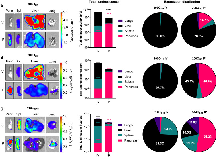
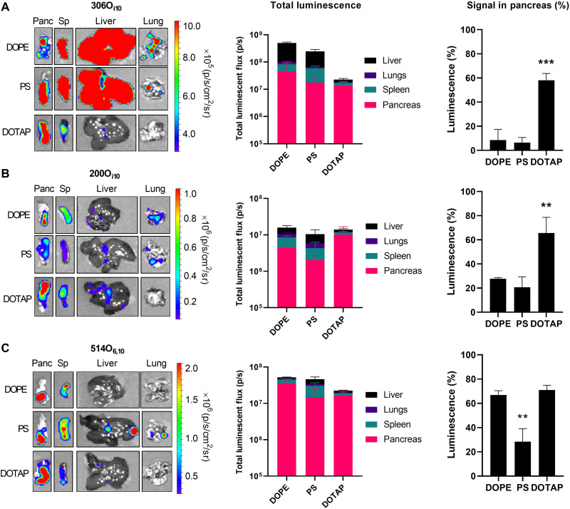
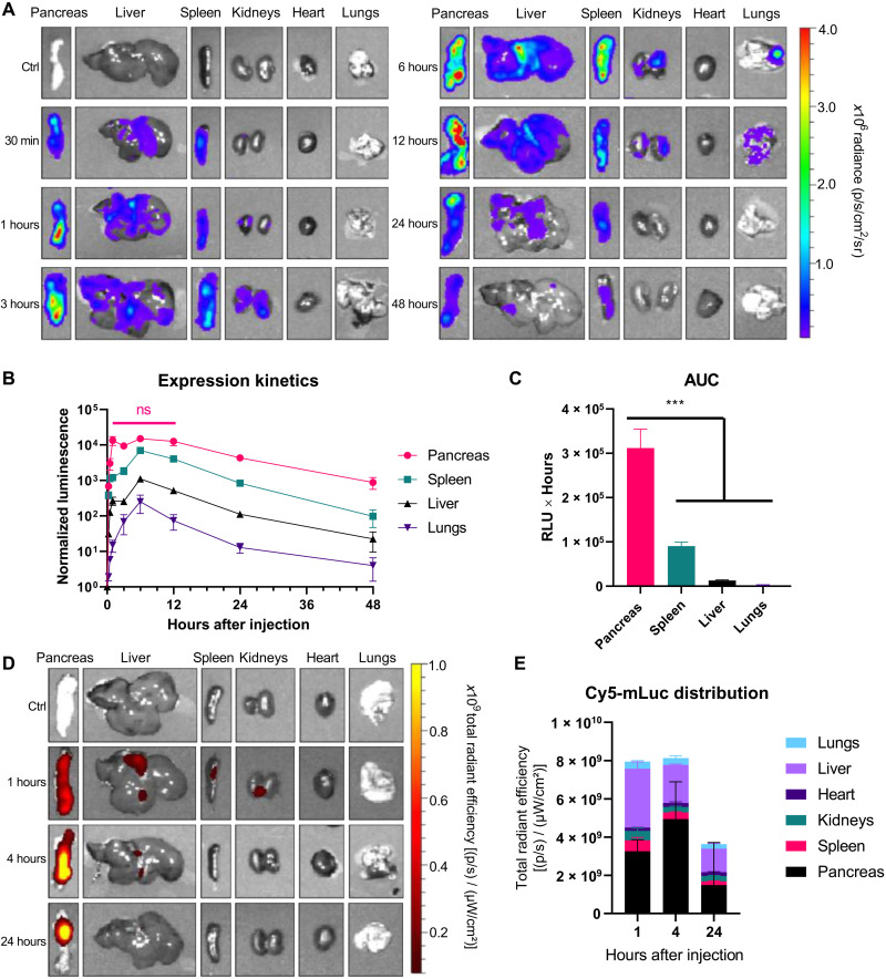
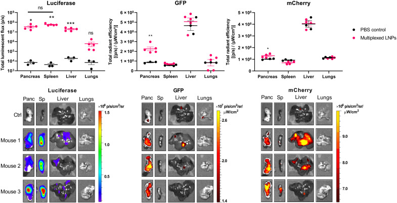
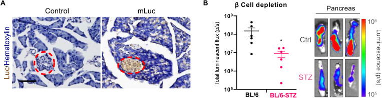
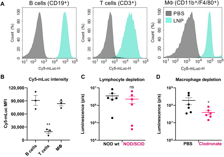
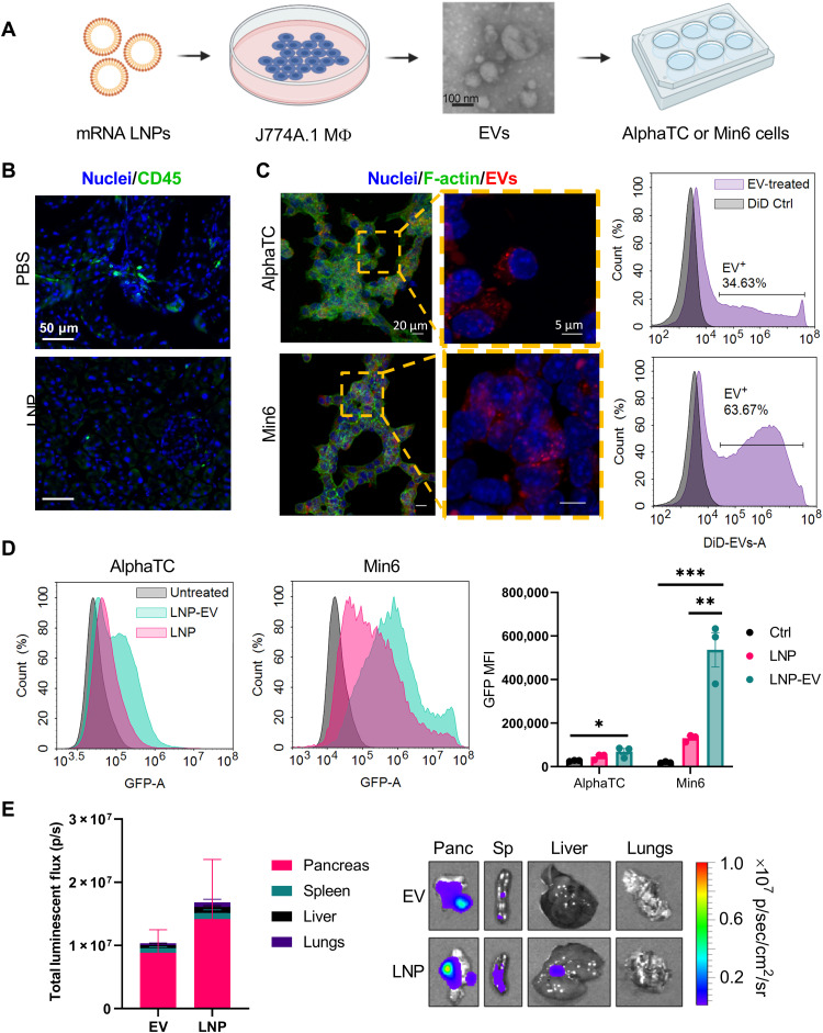
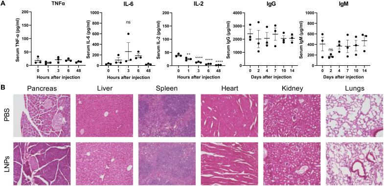
Systemic messenger RNA (mRNA) delivery to organs outside the liver, spleen, and lungs remains challenging. To overcome this issue, we hypothesized that altering nanoparticle chemistry and administration routes may enable mRNA-induced protein expression outside of the reticuloendothelial system. Here, we describe a strategy for delivering mRNA potently and specifically to the pancreas using lipid nanoparticles. Our results show that delivering lipid nanoparticles containing cationic helper lipids by intraperitoneal administration produces robust and specific protein expression in the pancreas. Most resultant protein expression occurred within insulin-producing β cells. Last, we found that pancreatic mRNA delivery was dependent on horizontal gene transfer by peritoneal macrophage exosome secretion, an underappreciated mechanism that influences the delivery of mRNA lipid nanoparticles. We anticipate that this strategy will enable gene therapies for intractable pancreatic diseases such as diabetes and cancer.


Figures

Fig. 1.
Fig. 1.. Intraperitoneal administration improves pancreatic mRNA delivery relative to intravenous injection.
LNPs containing mLuc were formulated using each of three ionizable lipidoids—(A) 306Oi10, (B) 200Oi10, or (C) 514O6,10—at a molar ratio of 35% lipidoid/16% DOPE/46.5% cholesterol/2.5% PEG-lipid and administered to C57BL/6 mice (mRNA at a dose of 0.5 mg/kg) (n = 3 mice per group). Three hours later, mice were injected with d-luciferin, euthanized, and dissected for ex vivo luminescence imaging using in vivo imaging system (IVIS). The left panels depict representative IVIS images of key organs, the middle panels quantify mLuc expression, the right panels illustrate the percentage of protein expression occurring per organ. Compared to intravenous (IV) delivery, intraperitoneal (IP) administration increased mRNA delivery for all formulations. Error bars represent SEM. Student’s t tests were used to compare intravenous and intraperitoneal delivery for each organ. ****P < 0.005 and ****P < 0.001. Data represent three biological replicates. Statistics are color-coded according to organ. Panc, pancreas; Sp, spleen.
Fig. 2.
Fig. 2.. The helper lipid DOTAP improves specificity for the pancreas by decreasing off-target protein expression.
LNPs containing mLuc were formulated using each of three different lipidoids—(A) 306Oi10, (B) 200Oi10, or (C) 514O6,10—in a molar ratio of 35% lipidoid/40% helper lipid/22.5% cholesterol/2.5% PEG-lipid and administered to C57BL/6 mice (mRNA at a dose of 0.5 mg/kg) (n = 3 mice per group). Three hours later, mice were injected with d-luciferin, euthanized, and dissected for ex vivo luminescence imaging using IVIS. The left panels depict representative IVIS images of key organs, the middle panels quantify mLuc expression, the right panels illustrate the percentage of protein expression occurring in the pancreas. Error bars represent SEM. Analysis of variance (ANOVA) with post hoc Tukey-Kramer was used to compare the fraction of total signal that occurred within the pancreas across helper lipids. **P < 0.01 and ***P < 0.005.
Fig. 3.
Fig. 3.. Protein expression in the pancreas persists for at least 48 hours following mRNA LNP injection.
LNPs containing (A to C) mLuc or (D and E) Cy5-mLuc were formulated using the lipidoid 306Oi10 in a molar ratio of 35% lipidoid/40% DOTAP/22.5% cholesterol/2.5% PEG-lipid and administered to C57BL/6 mice (mRNA at a dose of 0.5 mg/kg) (n = 3 mice per group). Unlabeled mLuc (A to C) allows the detection of functional translated protein, while Cy5-mLuc (D and E) is used to detect the presence of mRNA molecules. At the indicated times, mice were injected with d-luciferin, euthanized, and dissected for ex vivo luminescence (A to C) or fluorescence (D and E) imaging using IVIS. Error bars represent SEM. In (C), ANOVA with post hoc Tukey-Kramer was used to compare the area under the curve (AUC) for each organ; ***P < 0.005. Ctrl, control.
Fig. 4.
Fig. 4.. Multiple mRNAs can be simultaneously delivered to the pancreas.
LNPs containing mRNA encoding firefly luciferase, GFP, and mCherry were formulated using the lipidoid 306Oi10 in a molar ratio of 35% lipidoid/40% DOTAP/22.5% cholesterol/2.5% PEG-lipid and administered to C57BL/6 mice (mRNA at a dose of 0.6 mg/kg) (0.2 mg/kg each mRNA, n = 5 mice per group). Six hours later, mice were injected with d-luciferin, euthanized, and dissected for ex vivo luminescence and fluorescence imaging using IVIS. Error bars represent SEM. Student’s t test was used to compare signal from control untreated mice versus mice receiving multiplexed LNPs for each organ. *P < 0.05, **P < 0.01, and ***P < 0.005. ns, not significant.
Fig. 5.
Fig. 5.. LNPs transfect primarily pancreatic islets.
(A) LNPs containing mLuc were injected intraperitoneally into mice [mRNA (0.5 mg/kg)]. Six hours later, mice were euthanized, and pancreata were fixed, frozen, and sectioned onto slides. Slides were stained for luciferase by immunohistochemistry and counterstained with hematoxylin. Islets are within the red dashed ovals. Scale bar, 100 μm. Additional images are shown in fig. S6. (B) Depleting β cells in C57BL/6 mice with STZ reduces total mLuc delivery to the pancreas. Error bars represent SEM; *P = 0.036 by Student’s t test. The images on the right represent IVIS luminescence images of three independent replicates of control or STZ mice treated with mLuc LNPs.
Fig. 6.
Fig. 6.. Peritoneal macrophages contribute to pancreatic mRNA delivery following intraperitoneal administration.
(A) Mice were injected intraperitoneally with LNPs containing Cy5-mLuc (0.5 mg/kg). Immediately after injection, mice were euthanized, and peritoneal wash was collected for flow cytometry analysis. Cy5-mLuc is associated with B cells, T cells, and CD11b+/F4/80+ macrophages. (B) Median fluorescence intensity (MFI) of Cy5-mLuc in B cells, T cells, and CD11b+/F4/80+ macrophages. (C) Lymphocyte trafficking does not facilitate mRNA delivery to the pancreas. Wild-type (wt) NOD mice or NOD/SCID mice that lack mature lymphocytes were intraperitoneally injected with LNPs containing mLuc or Cy5-mLuc (0.5 mg/kg) and euthanized for ex vivo IVIS analysis 3 hours later. There were no differences in either Luc expression or Cy5 distribution to the pancreas resulting from lymphocyte depletion. (D) Mice were injected intraperitoneally with PBS (control) or clodronate liposomes to deplete macrophages. Forty-eight hours later, mice were injected intraperitoneally with mLuc-LNPs, and luminescence in the pancreas was quantified by IVIS. Transfection efficacy decreased in macrophage-depleted mice. Error bars represent SEM. *P < 0.05 and **P < 0.005 by one-way ANOVA with post hoc Tukey’s test.
Fig. 7.
Fig. 7.. EVs isolated from LNP-treated macrophages transfect pancreatic islet cells.
(A) To determine the role of EVs in pancreatic mRNA delivery, J774A.1 macrophages were treated with mRNA LNPs. 48 hours later, EVs were isolated from the supernatant by size exclusion chromatography (SEC) and characterized by electron microscopy (additional characterization in the Supplementary Materials). Purified EVs (20 μg) were then added to AlphaTC Clone 9 α cells or Min6 β cells in culture. (B) CD45 staining of mouse pancreas sections indicated no immune cell infiltration following LNP injection [mLuc (0.5 mg/kg), 3 hours after injection]. Additional replicates are shown in fig. S7. (C) Uptake of DiD-labeled EVs by AlphaTC and Min6 cells was assessed by confocal microscopy and flow cytometry. In the flow cytometry histograms, the control sample represents a control for residual DiD after exosome staining. (D) By flow cytometry, EVs from J774A.1 macrophages treated with mGFP mRNA-LNPs induce greater GFP expression in AlphaTC and Min6 cells than LNPs. AlphaTC and Min6 cells received the same LNP dose as the J774A.1 macrophages. (E) Peritoneal macrophages were isolated from mice and transfected with LNPs ex vivo. EVs were then isolated from the macrophages and injected intraperitoneally into mice, inducing pancreatic protein expression. Error bars represent SEM. *P < 0.05, **P < 0.01, and ***P < 0.001 by one-way ANOVA with post hoc Tukey’s test.
Fig. 8.
Fig. 8.. Pancreas-targeting LNPs are tolerated by mice.
In these studies, mice were treated with mLuc mRNA-LNPs (0.5 mg/kg), and serum or tissue samples were collected for analysis at the indicated time points. (A) By enzyme-linked immunosorbent assay, there are no significant increases in systemic inflammatory cytokines, IgG, or IgM after LNP treatment. (B) H&E staining indicates no signs of tissue damage 8 days after mRNA-LNP treatment. Images were collected at ×20 magnification.

Similar articles

Cited by

References

    1. N. Chaudhary, D. Weissman, K. A. Whitehead, mRNA vaccines for infectious diseases: Principles, delivery and clinical translation. Nat. Rev. Drug Discov. 20, 817–838 (2021). - PMC - PubMed
    1. N. Pardi, M. J. Hogan, R. S. Pelc, H. Muramatsu, H. Andersen, C. R. De Maso, K. A. Dowd, L. L. Sutherland, R. M. Scearce, R. Parks, W. Wagner, A. Granados, J. Greenhouse, M. Walker, E. Willis, J.-S. Yu, C. E. McGee, G. D. Sempowski, B. L. Mui, Y. K. Tam, Y.-J. Huang, D. Vanlandingham, V. M. Holmes, H. Balachandran, S. Sahu, M. Lifton, S. Higgs, S. E. Hensley, T. D. Madden, M. J. Hope, K. Karikó, S. Santra, B. S. Graham, M. G. Lewis, T. C. Pierson, B. F. Haynes, D. Weissman, Zika virus protection by a single low-dose nucleoside-modified mRNA vaccination. Nature 543, 248–251 (2017). - PMC - PubMed
    1. M. Brazzoli, D. Magini, A. Bonci, S. Buccato, C. Giovani, R. Kratzer, V. Zurli, S. Mangiavacchi, D. Casini, L. M. Brito, E. De Gregorio, P. W. Mason, J. B. Ulmer, A. J. Geall, S. Bertholet, Induction of broad-based immunity and protective efficacy by self-amplifying mRNA vaccines encoding influenza virus hemagglutinin. J. Virol. 90, 332–344 (2016). - PMC - PubMed
    1. M. J. Mulligan, K. E. Lyke, N. Kitchin, J. Absalon, A. Gurtman, S. Lockhart, K. Neuzil, V. Raabe, R. Bailey, K. A. Swanson, P. Li, K. Koury, W. Kalina, D. Cooper, C. Fontes-Garfias, P.-Y. Shi, Ö. Türeci, K. R. Tompkins, E. E. Walsh, R. Frenck, A. R. Falsey, P. R. Dormitzer, W. C. Gruber, U. Şahin, K. U. Jansen, Phase I/II study of COVID-19 RNA vaccine BNT162b1 in adults. Nature 586, 589–593 (2020). - PubMed
    1. L. A. Jackson, E. J. Anderson, N. G. Rouphael, P. C. Roberts, M. Makhene, R. N. Coler, M. P. M. Cullough, J. D. Chappell, M. R. Denison, L. J. Stevens, A. J. Pruijssers, A. M. Dermott, B. Flach, N. A. Doria-Rose, K. S. Corbett, K. M. Morabito, S. O’Dell, S. D. Schmidt, P. A. Swanson II, M. Padilla, J. R. Mascola, K. M. Neuzil, H. Bennett, W. Sun, E. Peters, M. Makowski, J. Albert, K. Cross, W. Buchanan, R. Pikaart-Tautges, J. E. Ledgerwood, B. S. Graham, J. H. Beigel; mRNA- Study Group , An mRNA vaccine against SARS-CoV-2 — Preliminary report. N. Engl. J. Med. 383, 1920–1931 (2020). - PMC - PubMed