. 2023 Jan 27;9(4):eadd4969.
doi: 10.1126/sciadv.add4969. Epub 2023 Jan 27.

SUMOylation-mediated PSME3-20 S proteasomal degradation of transcription factor CP2c is crucial for cell cycle progression

Affiliations

SUMOylation-mediated PSME3-20 S proteasomal degradation of transcription factor CP2c is crucial for cell cycle progression

Seung Han Son et al. Sci Adv. .

Abstract

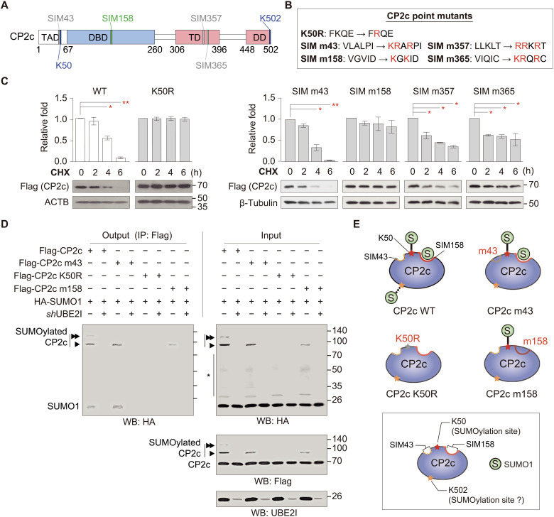
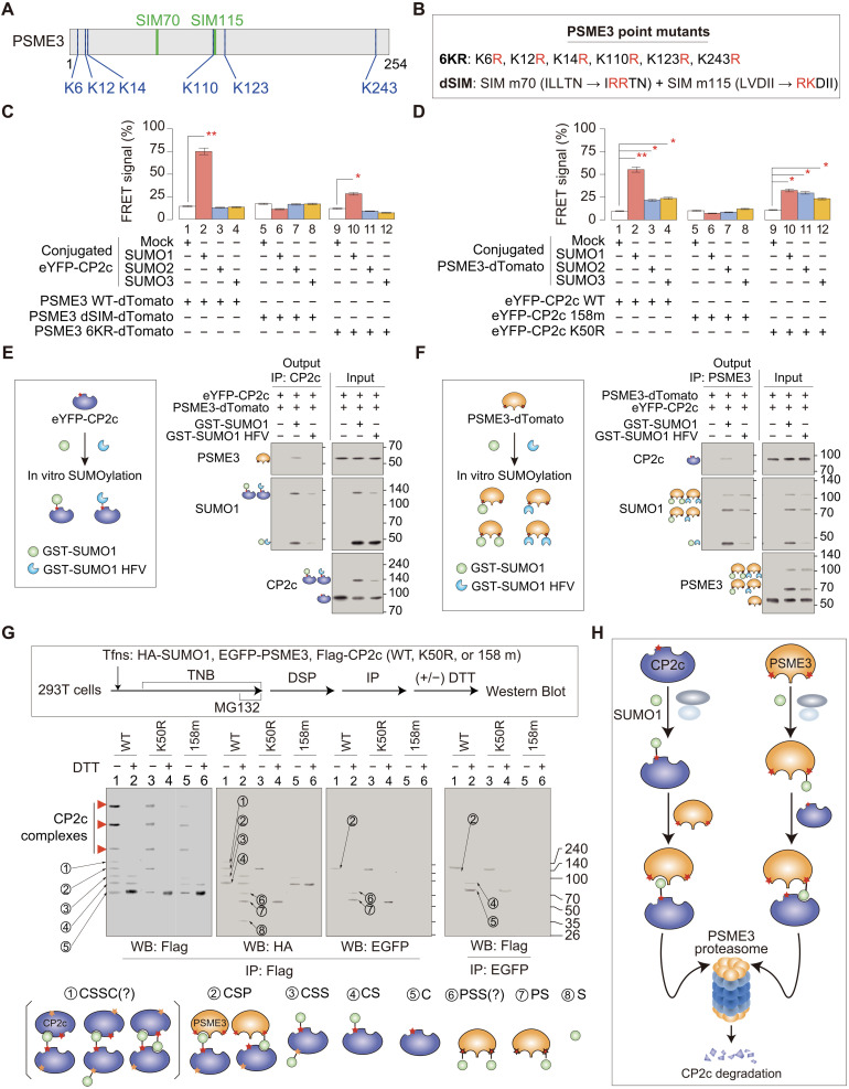
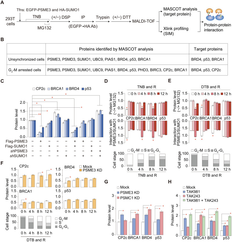
Transcription factor CP2c (also known as TFCP2, α-CP2, LSF, and LBP-1c) is involved in diverse ubiquitous and tissue/stage-specific cellular processes and in human malignancies such as cancer. Despite its importance, many fundamental regulatory mechanisms of CP2c are still unclear. Here, we uncover an unprecedented mechanism of CP2c degradation via a previously unidentified SUMO1/PSME3/20S proteasome pathway and its biological meaning. CP2c is SUMOylated in a SUMO1-dependent way, and SUMOylated CP2c is degraded through the ubiquitin-independent PSME3 (also known as REGγ or PA28)/20S proteasome system. SUMOylated PSME3 could also interact with CP2c to degrade CP2c via the 20S proteasomal pathway. Moreover, precisely timed degradation of CP2c via the SUMO1/PSME3/20S proteasome axis is required for accurate progression of the cell cycle. Therefore, we reveal a unique SUMO1-mediated uncanonical 20S proteasome degradation mechanism via the SUMO1/PSME3 axis involving mutual SUMO-SIM interaction of CP2c and PSME3, providing previously unidentified mechanistic insights into the roles of dynamic degradation of CP2c in cell cycle progression.


Figures

Fig. 1.
Fig. 1.. SUMO1, UBE2I, and PIAS1 interact with CP2c and are involved in CP2c degradation probably through CP2c SUMOylation.
(A) SUMO1, UBE2I, and PIAS1 are identified as CP2c-interacting proteins in the yeast two-hybrid assay. A mouse CP2c protein region used as a bait for yeast two-hybrid is schematically represented along with putative CP2c functional domains (top). The β-galactosidase activities are by colony filter lift assays (bottom). The yeast strain EGY48 was cotransformed with pLexA-CP2c (amino acids 306 to 502) and pB42AD-SUMO1 (1 to 101 amino acids), UBE2I (1 to 158 amino acids), and PIAS1-1 (5 to 651 amino acids). The pLexA-p53/pB42AD-T antigen and pLexA-p53/pB42AD were used as positive and negative controls, respectively. TAD, transcriptional activation domain; DBD, DNA binding domain; TD, tetramerization domain; DD, dimerization domain. (B) Representative co-IPs (n = 2) showing a direct interaction between CP2c and SUMO1, UBE2I, or PIAS1 in vivo. (C) Representative Western blot analyses (n = 3) showing CP2c degradation by the SUMOylation system. CP2c is degraded by ectopic SUMO1 expression. (D and E) SUMO1-mediated CP2c degradation was revealed by measuring CP2c protein half-life with pulse-chase metabolic labeling assays in K562 cells (D) and by measuring the expression level of CP2c protein in 293T cells under the CHX treatment (E). Various SUMO1 constructs were transiently transfected: mock, no transfection; shmSUMO1, mutant short hairpin SUMO1 RNA; shSUMO1, short hairpin SUMO1 RNA; EGFP-SUMO1, EGFP-tagged SUMO1. Data are means ± SD; n = 2. *P < 0.05; **P < 0.01. (F) Representative Western blot analyses (n = 3) show that CP2c degradation by SUMO1 requires UBE2I and PIAS1. (G) Representative Western blot (n = 2) showing CP2c SUMOylation status by in vitro conjugation assay of SUMO1, SUMO2, or SUMO3. See also fig. S1.
Fig. 2.
Fig. 2.. Identification of CP2c SUMOylation- and degradation-related structural signatures in the CP2c protein.
(A) Schematic drawing of CP2c protein showing putative SUMOylation sites and SIMs predicted by a GPS-SUMO 1.0 program. (B) CP2c constructs with point mutations in the putative SUMOylation and SIM sites. (C) CP2c protein stability tests of each CP2c mutation. Flag-tagged CP2c WT and various mutants were transiently transfected to 293T cells with HA-SUMO1, and the time-dependent CP2c protein levels were quantified by Western blot. Data are means ± SD; n = 2. *P < 0.05; **P < 0.01. (D) Representative co-IPs (n = 3) showing UBE2I-dependent CP2c SUMOylation phenomenon and the SUMO1 binding and SUMOylation status of each CP2c mutant. It is worth noting that MG132 (50 μM) and NEM (25 mM) were treated in cells for 12 hours before cell harvest and during cell extract preparation, respectively, to enhance SUMOylation signals by preventing CP2c degradation and deSUMOylation. An asterisk indicates other cellular SUMOylated proteins. (E) Schematic representation of CP2c SUMOylation sites and SIMs responsible for CP2c SUMOylation and degradation.
Fig. 3.
Fig. 3.. The SUMOylated CP2c is degraded through the ubiquitin- and STUbL-independent PSME3 proteasome system.
(A) CP2c is degraded via the endogenous SUMO conjugation machinery in a ubiquitin-independent manner in vivo. TAK981 and TAK243, specific inhibitors for the SUMOylation and ubiquitination system, respectively, were used to demonstrate which endogenous system is involved in the degradation of endogenous CP2c in cells. A nonconjugatable SUMO1 form (SUMO1 ∆GG) was also used as a control. n = 2. See also fig. S4 for additional data. (B) Representative co-IPs (n = 3) demonstrate that CP2c degradation occurs in a STUbL-independent manner. Both siRNF4 and siRNF111 were added to the MDA-MB-231 cells to inhibit endogenous STUbLs. TGF-β (1 ng/ml for 6 hours before cell harvest) was added to elicit the RNF111-dependent ubiquitination and degradation condition of poly-SUMOylated substrates, SKI and SKIL. See also fig. S5 for a detailed demonstration of the effects of the RNF4 and RNF111 STUbLs on the degradation of SUMOylated CP2c. (C) Representative Western blots (n = 2) show that CP2c degradation requires PSME3 (11S subunit) (left) but not PSMC1 (19S subunit) (right). Doxycycline-inducible PSME3 and PSMC1 KD constructs were transiently transfected with EGFP-SUMO1 or His-ubiquitin or in combination to 293T cells, and the doxycycline-dependent CP2c degradation was monitored. *, other SUMOylated or ubiquitinated proteins. (D) Representative Western blots (n = 3) showing the markedly reduced CP2c stability in the SUMO1 and PSME3 cotransfected cells. (E) Representative co-IPs (n = 2) showing binding of PSME3 to endogenous CP2c in UBE2I- and PIAS1-dependent manners. MG132 (10 μM) was treated in cells for 6 hours before cell harvest. (F) Representative Western blots (n = 2) show that SUMO1-mediated SUMOylation of CP2c is sufficient for degradation by PSME3 (the 20S proteasome) in vitro. Colored arrows and dots represent BRCA1- or CP2c-interacted or BRCA1- or CP2c-conjugated factors, respectively.
Fig. 4.
Fig. 4.. CP2c and PSME3 recognize each other through mutual SUMO-SIM interactions.
(A) Schematic representation of PSME3 showing putative SUMOylation and SIM sites. (B) Various PSME3 mutant constructs with point mutations in the putative SUMOylation and SIM sites. (C and D) Relative mean FRET percentages between the eYFP-CP2c WT with or without SUMO conjugation and the non-SUMOylated PSME3 WT, dSIM, or 6KR-dTomato, respectively (C), and between the PSME WT-dTomato with or without SUMO conjugation and the non-SUMOylated eYFP-CP2c WT, 158m, or K50R, respectively (D). n = 3. See fig. S7 (A to F) for the original data. (E and F) Representative co-IP assays (n = 2) show that CP2c and PSME3 recognize each other through the direct CP2c-SUMO1-PSME3 interaction. Epitope-tagged CP2c (E) or PSME3 (F) were subjected to the in vitro SUMOylation reaction using WT or the SUMO1 HFV mutant, and the resulting samples were incubated with PSME3 or CP2c, respectively, to see specific interactions using co-IPs. (G) Representative DSP XL-IP analyses (n = 3) identifying PSME3-CP2c protein subcomplexes in the reactions containing WT or CP2c mutants. The schematic experimental procedure (top) and the expected protein complex models (marked by the circled letters in the Western blots; bottom) are depicted. DSP, dithiobis (succinimidyl propionate); DTT, dithiothreitol; FRET, fluorescence resonance energy transfer; TNB, thymidine-nocodazole block; XL-IP, crosslinking immunoprecipitation. (H) Schematic model showing CP2c degradation through two ubiquitin-independent PSME3 proteasome pathways. CP2c degradation could occur by the coupled interactions of CP2c and PSME3 through bindings between SUMO in one protein and SIM in the other protein.
Fig. 5.
Fig. 5.. CP2c degradation through the SUMO1/PSME3/20S proteasome axis is crucial for proper cell cycle progression.
(A and B) Confocal microscopy showing the CP2c colocalization with PSME3 at the G2-M phase of the cell cycle. The CP2c eYFP-tagged CP2c and the dTomato-tagged PSME3 were transfected into 293T cells, and the synchronized cells were prepared by the TNB & R protocol (A) or by the DTB & R protocol (B). FRET signals were considered as colocalization. Data are means ± SD; n = 2. (C) Representative Western blots of proteins (left) and the quantified expression (right) in 293T cells after a thymidine block and release (TB & R). Data are means ± SD of duplicated experiments; *P < 0.05. The percentage of cell populations is shown at the bottom. (D) Representative co-IPs (n = 2) showing a cell cycle stage–dependent distribution of SUMOylated CP2c and PSME3 in the CP2c-PSME3 complexes. See also fig. S9 for additional data. (E) Cell cycle distribution profiles show the effects of the CP2c mutation in either SUMOylation or SIM on cell cycle progression. n = 2, *P < 0.05. See fig. S11 (A and B) for the overlay histograms of the original cell cycle analyses and similar analyses using CP2c dominant negative (62). (F) Schematic depiction of two CP2c degradation mechanisms during cell cycle progression. Red-colored asterisks represent SUMOylation sites, whereas grooves in the protein represent SIMs.
Fig. 6.
Fig. 6.. The SUMO1/PSME3/20S proteasome system is also involved in the cell cycle–dependent degradation of some other nuclear factors.
(A and B) Experimental scheme identifying the cellular proteins simultaneously interacting with both SUMO1 and PSME3 (A) and the identified proteins (B). A protein was scored as positive when it appeared repeatedly in duplicated experiments. See also fig. S12A. (C) Western blot testing the SUMO1/PSME3 proteasome–dependent protein degradation in 293T cells, where various constructs were transiently transfected. See also fig. S12B. (D and E) Cell stage–dependent protein levels and protein interactions with SUMO1 and PSME3 were measured in cells obtained by a TNB & R (D) or a DTB & R (E) protocol in the presence or absence of MG132 (see also fig. S12, C and D). (F) Cell stage–specific protein degradation profiles in cell stage–synchronized PSME3 KD cells (see also fig. S12E). (G and H) Tests discriminating two mechanisms of protein degradation, a ubiquitin-dependent 26S proteasome pathway, and a SUMO1/PSME3 proteasome pathway. Cellular protein levels were quantified in PSME3 or PSMC1 KD 293T cells (G) or in cells where SUMOylation and/or ubiquitination were inhibited (H) (see also fig. S12, E and F). All of the data in each panel obtained from duplicated experiments are shown as means ± SD. *P < 0.05; **P < 0.01.

Similar articles

Cited by

References

    1. Kang H. C., Chae J. H., Lee Y. H., Park M.-A., Shin J. H., Kim S.-H., Ye S.-K., Cho Y. S., Fiering S., Kim C. G., Erythroid cell-specific α-globin gene regulation by the CP2 transcription factor family. Mol. Cell. Biol. 25, 6005–6020 (2005). - PMC - PubMed
    1. Rodda S., Sharma S., Scherer M., Chapman G., Rathjen P., CRTR-1, a developmentally regulated transcriptional repressor related to the CP2 family of transcription factors. J. Biol. Chem. 276, 3324–3332 (2001). - PubMed
    1. Wilanowski T., Tuckfield A., Cerruti L., O’Connell S., Saint R., Parekh V., Tao J., Cunningham J. M., Jane S. M., A highly conserved novel family of mammalian developmental transcription factors related to Drosophila grainyhead. Mech. Dev. 114, 37–50 (2002). - PubMed
    1. Yoon J. B., Li G., Roeder R. G., Characterization of a family of related cellular transcription factors which can modulate human immunodeficiency virus type 1 transcription in vitro. Mol. Cell. Biol. 14, 1776–1785 (1994). - PMC - PubMed
    1. Jane S. M., Nienhuis A. W., Cunningham J. M., Hemoglobin switching in man and chicken is mediated by a heteromeric complex between the ubiquitous transcription factor CP2 and a developmentally specific protein. EMBO J. 14, 97–105 (1995). - PMC - PubMed