. 2023 Jan 27;9(4):eadd2873.
doi: 10.1126/sciadv.add2873. Epub 2023 Jan 27.

Conserved meiotic mechanisms in the cnidarian Clytia hemisphaerica revealed by Spo11 knockout

Affiliations

Conserved meiotic mechanisms in the cnidarian Clytia hemisphaerica revealed by Spo11 knockout

Catriona Munro et al. Sci Adv. .

Abstract

During meiosis, DNA recombination allows the shuffling of genetic information between the maternal and paternal chromosomes. Recombination is initiated by double-strand breaks (DSBs) catalyzed by the conserved enzyme Spo11. How this crucial event is connected to other meiotic processes is unexpectedly variable depending on the species. Here, we knocked down Spo11 by CRISPR in the jellyfish Clytia hemisphaerica. Germ cells in Clytia Spo11 mutants fail to assemble synaptonemal complexes and chiasmata, and in consequence, homologous chromosome pairs in females remain unassociated during oocyte growth and meiotic divisions, creating aneuploid but fertilizable eggs that develop into viable larvae. Clytia thus shares an ancient eukaryotic dependence of synapsis and chromosome segregation on Spo11-generated DSBs. Phylogenetically, Clytia belongs to Cnidaria, the sister clade to Bilateria where classical animal model species are found, so these results provide fresh evolutionary perspectives on meiosis regulation.


Figures

Fig. 1.
Fig. 1.. Phylogenetic placement, life cycle, and gonad organization of C. hemisphaerica.
(A) Evolutionary relationships among major animal meiosis models. Silhouettes from PhyloPic.org, S. mediterranea: credit to Noah Schlottman, C. hemisphaerica: credit to Joseph Ryan, photo by Patrick Steinmetz, under a share-alike license https://creativecommons.org/licenses/by-sa/3.0/. (B) Life cycle of C. hemisphaerica. The polyp colony propagates asexually for many years in the laboratory. Specialized polyps called gonozooids release jellyfish continuously. Sexually mature jellyfish release eggs or sperm daily in response to dark-light transitions. The embryo develops into a “planula” larva, which settles and metamorphoses to form a primary polyp. To achieve gene knockouts, CRISPR-Cas9 is injected in the mature egg immediately before fertilization. (C) Development and organization of the male and female gonad from the first day of jellyfish release to sexual maturity. GC, gastric cavity. (D) Maximum intensity projection of a confocal image stack of a 3-day-old jellyfish stained with anti-Piwi. Go, gonad; Ra, radial canal; M, manubrium/mouth; Ten, tentacle bulb; Cir, circular canal. Scale bar, 100 μm. A similar projection from a 3-day-old gonad is shown on the right. Scale bar, 10 μm. (E) Confocal section showing oocytes at mixed meiotic stages within the gonad of a 1-week-old female jellyfish stained with telomere FISH (magenta) and Hoechst (cyan). Scale bar, 10 μm. Inset shows close-up of representative nuclei at different prophase I stages; L/Z, leptotene/zygotene; P, pachytene; D, diplotene. Arrow indicates telomere bouquet. Scale bars, 5 μm.
Fig. 2.
Fig. 2.. Meiotic progression in Clytia gonads.
Prophase I stages in females and males stained with anti-Sycp1 (yellow), anti-Sycp3 (magenta), and Hoechst dye (cyan). L, leptotene; Z, zygotene; P, pachytene; EP, early pachytene; LP, late pachytene; D, diplotene; GC, gastric cavity; S, spermatid/spermatozoa. (A) Confocal section of a 1-week-old female ovary. Scale bar, 10 μm; right inset is a close-up of representative prophase I stages. Scale bars, 5 μm. (B) Confocal section of a 3-week-old male gonad. Scale bar, 10 μm. (C and D) Zoom on the different confocal sections of a 3-week-old male gonad, highlighting leptotene/zygotene and pachytene stages. Scale bars, 10 μm.
Fig. 3.
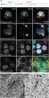
Fig. 3.. Spo11 is required for synapsis in C. hemisphaerica males and females.
(A) Diagram of the Spo11 exons indicating the target sites of CRISPR-Cas9 guides. (B) Confocal planes of wild-type and Spo11 mutant pachytene and pachytene-like nuclei stained with anti-Sycp1, anti-Sycp3, and Hoechst. Scale bars, 5 μm. (C). Transmission electron microscope (TEM) image of a wild-type SC. Scale bar, 0.5 μm. (D) TEM image of a Spo11 mutant (P16) axial element. Counts are in table S1. Scale bar, 0.5 μm.
Fig. 4.
Fig. 4.. Spo11 is required for crossovers in C. hemisphaerica.
(A) Diagram of oocyte morphology during the 24 hours after spawning; stage I oocytes undergo rapid growth and displacement of the germinal vesicle (GV) and then, at stage III (fully grown), progressive condensation of chromosomes within the GV. (B) Percentage of univalent and bivalent chromosomes counted in Spo11 mutant (P20 and P16) and wild-type fully grown oocytes (n = 10 oocytes). (C) Confocal images of stage III oocytes stained with Hoechst dye (purple) and anti–CenH3-1 (green). Left column images are of oocytes at an early stage of chromosome condensation (maximum projection of several z-stacks); right column images are of oocytes at a later stage of chromosome condensation (maximum intensity projections of z-stacks to show all chromosome pairs). Top left: Wild-type oocyte nucleus showing two homologous chromosomes with one chiasma (white arrow). Top right: Wild-type oocyte nucleus showing 15 pairs of chromosomes with one or two chiasma per pair. Bottom left: Mutant oocyte nucleus showing a single univalent (white arrowhead), with two other univalent arms in the top left and bottom right. Bottom right: Maximum intensity projection of a mutant oocyte nucleus showing 28 univalents and no bivalents (two chromatids likely obscured). Scale bars, 5 μm.
Fig. 5.
Fig. 5.. Meiotic division defects in Spo11 mutant oocytes.
DNA is stained with Hoechst dye (blue), microtubules are stained with anti–tyrosinated tubulin (green), and centromeres are stained with anti–CenH3-1 (magenta). Dotted lines contour the oocyte surface. (A) Wild-type meiotic divisions. From left to right: metaphase I (MI), first polar body (1st PB) emission, metaphase II (MII), and second polar body (2nd PB). White arrowheads indicate metaphase plates. Scale bars, 5 μm. (B) Spo11 mutant meiotic divisions. From left to right: metaphase I (MI’), white arrowhead indicates where the metaphase plate should be; failed emission of the first polar body; metaphase II (MII’); Spo11 mutant oocyte at the time of second polar body emission. Scale bars, 5 μm. (C) Examples of Spo11 mutants during meiosis II, following a failed emission of the first polar body. Scale bars, 5 μm. (D) Wild-type nucleus after meiosis is complete. DNA is stained with Hoechst dye (blue), and centromeres are stained with anti–CenH3-1 (magenta). Scale bar, 5 μm. (E) Three representative Spo11 mutant nuclei after meiosis are complete. DNA is stained with Hoechst dye (blue), and centromeres are stained with anti–CenH3-1 (magenta). Scale bars, 5 μm. (F) Metaphase spread of wild-type embryos stained with Hoechst dye. Scale bar, 5 μm. (G) Metaphase spreads of Spo11 mutant embryos stained with Hoechst dye. Scale bars, 5 μm.
Fig. 6.
Fig. 6.. Summary of meiotic progression from leptotene to the first meiotic division and postmeiosis in wild-type and Spo11 mutants.
DNA (blue), Sycp3 (magenta/pink), Sycp1 (yellow), telomeres (purple), and microtubules (gray).

References

    1. D. Zickler, N. Kleckner, Recombination, pairing, and synapsis of homologs during meiosis. Cold Spring Harb. Perspect. Biol. 7, a016626 (2015). - PMC - PubMed
    1. D. Zickler, N. Kleckner, A few of our favorite things: Pairing, the bouquet, crossover interference and evolution of meiosis. Semin. Cell Dev. Biol. 54, 135–148 (2016). - PMC - PubMed
    1. S. L. Page, R. S. Hawley, The genetics and molecular biology of the synaptonemal complex. Annu. Rev. Cell Dev. Biol. 20, 525–558 (2004). - PubMed
    1. U. Goodenough, J. Heitman, Origins of eukaryotic sexual reproduction. Cold Spring Harb. Perspect. Biol. 6, a016154 (2014). - PMC - PubMed
    1. A. J. MacQueen, A. Hochwagen, Checkpoint mechanisms: The puppet masters of meiotic prophase. Trends Cell Biol. 21, 393–400 (2011). - PubMed