. 2023 Apr 13;141(15):1871-1883.
doi: 10.1182/blood.2022017074.

Factor XII contributes to thrombotic complications and vaso-occlusion in sickle cell disease

Affiliations

Factor XII contributes to thrombotic complications and vaso-occlusion in sickle cell disease

Erica M Sparkenbaugh et al. Blood. .

Abstract

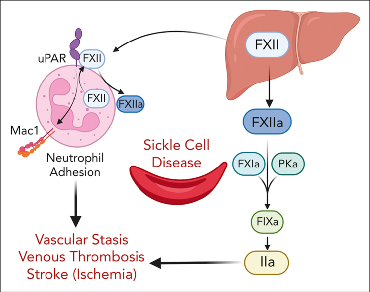
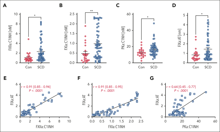
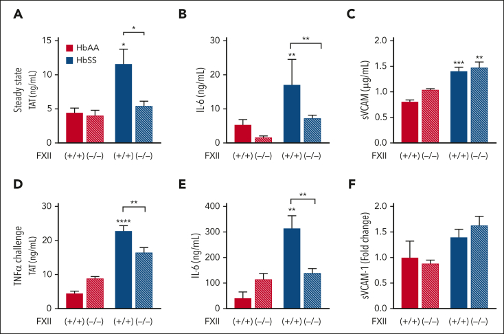
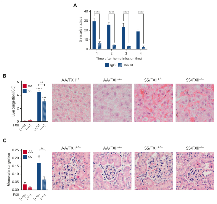
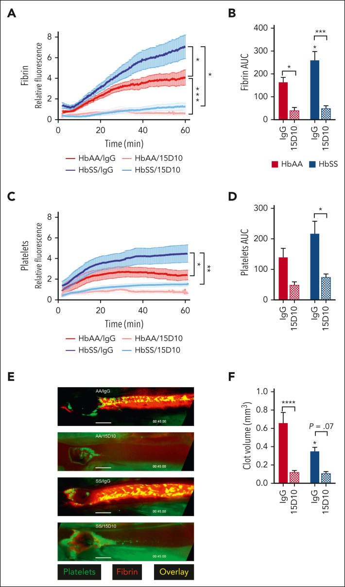
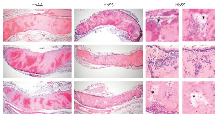
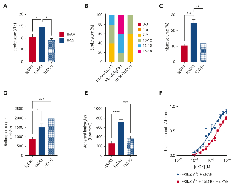
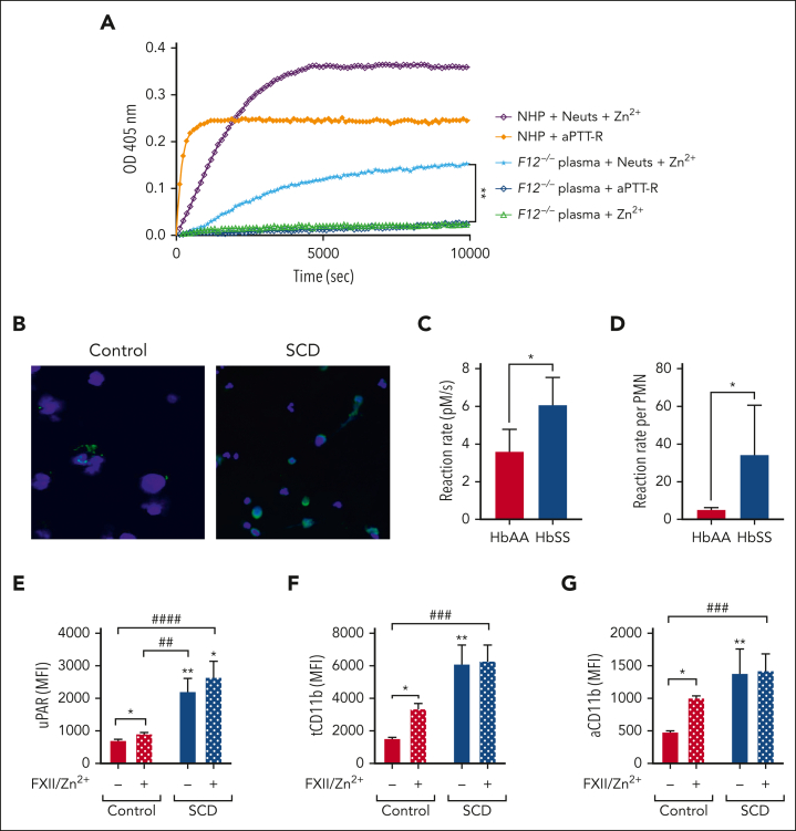
A hypercoagulable state, chronic inflammation, and increased risk of venous thrombosis and stroke are prominent features in patients with sickle cell disease (SCD). Coagulation factor XII (FXII) triggers activation of the contact system that is known to be involved in both thrombosis and inflammation, but not in physiological hemostasis. Therefore, we investigated whether FXII contributes to the prothrombotic and inflammatory complications associated with SCD. We found that when compared with healthy controls, patients with SCD exhibit increased circulating biomarkers of FXII activation that are associated with increased activation of the contact pathway. We also found that FXII, but not tissue factor, contributes to enhanced thrombin generation and systemic inflammation observed in sickle cell mice challenged with tumor necrosis factor α. In addition, FXII inhibition significantly reduced experimental venous thrombosis, congestion, and microvascular stasis in a mouse model of SCD. Moreover, inhibition of FXII attenuated brain damage and reduced neutrophil adhesion to the brain vasculature of sickle cell mice after ischemia/reperfusion induced by transient middle cerebral artery occlusion. Finally, we found higher FXII, urokinase plasminogen activator receptor, and αMβ2 integrin expression in neutrophils of patients with SCD compared with healthy controls. Our data indicate that targeting FXII effectively reduces experimental thromboinflammation and vascular complications in a mouse model of SCD, suggesting that FXII inhibition may provide a safe approach for interference with inflammation, thrombotic complications, and vaso-occlusion in patients with SCD.


Conflict of interest statement

Conflict-of-interest disclosure: R.P. and E.M.S. have received research funding from CSL. A.G. and M.W. are employees of Aronora, Inc. J.D.B. and G.M.V. have received research funding from Omeros, CSL Behring, Hillhurst Biopharmaceuticals, and Astellas/Mitobridge. J.D.B. is a consultant for Astellas/Mitobridge. G.M.V. is a consultant for Sanofi and Astellas/Mitobridge. The remaining authors declare no competing financial interests.

Figures

None
Graphical abstract
Figure 1.
Figure 1.
Markers of contact and intrinsic pathway activation are elevated in patients with SCD compared with that in healthy controls. Plasma from healthy controls (n = 23) and patients with SCD (n = 53) was assayed for (A) FXIIa-C1INH, (B) FXIa:C1INH, (C) PKa:C1INH, and (D) FIXa-AT complexes. Data are presented as mean ± SEM and analyzed by Student t test. ∗P < .05, ∗∗P < .01. Correlations between (E) FXIIa:C1INH, (F) FXIa:C1INH, and (G) PKa:C1INH with FIXa-AT were calculated and analyzed by linear regression. Mean r with range and P value is reported on graphs. AT, antithrombin.
Figure 2.
Figure 2.
Nonhematopoietic FXII contributes to increased thrombin generation and inflammation in HbSS mice at steady state and after TNFα challenge. FXII−/− and FXII+/+ mice were irradiated and received transplantation with HbAA or HbSS BM. Four months later, plasma was collected for analysis of (A) TAT complexes, (B) IL-6, and (C) sVCAM-1. In a separate study, all mice were treated with TNFα (2 μg/kg, intraperitoneally). Five hours later, plasma was collected for analysis of (D) TAT complexes, (E) IL-6, and (F) sVCAM-1. Data are represented as mean ± SEM, n = 10 to 26 per group. ∗P < .05, ∗∗P < .01, and ∗∗∗∗P < .0001 by two-way analysis of variance (ANOVA) and Tukey post-hoc test. Asterisks above bars indicate significance within the same FXII genotype. Asterisks above lines indicate difference between Hb genotype within FXII genotype.
Figure 3.
Figure 3.
FXII contributes to vascular stasis and congestion in HbSS mice. (A) Townes HbSS mice were implanted with dorsal skinfold chambers. Using intravital microscopy, between 20 and 25 subcutaneous venules were selected and mapped. Mice were treated with IgG or 15D10 (10 mg/kg, IV) 30 minutes before challenge with stroma-free Hb (1 μmol/kg, IV). The preselected venules were marked as flowing or static at 1, 2, 3, and 4 hours after Hb infusion, and the percentage static venules was calculated. n = 4 mice per group, data represent mean ± SEM. ∗∗∗∗P < .0001 vs IgG/SS at each time point by two-way ANOVA. Paraffin sections of livers and kidneys from AA/FXII+/+, AA/FXII−/−, SS/FXII+/+, and SS/FXII−/− mice 4 months after BM transplantation were stained with hematoxylin and eosin. (B) Sinusoidal congestion and (C) glomerular congestion were scored by blinded pathologists. Representative images are shown; n = 4 to 8 mice per group, data represent mean ± SEM. ∗∗P < .01, ∗∗∗P < .001, ∗∗∗∗P < .0001 by two-way ANOVA and Tukey post-hoc test. Asterisks above bar represent difference from HbAA, asterisks above line indicate difference between FXII genotype. Red staining on representative images showed in panels B and C demonstrates presence of RBC within congested vessels. ANOVA, analysis of variance.
Figure 4.
Figure 4.
FXII(a) inhibition attenuates femoral vein thrombosis induced by electrolytic injury. Townes HbAA and HbSS mice were treated with IgG or 15D10 (5 mg/kg, IV) 30 minutes before electrolytic injury to the femoral vein. Quantification of (A) relative fibrin intensity over time and (B) the total fibrin fluorescence measured by AUC. Quantification of (C) relative platelet intensity over time and (D) the total platelet fluorescence measured by AUC. Data represent mean ± SEM; n = 6 to 10 mice per group. (E) Representative images of fibrin (red) and platelet (green) accumulation taken 45 minutes after electrolytic injury. (F) Histomorphometric quantification of clot volume. Data represent mean ± SEM of clot volume (mm3); n = 6 to 10 mice per group. ∗P < .05, ∗∗P < .01, ∗∗∗P < .001 by two-way ANOVA and Tukey post-hoc test. ANOVA, analysis of variance; AUC, area under the curve.
Figure 5.
Figure 5.
SCD affects morphology of venous clots. Representative, hematoxylin and eosin–stained images of femoral vein clots harvested from HbAA and HbSS mice 1 hour after electrolytic injury, (original magnification ×4). In the clots, areas rich in RBCs stain dark red whereas areas of fibrin and platelets stain light pink. In the enlarged images on the right, asterisks denote acellular empty spaces within the clots; dark blue staining denotes inflammatory cells within the clots.
Figure 6.
Figure 6.
FXII inhibition attenuates stroke severity in HbSS mice after tMCAO. Quantification of (A) stroke score, (B) proportion of stroke severity, (C) and brain infarct in HbAA and HbSS mice subjected to brain ischemia/reperfusion injury after treatment with IgG or 15D10 antibodies (10 mg/kg, IV). Intravital microscopy analysis was performed to assess the number of (D) rolling and (E) adherent leukocytes. Data represent mean ± SEM; n = 6 mice per group. ∗P < .05, ∗∗P < .01, ∗∗∗P < .001 by one-way ANOVA and Tukey post-hoc test. (F) Microscale thermophoresis was performed to measure direct binding between uPAR and FXII, in the absence (blue line) or presence (red line) of 15D10. Recombinant His-tagged murine FXII was fluorescently labeled with RED-tris-nitrilotriacetic acid and subsequently incubated with 15 μM Zn2+ and serially diluted murine uPAR. Where indicated, 1 μM of 15D10 was added to the reaction mixture. Initial fluorescence intensity of RED-FXII was used to normalize fluorescence changes (ΔFnorm representing the bound fraction). Binding constants (Kd) over time were determined for FXII-uPAR based on triplicate measurements of n = 3 individual experiments. ANOVA, analysis of variance.
Figure 7.
Figure 7.
Neutrophil-derived FXII contributes to local FXIIa formation and enhanced neutrophil integrin activation in SCD. (A) Normal human plasma (NHP) or FXII-deficient plasma (F12−/− plasma) were incubated with aPTT-R or neutrophils supplemented with 15 μM Zn2+. The generation of FXIIa was determined by monitoring the cleavage of S2302 (200 μM) over time. NHP incubated with aPTT-R was used as positive control. n = 3 individual experiments, analyzed in triplicate. (B) Visualization of FXII (green) and 4′,6-diamidino-2-phenylindole (blue) in neutrophils isolated from healthy controls (AA) and patients with SCD. Images shown are representative of 3 individual experiments. Images shown at ×20 original magnification, scale: 10 μm. Mouse HbAA and HbSS neutrophils (n = 3-4, analyzed in duplicate) were assessed for FXIIa activity (S2302 cleavage) over 4 hours. Reaction rate of FXIIa activity was calculated in pM/s (C) per 250 000 neutrophils or (D) multiplied by the total number of circulating neutrophils in each individual mouse before isolation (pM/s × polymorphonuclear leukocyte [PMN]). Data represent mean ± SEM; ∗P < .05 vs HbAA by Student t test. Flow cytometry analysis of (E) uPAR, (F) total αMβ2 integrin, and (G) active αMβ2 integrin surface expression on untreated (Veh) and FXII/Zn2+–stimulated neutrophils isolated from healthy controls (Con) and patients with SCD. Data represent mean ± SEM; n = 4 to 9; ∗P < .05, ∗∗P < .01, ∗∗∗P < .001 by Kruskal-Wallis one-way analysis of variance. aPTT-R, aPTT reagent.

Comment in

Similar articles

Cited by

References

    1. Hebbel RP, Osarogiagbon R, Kaul D. The endothelial biology of sickle cell disease: inflammation and a chronic vasculopathy. Microcirculation. 2004;11(2):129–151. - PubMed
    1. Kato GJ, Piel FB, Reid CD, et al. Sickle cell disease. Nat Rev Dis Primers. 2018;4:18010. - PubMed
    1. Kavanagh PL, Fasipe TA, Wun T. Sickle cell disease: a review. JAMA. 2022;328(1):57–68. - PubMed
    1. Sparkenbaugh E, Pawlinski R. Interplay between coagulation and vascular inflammation in sickle cell disease. Br J Haematol. 2013;162(1):3–14. - PMC - PubMed
    1. Naik RP, Streiff MB, Haywood C, Jr., Nelson JA, Lanzkron S. Venous thromboembolism in adults with sickle cell disease: a serious and under-recognized complication. Am J Med. 2013;126(5):443–449. - PMC - PubMed

Publication types