. 2023 Mar 2;31(3):265-281.e7.
doi: 10.1016/j.str.2023.01.001. Epub 2023 Jan 26.

Peptides from human BNIP5 and PXT1 and non-native binders of pro-apoptotic BAK can directly activate or inhibit BAK-mediated membrane permeabilization

Affiliations

Peptides from human BNIP5 and PXT1 and non-native binders of pro-apoptotic BAK can directly activate or inhibit BAK-mediated membrane permeabilization

Fiona Aguilar et al. Structure. .

Abstract

Apoptosis is important for development and tissue homeostasis, and its dysregulation can lead to diseases, including cancer. As an apoptotic effector, BAK undergoes conformational changes that promote mitochondrial outer membrane disruption, leading to cell death. This is termed "activation" and can be induced by peptides from the human proteins BID, BIM, and PUMA. To identify additional peptides that can regulate BAK, we used computational protein design, yeast surface display screening, and structure-based energy scoring to identify 10 diverse new binders. We discovered peptides from the human proteins BNIP5 and PXT1 and three non-native peptides that activate BAK in liposome assays and induce cytochrome c release from mitochondria. Crystal structures and binding studies reveal a high degree of similarity among peptide activators and inhibitors, ruling out a simple function-determining property. Our results shed light on the vast peptide sequence space that can regulate BAK function and will guide the design of BAK-modulating tools and therapeutics.

Keywords: BAK activation; BAK inhibitor; BH3 peptides; BH3 profiling; apoptosis; binding kinetics; peptide design.


Conflict of interest statement

Declaration of interests The authors declare no competing interests.

Figures

Figure 1.
Figure 1.. Model of BAK activation.
A BH3 segment (dark green) from an activating protein binds to monomeric BAK (grey) (PDB 2IMS), as observed in structure PDB 5VX0. Binding triggers release of the BAK “latch” (α6-α8) (light grey) from the “core” (α2-α5) (dark grey), as observed in domain-swapped structures of BAK (PDB 5VWV), as well as the disengagement of helix α1 (not shown). Subsequent conformational changes lead to a “BH3:groove homodimer” (PDB 7K02).
Figure 2.
Figure 2.. Three methods used to discover peptide binders of BAK.
A) The structure of BAK bound to BIM-h3Glg (PDB 5VX0) was used to generate BK3 (light blue). “X” is a non-natural amino acid. Peptide positions are labeled using a heptad notation, abcdefg, where a and d positions are hydrophobic. B) Cartoon of the yeast surface display assay. Peptide expression and binding to BAK tetramers were quantified using allophycocyanin (APC) and streptavidin-phycoerythrin (SAPE), respectively. Peptides were assigned as high-binding (green), medium-binding (light orange), low-binding (dark orange), or non-binding (red). Image made using Biorender.com. C) BH3-like regions reported by DeBartolo et al. were scored for binding to pro-apoptotic BAK using dTERMen. Peptides with low energy and those that bound all five antiapoptotic BCL-2 proteins are indicated in blue.
Figure 3.
Figure 3.. Peptides dM2, dF8, dM4, BNIP5, and PXT1 function as BAK activators.
A) Peptides tested for activation. Hydrophobic residue positions are orange. Dye-containing liposomes were incubated with 300 nM BAKΔC25-His6 (indicated as “BAK” in figure panels) and BAK-binding peptides. B) Non-native peptides were compared to 1.25 μM BIM-RT as a positive control (open bars). Plots show fluorescence signal at 1.5 hours. C) Peptides from BNIP5 and PXT1 were compared to 500 nM BIM-RT as a positive control (open bars). Plots show fluorescence signal at 1 hour. In all panels, error bars indicate standard deviations over three replicates.
Figure 4.
Figure 4.. Inhibitor peptides block BAK activation by BIM-RT.
A) Inhibitor peptide sequences. Hydrophobic residue positions are orange. B) Liposomes with 300 nM BAKΔC25-His6 (shown as BAK in the figure) plus 200 nM BIM-RT were incubated with dF2, BK3, dF3, or dF4. BIM-h3Pc-RT peptide served as a positive control for inhibition . Plots show fluorescence signals at 1.5 hours. C) Liposomes with 300 nM BAKΔC25-His6 (shown as BAK in the figure) plus 500 nM BIM-RT were incubated with dF7. Plots show fluorescence signals at 1.5 hours. In all panels, error bars indicate standard deviations over three replicates.
Figure 5.
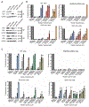
Figure 5.. Non-native peptides and peptides from human BNIP5 and PXT1 induce BAK-mediated membrane permeabilization in cells.
Percent cytochrome-c release was measured by BH3 profiling in permeabilized HeLa cells including WT, BAK-only, BAX-only, and BAK/BAX siRNA cells. A) Western blots performed after 72-hour siRNA treatment of HeLa cells show levels of BAK and BAX, with GAPDH as a loading control. Replicates are labeled as #1, #2, and #3. B) Non-native peptides dM2 (navy blue), dM4 (red), and dF8 (orange). C) BH3 peptides from BIM (light blue), BID (grey), and PUMA (brown) plus peptides from BNIP5 (green) and PXT1 (olive). Data are from three independent experiments, and error bars indicate standard deviations over three replicates.
Figure 6.
Figure 6.. Peptides dF2, dF3, and dM2 bind to BAK similarly to other inhibitors and activators.
A) BAK (grey) bound to dF2 (purple; 8CZF), dF3 (light pink; 8CZG), and dM2 (pale green; 8CZH). Inhibitors are in shades of purple and pink and activators are in shades of green. B) (Top) BAK bound to dF2 and dF3. BAK-contacting residues are shown with sticks at right. (Bottom) BAK bound to inhibitors containing non-natural amino acids including BIM-h3Glg (hot pink; 5VX0) and BIM-h3Pc (light magenta; 5VWZ) compared with dF2 and dF3 complexes. C) BAK bound to dM2 (pale green) and previously published activators M3W5_BID (lime green; 7M5B) and W3W5_BID (forest; 7M5A). D) Comparison of binding modes of dM2, dF2, and dF3. Further comparisons are in Figure S8.
Figure 7.
Figure 7.. Peptides dF2, dF3, and dM2 make similar polar contacts with BAK.
A) and B) Inhibitors dF2 (purple) and dF3 (pink) make similar polar contacts involving residues at peptide positions 3b, 3g, 4b, 4d; 2b hydrogen bonds with BAK only in the BAK:dF3 complex. C) Activator dM2 (green) makes many of the same polar contacts seen for inhibitors. In peptide dM2, position 4d is an alanine and thus cannot form the helix capping-like interaction made by Lys at 4d in dF2 and dF3.
Figure 8.
Figure 8.. Binding rate constants and affinities for activator and inhibitor peptides.
Inhibitors are in purple and activators are in green. (A) Association (kon) and (B) dissociation (koff) rate constants determined by BLI. At least three replicate measurements for each peptide are indicated. C) Kd values computed as the ratio of average koff/kon values measured in A and B, unless otherwise indicated (see Figures S12 and S13). D) Equilibrium Kd values measured using fluorescence polarization. Replicates consisted of dF4 n=3, dF3 n=5, dF2 n=5, BK3 n=10, dF7 n=5, BNIP5 n=4, dF8 n=4, dM2 n=8, dM4=3, and PXT1=3.
Figure 9.
Figure 9.. Free energy diagrams for BH3 peptide activation or inhibition of BAK.
Crystal structures are used to illustrate steps in activation, with the BAK core in dark grey and the latch in light grey. BAK monomer is placed at higher energy compared to the BH3:groove homodimer. The structure of the transition state is unknown but may resemble an unlatched conformation of BAK (represented by 4U2U). A) The transition state can be stabilized by binding an activator (green). B) Monomeric BAK can be stabilized by binding an inhibitor (purple). C) For activation by antibody 7D10, the free energy of the complex between BAK and 7D10 is lower than the unbound state, but the displacement of the α1-α2 loop destabilizes the core-latch globular core, which decreases the energy barrier for activation.

Similar articles

Cited by

References

    1. Chittenden T, Harrington EA, O’Connor R, Remington C, Lutz RJ, Evan GI, and Guild BC (1995). Induction of apoptosis by the Bcl-2 homologue Bak. Nature 374, 733–736. 10.1038/374733a0. - DOI - PubMed
    1. Kerr JFR, Wyllie AH, and Currie AR (1972). Apoptosis: A Basic Biological Phenomenon with Wideranging Implications in Tissue Kinetics. Brit J Cancer 26, 239–257. 10.1038/bjc.1972.33. - DOI - PMC - PubMed
    1. Lindsten T, Ross AJ, King A, Zong W-X, Rathmell JC, Shiels HA, Ulrich E, Waymire KG, Mahar P, Frauwirth K, et al. (2000). The Combined Functions of Proapoptotic Bcl-2 Family Members Bak and Bax Are Essential for Normal Development of Multiple Tissues. Mol Cell 6, 1389–1399. 10.1016/s1097-2765(00)00136-2. - DOI - PMC - PubMed
    1. Singh R, Letai A, and Sarosiek K (2019). Regulation of apoptosis in health and disease: the balancing act of BCL-2 family proteins. Nat Rev Mol Cell Bio 20, 175–193. 10.1038/s41580-018-0089-8. - DOI - PMC - PubMed
    1. Takeuchi O, Fisher J, Suh H, Harada H, Malynn BA, and Korsmeyer SJ (2005). Essential role of BAX,BAK in B cell homeostasis and prevention of autoimmune disease. Proc National Acad Sci 102, 11272–11277. 10.1073/pnas.0504783102. - DOI - PMC - PubMed

Publication types

MeSH terms