

A spatiotemporal drug release scaffold with antibiosis and bone regeneration for osteomyelitis
- PMID: 36706987
- PMCID: PMC10704079
- DOI: 10.1016/j.jare.2023.01.019
A spatiotemporal drug release scaffold with antibiosis and bone regeneration for osteomyelitis
Abstract
Introduction: Scaffolds loaded with antibacterial agents and osteogenic drugs are considered essential tools for repairing bone defects caused by osteomyelitis. However, the simultaneous release of two drugs leads to premature osteogenesis and subsequent sequestrum formation in the pathological situation of unthorough antibiosis.
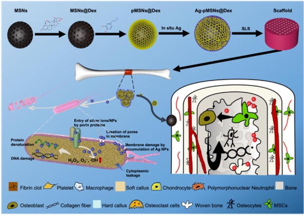
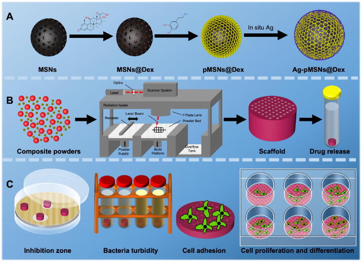
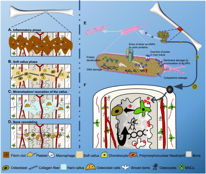
Objectives: In this study, a spatiotemporal drug-release polydopamine-functionalized mesoporous silicon nanoparticle (MSN) core/shell drug delivery system loaded with antibacterial silver (Ag) nanoparticles and osteogenic dexamethasone (Dex) was constructed and introduced into a poly-l-lactic acid (PLLA) scaffold for osteomyelitis therapy.
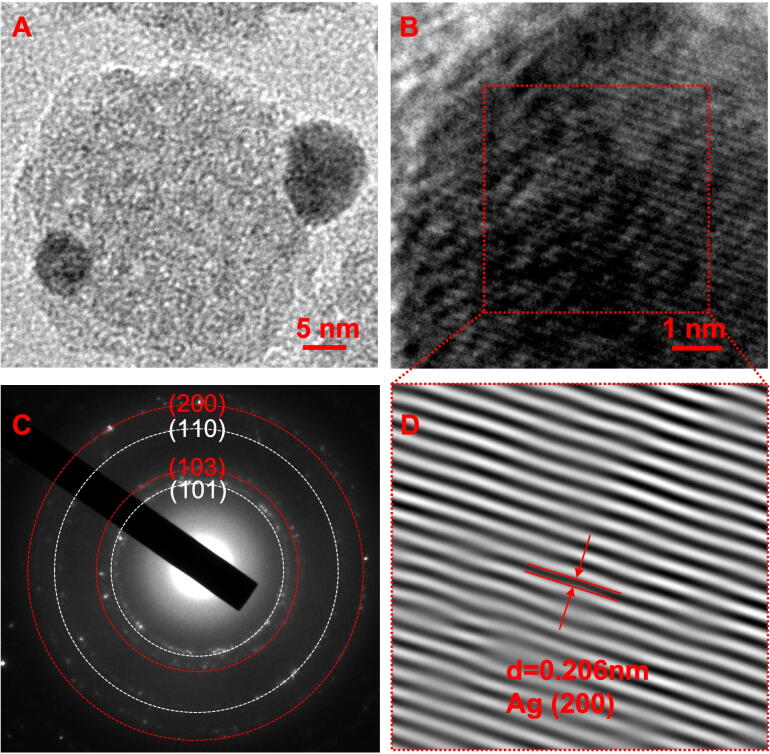
Methods: MSNs formed the inner core and were loaded with Dex through electrostatic adsorption (MSNs@Dex), and then polydopamine was used to seal the core through the self-assembly of dopamine as the outer shell (pMSNs@Dex). Ag nanoparticles were embedded in the polydopamine shell via an in situ growth technique. Finally, the Ag-pMSNs@Dex nanoparticles were introduced into PLLA scaffolds (Ag-pMSNs@Dex/PLLA) constructed by selective laser sintering (SLS).
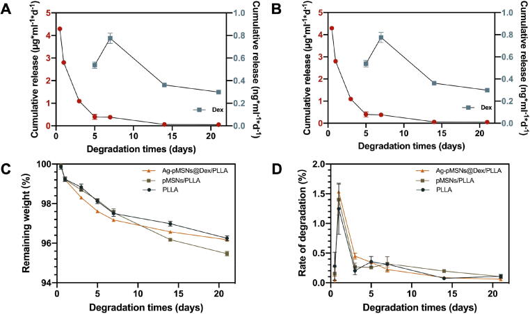
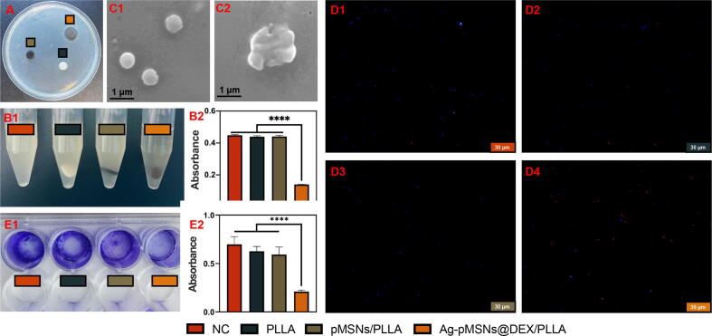
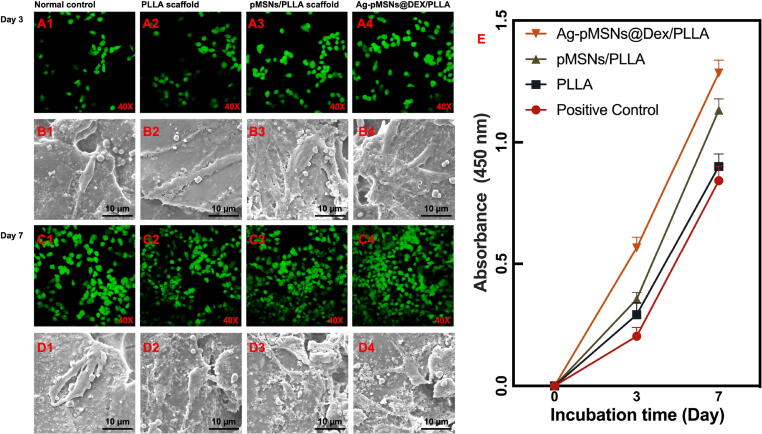
Results: The Ag-pMSNs@Dex/PLLA scaffold released Ag+ at the 12th hour, followed by the release of Dex starting on the fifth day. The experiments verified that the scaffold had excellent antibacterial performance against Escherichia coli and Staphylococcus aureus. Moreover, the scaffold significantly enhanced the osteogenic differentiation of mouse bone marrow mesenchymal stem cells.
Conclusion: The findings suggested that this spatiotemporal drug release scaffold had promising potential for osteomyelitis therapy.
Keywords: Antibiosis; Bone regeneration; Core/shell drug delivery system; Osteomyelitis; Selective laser sintering; Spatiotemporal drug release.
Copyright © 2023. Production and hosting by Elsevier B.V.
Conflict of interest statement
Declaration of Competing Interest The authors declare that they have no known competing financial interests or personal relationships that could have appeared to influence the work reported in this paper.
Figures


References
-
- Feng P., Kong Y., Liu M., Peng S., Shuai C. Dispersion strategies for low-dimensional nanomaterials and their application in biopolymer implants. Materials Today. Nano. 2021;15 doi: 10.1016/j.mtnano.2021.100127. - DOI
-
- R, Schnettler, K, Emara, D, Rimashevskij, R, Diap, A, Emara, J, Franke, et al. in Basic Techniques for Extremity Reconstruction: External Fixator Applications According to Ilizarov Principles (eds Mehmet Çakmak et al.) 605-628 (Springer International Publishing, 2018).
Publication types
MeSH terms
Substances
LinkOut - more resources
Full Text Sources
Research Materials
Miscellaneous
