

Unsuspected gastric glomus tumour
- PMID: 36707101
- PMCID: PMC9884849
- DOI: 10.1136/bcr-2022-253020
Unsuspected gastric glomus tumour
Abstract
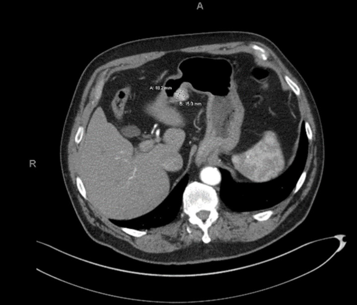
Gastric glomus tumours (GGTs) are rare predominantly benign, mesenchymal neoplasms that commonly arise from the muscularis or submucosa of the gastric antrum and account for <1% of gastrointestinal soft-tissue tumours. Historically, GGT has been difficult to diagnose preoperatively due to the lack of unique clinical, endoscopic and CT features. We present a case of an incidentally identified GGT in an asymptomatic man that was initially considered a neuroendocrine tumour (NET) by preoperative fine-needle aspiration biopsy with focal synaptophysin reactivity. An elective robotic distal gastrectomy and regional lymphadenectomy were performed. Postoperative review by pathology confirmed the diagnosis of GGT. GGTs should be considered by morphology as a differential diagnosis of gastric NET on cytology biopsy, especially if there is focal synaptophysin reactivity. Additional staining for SMA and BRAF, if atypical/malignant, can help with this distinction. Providers should be aware of the biological behaviour and treatment of GGTs.
Keywords: Gastric cancer; Pathology; Stomach and duodenum; Surgical oncology.
© BMJ Publishing Group Limited 2023. No commercial re-use. See rights and permissions. Published by BMJ.
Conflict of interest statement
Competing interests: None declared.
Figures


Similar articles
-
Diagnostic utility of endoscopic ultrasound-guided fine-needle aspiration biopsy for glomus tumor of the stomach.World J Gastroenterol. 2015 Jun 14;21(22):7052-8. doi: 10.3748/wjg.v21.i22.7052. World J Gastroenterol. 2015. PMID: 26078584 Free PMC article.
-
Gastrointestinal Glomus Tumors: A Single Institution, 20-Year Retrospective Study.J Surg Res. 2023 Mar;283:982-991. doi: 10.1016/j.jss.2022.10.070. Epub 2022 Dec 10. J Surg Res. 2023. PMID: 36915027
-
[Gastric glomus tumors expressing synaptophysin: clinicopathologic and immunohistochemical analyses].Zhonghua Bing Li Xue Za Zhi. 2017 Nov 8;46(11):756-759. doi: 10.3760/cma.j.issn.0529-5807.2017.11.004. Zhonghua Bing Li Xue Za Zhi. 2017. PMID: 29136687 Chinese.
-
A case of malignant gastric glomus tumor and literature review: A case report.Medicine (Baltimore). 2024 Aug 9;103(32):e39208. doi: 10.1097/MD.0000000000039208. Medicine (Baltimore). 2024. PMID: 39121329 Free PMC article. Review.
-
Clinicopathologic features of gastric glomus tumor: A report of 15 cases and literature review.Pathol Oncol Res. 2023 Jan 9;28:1610824. doi: 10.3389/pore.2022.1610824. eCollection 2022. Pathol Oncol Res. 2023. PMID: 36699621 Free PMC article. Review.
Cited by
-
Case report: One case of precise resection of gastric glomus tumor by gastroscopy combined with laparoscopy.Front Oncol. 2025 Jan 7;14:1501442. doi: 10.3389/fonc.2024.1501442. eCollection 2024. Front Oncol. 2025. PMID: 39839772 Free PMC article.
References
-
- Fletcher CDM, ed. Pathology and genetics of tumours of soft tissue and bone. Lyon: IARC Press, 2002.
Publication types
MeSH terms
Substances
LinkOut - more resources
Full Text Sources
Medical
Research Materials
Miscellaneous