. 2023 Sep;72(9):1774-1782.
doi: 10.1136/gutjnl-2022-327909. Epub 2023 Jan 27.

Combining ferroptosis induction with MDSC blockade renders primary tumours and metastases in liver sensitive to immune checkpoint blockade

Affiliations

Combining ferroptosis induction with MDSC blockade renders primary tumours and metastases in liver sensitive to immune checkpoint blockade

Claire Conche et al. Gut. 2023 Sep.

Abstract

Objective: Investigating the effect of ferroptosis in the tumour microenvironment to identify combinatory therapy for liver cancer treatment.

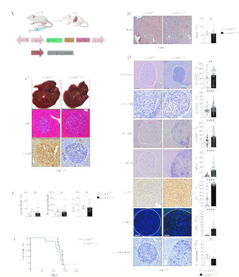
Design: Glutathione peroxidase 4 (GPx4), which is considered the master regulator of ferroptosis, was genetically altered in murine models for hepatocellular carcinoma (HCC) and colorectal cancer (CRC) to analyse the effect of ferroptosis on tumour cells and the immune tumour microenvironment. The findings served as foundation for the identification of additional targets for combine therapy with ferroptotic inducer in the treatment of HCC and liver metastasis.

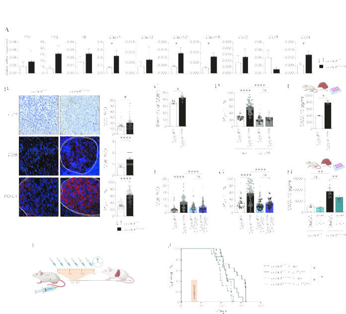
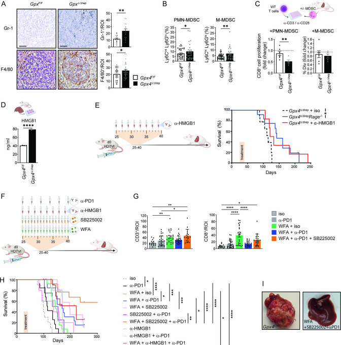
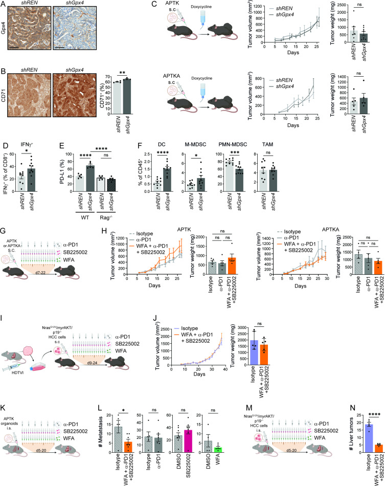
Results: Surprisingly, hepatocyte-restricted GPx4 loss does not suppress hepatocellular tumourigenesis. Instead, GPx4-associated ferroptotic hepatocyte death causes a tumour suppressive immune response characterised by a CXCL10-dependent infiltration of cytotoxic CD8+ T cells that is counterbalanced by PD-L1 upregulation on tumour cells as well as by a marked HMGB1-mediated myeloid derived suppressor cell (MDSC) infiltration. Blocking PD-1 or HMGB1 unleashes T cell activation and prolongs survival of mice with Gpx4-deficient liver tumours. A triple combination of the ferroptosis inducing natural compound withaferin A, the CXCR2 inhibitor SB225002 and α-PD-1 greatly improves survival of wild-type mice with liver tumours. In contrast, the same combination does not affect tumour growth of subcutaneously grown CRC organoids, while it decreases their metastatic growth in liver.

Conclusion: Our data highlight a context-specific ferroptosis-induced immune response that could be therapeutically exploited for the treatment of primary liver tumours and liver metastases.

Keywords: adenocarcinoma; cell death; colorectal cancer; immunotherapy; liver.


Conflict of interest statement

Competing interests: None declared.

Figures

Figure 1
Figure 1
Ferroptotic induction in a murine model of HCC is not sufficient to limit tumour growth. (A) Scheme of HCC murine model with HTVI of oncogenic NrsasG12V and myristoylated-AKT together with sleeping beauty (SB). (B) Nras immunohistochemical analysis in livers of Gpx4 F/F and Gpx4 Δ/Δhep mice 7 days after HTVI (n≥10). (C) Representative images of multifocal HCC and H&E and Gpx4-IHC staining of livers of Gpx4 F/F and Gpx4 Δ/Δhep mice 25 days after HTVI. (D) Immunohistochemical analysis of 8-OHdG+ (n≥=73), p-H2AX+ (n≥80), 4-HNE+ (n≥99), MDA+ (n≥59), CD71+ (n≥69), cleaved-caspase 3+ (n≥11) and TUNEL+ (n≥=30) cells in liver tumours of Gpx4 F/F and Gpx4 Δ/Δhep mice. (E) Quantification of tumour area percentage to the liver and number of tumours per liver from H&E-stained serial sections. Immunohistochemical analysis of BrdU incorporation in tumours (n≥13). (F) Survival of Gpx4 F/F and Gpx4 Δ/Δhep mice after HTVI (n=9), p=0176 by log-rank (Mantel-Cox) test. (A–E) Scale bars=100 µm. Data are mean±SEM, n.s. not significant, **p≤0.01, ****p≤0.0001 by t-test. HCC, hepatocellular carcinoma; HTVI, hydrodynamic tail vein injection; IR/DR, inverted repeats and direct repeats; IRES, internal ribosome entry site.
Figure 2
Figure 2
Ferroptosis in HCC induces immune reaction with T cell activation dampened by PD-L1 upregulation. (A) Gene expression quantification by quantitative RT-PCR in tumours from Gpx4 F/F and Gpx4 Δ/Δhep mice 25 days after HTVI (n=5). (B) Immunohistochemical analysis of CD3+ (n=35), CD8 (n≥36), PD-L1+ (n=38) cells in HCC tumours of Gpx4 F/F and Gpx4 Δ/Δhep mice after HTVI. Scale bars=100 µm. (C) Flow cytometry analysis of IFNγ expression in CD8 T cells from liver of Gpx4 F/F and Gpx4 Δ/Δhep tumour bearing mice after PMA/Ionomycin stimulation (n≥4). (D) Immunofluorescence quantification of PD-L1 expression in HCC tumours from Gpx4 F/F and Gpx4 Δ/Δhep mice treated with depleting α-CD8 or Rat IgG2b, κ antibodies (n≥42). (E) CXCL10 release by primary hepatocytes from Gpx4 F/F and Gpx4 Δ/Δhep mice ex vivo for 4 hours (n=2). (F, G) Immunofluorescence quantification of CD8+ T cells infiltration (F) and PD-L1 expression (G) in HCC tumours from Gpx4 F/F, Gpx4 Δ/Δhep, Gpx4 F/F CXCR3 -/-, Gpx4 Δ/Δhep CXCR3 -/- mice (n≥40). (H) CXCL10 secretion by Gpx4 F/F and Gpx4 Δ/Δhep primary hepatocytes ex vivo treated with 10 µM of C176 and C178 or DMSO for 4 hour (n≥6). (I) Treatment scheme for i.p. injections, of α-PD-1 or isotype (Rat IgG2a,κ). (J) Survival of Gpx4 F/F+ isotype (Rat IgG2a,κ) (n=10), Gpx4 F/F + α-PD-1 (n=13), Gpx4 Δ/Δhep + isotype (Rat IgG2a,κ) (n=9) and Gpx4 Δ/Δhep +α-PD-1 (n=14) mice with HCC tumours. (A–H) Data are mean±SEM, n.s not significant *p≤0.05, **p≤0.01, ****p≤0.0001 by t-test (A–C, E) or one-way ANOVA with Šídák’s multiple comparisons test (D, F–H) of the indicated pairs or by log-rank (Mantel-Cox) test (J). ANOVA, analysis of variance; HTVI, hydrodynamic tail vein injection; PMA, phorbol myristate acetate.
Figure 3
Figure 3
T cell activation and MDSC tumour infiltration induced by ferroptosis serve as targets for combinatory therapy for HCC. (A) Immunohistochemical analysis of Gr-1+ (n=15) and F4/80+ (n=35) cells in Gpx4 F/F and Gpx4 Δ/Δhep HCC tumours. scale bars=100 µm. (B) Flow cytometry quantification of the percentages of PMN-MDSC and M-MDSC within the CD45+infiltrates of livers from Gpx4 F/F and Gpx4 Δ/Δhep HCC tumour-bearing mice. (n≥22). (C) Immunosuppression of MDSC isolated from livers of Gpx4 F/F and Gpx4 Δ/Δhep tumour bearing mice on CD8+ T cell proliferation. Fold change of the percentages of divided CD8+ T cells with MDSC to CD8+ T cells without MDSC (n≥5). (D) HMGB1 release by primary hepatocytes from Gpx4 F/F and Gpx4 Δ/Δhep mice ex vivo for 4 hours (n=2). (E) Survival of Gpx4 Δ/Δhep mice treated with isotype Rat IgG2a,κ (as in 2J, n=9) or α-HMGB1 (n=6) and untreated Gpx4 Δ/Δhep/Rage -/- (n=12) mice after HTVI. (F) Treatment scheme with i.p. injections of 250 µg of α-PD-1 or isotype (Rat IgG2a,κ), 50 µg of α-HMGB1, 2.5 mg/kg of WFA or 1 mg/kg of SB225002. (G) Immunohistochemical analysis of CD3+ or CD8+ T cell infiltration in HCC tumours of Gpx4 F/F and Gpx4 Δ/Δhep mice>100 days after HTVI (n≥15). (H) Survival of WT mice after HTVI receiving Isotype (Rat IgG2a,κ) (n=10), α-PD-1 (n=13) (as in figure 2), WFA+isotype (Rat IgG2a,κ) (n=13), WFA + α-PD-1(n=14), WFA+SB225002 (n=12), SB225002 + α-PD-1 (n=13), WFA+SB225002 + α-PD-1 (n=14), α-HMGB1 (n=5), α-HMGB1 + α-PD1 (n=6) or WFA + α-HMGB1 + α-PD1 (n=12) treatments. (I) Pictures of livers of untreated Gpx4 F/F mice at end point and WT mice treated with WFA+SB225002+α-PD-1 >300 days after HTVI. (A–H) Data are mean±SEM, *p≤0.05, **p≤0.01, ***p≤0.001, ****p≤0.0001 by t-test (A–D) one-way ANOVA with Šídák’s’s multiple comparisons of all groups (G) or by log-rank (Mantel-Cox) test (E, H). ANOVA, analysis of variance; HCC, hepatocellular carcinoma; HTVI, hydrodynamic tail vein injection; MDSC, myeloid derived suppressor cell; WFA, withaferin A.
Figure 4
Figure 4
Ferroptosis effect on colorectal cancer. (A) Representative immunohistochemical Gpx4 staining of shREN or shGPX4 APTKA s.c tumours. (B) Immunohistochemical quantification of CD71+ cells in shREN and shGpx4 APTKA s.c. tumours (n=4). (C) Tumour volumes and weights at end point of shREN and shGpx4 APTK and ATPKA subcutaneous tumours (n≥5). (D) Flow cytometry analysis of IFNγ expression in CD8+ T cells from shREN and shGpx4 s.c. APTKA tumours, after ex vivo PMA/Ionomycin stimulation (n≥9). (E) Immunofluorescence quantification of PD-L1 expression in s.c shREN and shGpx4 APTKA tumours from WT or Rag-/- mice (n=5). (F) Percentages of DC, M-MDSC, PMN-MDSC in immune infiltrates of shREN and shGpx4 s.c. APTKA tumours analysed by flow cytometry (n≥9). (G) Treatment scheme after s.c. transplantation of APTK or APTKA organoids. (H) Tumour volumes and end point weights of s.c APTK and APTKA treated with the indicated compounds (n≥5). (I) Scheme for the isolation of NrasG12V /myrAKT/p19 -/- HCC cells followed by subcutaneous transplantation and treatments. (J) Tumour volumes and weights at end-point of subcutaneously transplanted NrasG12V /myrAKT/p19 -/- HCC cells with the indicated treatment. (K) Treatment scheme after intrasplenic (i.s.) injection of APTK organoids. (L) Numbers of liver metastasis after intrasplenic injection of APTK organoids and treatment as indicated (n≥4). (M) Treatment scheme after intrasplenic (i.s.) injection of NrasG12V /myrAKT/p19 -/- HCC cells. (N) Number of liver tumours after intrasplenic transplantation of NrasG12V /myrAKT/p19 -/- HCC cells and the indicated treatment (n≥5). (G, I, K, M) Scheme for i.p. injections, each represented by a syringe, with 250 µg of α-PD-1 or isotype (Rat IgG2a,κ), 2.5 mg/kg of Withaferin A (WFA) or 1 mg/kg of SB225002. (B–M) Data are mean±SEM, n.s not significant, *p≤0.05, ***p≤0.001, ****p≤0.0001 by t-test (B–F, J, L, N) one-way ANOVA with Šídák’s multiple comparisons test (H) of the indicated pairs. ANOVA, analysis of variance; DC, dendritic cell; HCC, hepatocellular carcinoma; MDSC, myeloid derived suppressor cell; PMA, phorbol myristate acetate; s.c., subcutaneous.

Comment in

References

    1. Dixon SJ, Lemberg KM, Lamprecht MR, et al. . Ferroptosis: an iron-dependent form of nonapoptotic cell death. Cell 2012;149:1060–72. 10.1016/j.cell.2012.03.042 - DOI - PMC - PubMed
    1. Fellmann C, Hoffmann T, Sridhar V, et al. . An optimized microRNA backbone for effective single-copy RNAi. Cell Rep 2013;5:1704–13. 10.1016/j.celrep.2013.11.020 - DOI - PubMed
    1. Chen X, Kang R, Kroemer G, et al. . Ferroptosis in infection, inflammation, and immunity. J Exp Med 2021;218:e20210518. 10.1084/jem.20210518 - DOI - PMC - PubMed
    1. Ursini F, Maiorino M, Valente M, et al. . Purification from pig liver of a protein which protects liposomes and biomembranes from peroxidative degradation and exhibits glutathione peroxidase activity on phosphatidylcholine hydroperoxides. Biochim Biophys Acta 1982;710:197–211. 10.1016/0005-2760(82)90150-3 - DOI - PubMed
    1. Friedmann Angeli JP, Krysko DV, Conrad M. Ferroptosis at the crossroads of cancer-acquired drug resistance and immune evasion. Nat Rev Cancer 2019;19:405–14. 10.1038/s41568-019-0149-1 - DOI - PubMed

Publication types

MeSH terms

Substances

LinkOut - more resources