. 2023 May;72(5):972-983.
doi: 10.1136/gutjnl-2022-328380. Epub 2023 Jan 27.

Quantification of the hepatitis B virus cccDNA: evidence-based guidelines for monitoring the key obstacle of HBV cure

Affiliations

Quantification of the hepatitis B virus cccDNA: evidence-based guidelines for monitoring the key obstacle of HBV cure

Lena Allweiss et al. Gut. 2023 May.

Abstract

Objectives: A major goal of curative hepatitis B virus (HBV) treatments is the reduction or inactivation of intrahepatic viral covalently closed circular DNA (cccDNA). Hence, precise cccDNA quantification is essential in preclinical and clinical studies. Southern blot (SB) permits cccDNA visualisation but lacks sensitivity and is very laborious. Quantitative PCR (qPCR) has no such limitations but inaccurate quantification due to codetection of viral replicative intermediates (RI) can occur. The use of different samples, preservation conditions, DNA extraction, nuclease digestion methods and qPCR strategies has hindered standardisation. Within the ICE-HBV consortium, available and novel protocols for cccDNA isolation and qPCR quantification in liver tissues and cell cultures were compared in six laboratories to develop evidence-based guidance for best practices.

Design: Reference material (HBV-infected humanised mouse livers and HepG2-NTCP cells) was exchanged for cross-validation. Each group compared different DNA extraction methods (Hirt extraction, total DNA extraction with or without proteinase K treatment (+PK/-PK)) and nuclease digestion protocols (plasmid-safe ATP-dependent DNase (PSD), T5 exonuclease, exonucleases I/III). Samples were analysed by qPCR and SB.

Results: Hirt and -PK extraction reduced coexisting RI forms. However, both cccDNA and the protein-free relaxed circular HBV DNA (pf-rcDNA) form were detected by qPCR. T5 and Exo I/III nucleases efficiently removed all RI forms. In contrast, PSD did not digest pf-rcDNA, but was less prone to induce cccDNA overdigestion. In stabilised tissues (eg, Allprotect), nucleases had detrimental effects on cccDNA.

Conclusions: We present here a comprehensive evidence-based guidance for optimising, controlling and validating cccDNA measurements using available qPCR assays.

Keywords: CHRONIC HEPATITIS; HEPATITIS B; LIVER; LIVER BIOPSY; REAL TIME PCR.


Conflict of interest statement

Competing interests: LA, BT, MY, CK, BQ, MLu, JL and MLe declare no conflict of interest. FZ: Consultancy for Aligos, Antios, Arbutus, Assembly, Blue Jay, Enanta, Enochian, Gilead, GSK, Zhimeng. HG: Consultancy for Aligos and Assembly; shareholder of Arbutus. UP: Consultancy for Aligos, Arbutus, Gilead, GSK, Sanofi, Vaccitech, VirBio. Research collaboration with Abbott and Roche. Share holder and board member of SCG Cell Therapy. MD: research collaboration with Gilead, MYR GmbH/Hepatera and HUMABS BioMed; consultancy for Gilead and Aligos. SPF: employment and shareholder Gilead Sciences.

Figures

Figure 1
Figure 1
The effect of different DNA extraction methods on cccDNA quantification by qPCR and SB in HBV-infected USG mouse liver tissue. (A) Schematic presentation of the experimental design used for the cross-validation. (B) SB analysis on non-digested DNA extracts using HBV DNA probes in three of the labs. (C) qPCR measurements of total HBV DNA and cccDNA in the DNA extracts. Bars depict the median and range across all four labs. (D) qPCR measurements of cccDNA shown separately for every lab, relative to the amount in the +PK DNA extractions. Bars depict the mean of duplicate measurements. +PK, total DNA extraction with proteinase K digestion; −PK, total DNA extraction without proteinase K digestion; cccDNA, covalently closed circular DNA; HBV, hepatitis B virus; Hirt, Hirt DNA extraction, which does not include a proteinase K digestion; qPCR, quantitative PCR; SB, Southern blot.
Figure 2
Figure 2
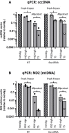
The effect of nuclease digestions on different DNA extracts in HBV-infected USG mouse liver tissue. The bar graphs depict qPCR measurements of total HBV DNA (A) and cccDNA (B) in all DNA extracts and labs without nuclease digestion (hatched bars) or after the indicated nuclease treatments. The bars depict the median and range of the values from three labs. Every lab performed the nuclease digestion in duplicates. The values (copies/PCR) after nuclease treatment were first normalised to the non-digested value in the respective DNA extract, then all values derived from Hirt or −PK DNA extracts were normalised to the +PK extract. (C) qPCR of mtDNA DNA via the mitochondrial gene ND2, depicted as arbitrary units normalised to the non-digested values in the respective DNA extract. (D) SB analysis in one of the labs using a second stably HBV-infected and untreated USG mouse and all three DNA extracts and four nuclease digestion conditions using HBV DNA probes (top panel) and densitometric analysis of the cccDNA band (lower panel). The ‘X’ denotes a sample that could not be quantified because of high background staining. The samples were run on two separate blots but identical amounts of non-digested DNA, with the help of which densitometry was normalised between blots. ND2, mitochondrially encoded NADH:ubiquinone oxidoreductase core subunit 2. +PK, total DNA extraction with proteinase K digestion; −PK, total DNA extraction without proteinase K digestion; cccDNA, covalently closed circular DNA; HBV, hepatitis B virus; qPCR, quantitative PCR; SB, Southern blot.
Figure 3
Figure 3
The pf-rcDNA is detected with the same efficiency as cccDNA by cccDNA-selective qPCR but is removed through T5 exonuclease treatment. Parallel SB (A) and qPCR (B) analysis in three stably HBV-infected USG mice (after 6 or 12 weeks of lamivudine treatment or untreated). Liver DNA was extracted with the −PK, digested with nucleases or left undigested before analysis. (A) SB using HBV DNA probes (top panel) and densitometric analysis of the pf-rcDNA (middle panel) and cccDNA band (lower panel). DNA amounts were normalised to ND2 via qPCR with human-specific primers before digestion to ensure loading of equal amounts of DNA derived from human cells. (B) qPCR measurements of total HBV DNA (grey bars) and cccDNA (checked bars) normalised to human hepatocytes via human HBB counts in the non-digested DNA extract. The cccDNA copy numbers/cell are depicted above each bar. (C) Preparative agarose gel using the same samples as in (A) and identical settings for the gel electrophoresis. The upper and lower bands were excised separately, DNA was extracted and used for qPCR analysis (total HBV DNA and cccDNA). The ratio of total HBV DNA copies to cccDNA copies was calculated for all samples derived from the upper SB band (top) and the lower band (bottom), respectively. +PK, total DNA extraction with proteinase K digestion; −PK, total DNA extraction without proteinase K digestion; cccDNA, covalently closed circular DNA; HBV, hepatitis B virus; Lam, lamivudine; NT, non-treated; qPCR, quantitative PCR; SB, Southern blot.
Figure 4
Figure 4
The effect of different DNA extraction methods and nuclease digestions on cccDNA quantification by qPCR and SB in HBV-infected HepG2-NTCP cells. (A–F) Frozen cell pellets harvested at day nine post infection were shipped to the participating labs. (A) qPCR measurements of total HBV DNA and cccDNA in the undigested DNA extracts. Bars depict the median and range across all four labs. (B) PCR measurements of cccDNA shown separately for every lab and relative to the amount in the+PK DNA extractions. Bars depict the mean of duplicate measurements. (C, D) qPCR measurements of total HBV DNA (C) and cccDNA (D) in all DNA extracts and labs either without nuclease digestion (hatched bars) or after the indicated nuclease treatments (performed in duplicates). The bars depict the median and range of the values from three labs. The values (copies/PCR) after nuclease treatment were first normalised to the non-digested value in the respective DNA extract, then all values derived from Hirt or −PK DNA extracts were normalised to the +PK extract. (E) qPCR of mtDNA DNA via the mitochondrial gene ND2, depicted as arbitrary units normalised to the non-digested values in the respective DNA extract. (F) SB using HBV DNA probes (top panel) and densitometric analysis of the cccDNA band (lower panel). The ‘X’ denotes a sample that could not be quantified because of high background staining. The samples were run on two separate blots with identical amounts of non-digested DNA, with the help of which densitometry was normalised between blots. +PK, total DNA extraction with proteinase K digestion; −PK, total DNA extraction without proteinase K digestion; cccDNA, covalently closed circular DNA; HBV, hepatitis B virus; qPCR, quantitative PCR; SB, Southern blot.
Figure 5
Figure 5
Nuclease treatment bears the risk of overdigesting cccDNA in Allprotect-stored samples. Comparison of nuclease digestion in fresh-frozen liver tissue or liver tissue preserved in Allprotect. Liver DNA was extracted using the −PK method from two untreated HBV-infected mice and two mice treated with siRNA targeting all HBV transcripts for 6 weeks. DNA was digested with PSD or T5 exonuclease and subjected to qPCR for cccDNA (A) and ND2 (B). Every dot depicts a single mouse, bars the median. +PK, total DNA extraction with proteinase K digestion; −PK, total DNA extraction without proteinase K digestion; cccDNA, covalently closed circular DNA; HBV, hepatitis B virus; qPCR, quantitative PCR; siRNA, small interfering RNA.
Figure 6
Figure 6
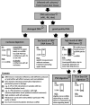
Schematic presentation of the main findings of the study. The flow chart summarises the experimental flow for cccDNA quantification and highlights optimal and suboptimal results for both qPCR and SB, including the major caveats. cccDNA, covalently closed circular DNA; qPCR, quantitative PCR; SB, quantitative PCR.

References

    1. WHO fact sheet hepatitis B. 2021. Available: https://www.who.int/news-room/fact-sheets/detail/hepatitis-b [Accessed 27 Jan 2022].
    1. Revill PA, Chisari FV, Block JM, et al. . A global scientific strategy to cure hepatitis B. Lancet Gastroenterol Hepatol 2019;4:545–58. 10.1016/S2468-1253(19)30119-0 - DOI - PMC - PubMed
    1. Marchetti AL, Guo H. New insights on molecular mechanism of hepatitis B virus covalently closed circular DNA formation. Cells 2020;9:2430. 10.3390/cells9112430 - DOI - PMC - PubMed
    1. Wei L, Ploss A. Mechanism of hepatitis B virus cccDNA formation. Viruses 2021;13:1463. 10.3390/v13081463 - DOI - PMC - PubMed
    1. Martinez MG, Boyd A, Combe E, et al. . Covalently closed circular DNA: the ultimate therapeutic target for curing HBV infections. J Hepatol 2021;75:706–17. 10.1016/j.jhep.2021.05.013 - DOI - PubMed

Publication types