. 2023 Jan 27;6(1):104.
doi: 10.1038/s42003-023-04468-z.

Kinetic fingerprinting of metabotropic glutamate receptors

Affiliations

Kinetic fingerprinting of metabotropic glutamate receptors

Taulant Kukaj et al. Commun Biol. .

Abstract

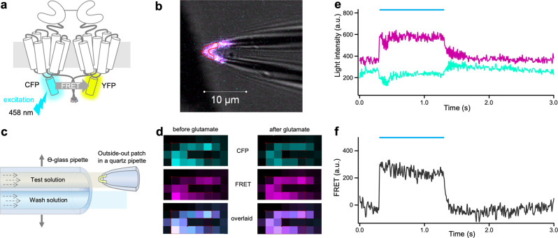
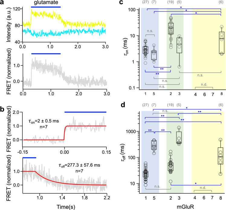
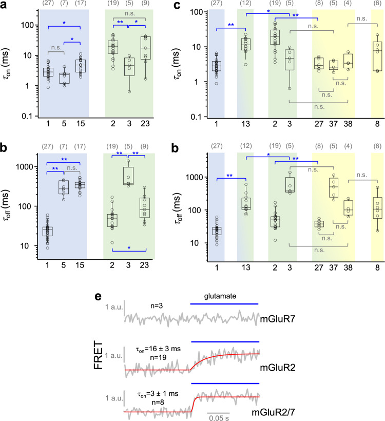
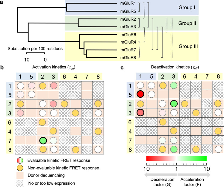
Dimeric metabotropic glutamate receptors (mGluRs) are abundantly expressed in neurons. In mammals, eight subunit isoforms, mGluR1-8, have been identified, forming the groups I, II, and III. We investigated receptor dimerization and kinetics of these mGluR isoforms in excised membrane patches by FRET and confocal patch-clamp fluorometry. We show that 5 out of 8 homodimeric receptors develop characteristic glutamate-induced on- and off-kinetics, as do 11 out of 28 heterodimers. Glutamate-responsive heterodimers were identified within each group, between groups I and II as well as between groups II and III, but not between groups I and III. The glutamate-responsive heterodimers showed heterogeneous activation and deactivation kinetics. Interestingly, mGluR7, not generating a kinetic response in homodimers, showed fast on-kinetics in mGluR2/7 and mGluR3/7 while off-kinetics retained the speed of mGluR2 or mGluR3 respectively. In conclusion, glutamate-induced conformational changes in heterodimers appear within each group and between groups if one group II subunit is present.


Conflict of interest statement

The authors declare no competing interests.

Figures

Fig. 1
Fig. 1. Donor dequenching after photobleaching the acceptor in homodimeric mGluRs.
a Cartoon of a dimeric mGluR. Each subunit contains an N-terminal ligand-binding domain (LBD) that is connected to a cysteine-rich domain (CRD) which is connected to a seven-helix transmembrane domain (7TMD). The cyan or yellow fluorescent protein, CFP, and YFP are included in the intracellular i2 loop. The quality control system of the GABAB receptor was used to control the composition of the receptors to be analyzed (see Methods). The principle of acceptor-bleaching induced donor dequenching is indicated. b Confocal micrograph of an oocyte membrane expressing mGluR2/7 before (top) and after photobleaching with the light of 514 nm (bottom). The membrane region with the bleached yellow signal is easily visible. c FRET efficiency for the representative example mGluR5 for donor dequenching by photobleaching of the acceptor. b.p., and a.p. means before and after photobleaching, respectively. The light intensity of the donor was increased by 23 ± 4%. d FRET efficiency of the donor was evoked by photobleaching the acceptor for the eight homodimeric mGluRs. The values were obtained from 3 to 7 cells. n.d., not determinable. The numbers of experiments are shown at the top in brackets.
Fig. 2
Fig. 2. Kinetic FRET measurements in outside-out patches.
a Cartoon illustrating the principle of FRET measurements by exciting the donor CFP at 458 nm and detecting the emissions of CFP and YFP. b Tip of a patch pipette carrying an outside-out patch containing a large number of labeled homodimeric mGluR1. c Scheme illustrating fast solution switches at the outside-out patch. A double-barreled Θ-glass pipette delivering two laminar streams of solutions is stepped by a piezo device. d Confocal images of a patch before and after adding glutamate. Left panels show signals coming from CFP, FRET, and overlaid channels before adding glutamate. The right panels show a decreased CFP signal, increased FRET signal, and a clear signal change in the overlaid channel, presumably due to a conformational change. e Fluorescence signal and glutamate-induced changes in time-dependent traces of YFP (magenta) and CFP (cyan) after correction for crosstalk and photobleaching. The light blue bar indicates the application of glutamate (1 mM). f Calculated FRET signal from e.
Fig. 3
Fig. 3. Activation and deactivation kinetics in homodimeric mGluRs.
a Time course of fluorescence changes for donor and acceptor in dimeric mGluR5 (top) and resulting FRET signal (bottom). Here and in the following, the blue bar indicates the application of 1 mM glutamate. Shown are representative photobleaching and cross-talk corrected traces of CFP (cyan) and YFP (yellow) as well as the corrected normalized FRET signal (gray). b Time courses of activation (top, τon) and deactivation (bottom, τoff) of an individual experiment (gray). The superimposed curves are best fits to average traces obtained from n = 7 individual traces each. c, d Activation (c) and deactivation (d) time constants for the mGluRs. Three mGluRs did not show evaluable time courses. n.d., not determinable. Significant differences are indicated (ANOVA followed by Turkey-Kramer posthoc test, see Methods: *p < 0.05, **p < 0.01). In c and d the numbers of experiments are shown at the top in brackets.
Fig. 4
Fig. 4. Kinetic responses of heterodimeric mGluRs.
a,b τon and τoff for two heterodimers in groups I and II. c, d τon and τoff for two heterodimers between group I and II and three heterodimers between group II and III. Homomeric data from Fig. 3 are shown for comparison. Significant differences are indicated (ANOVA followed by Turkey-Kramer posthoc test, see Methods: *p < 0.05, **p < 0.01). The computed p-values are provided in Supplementary Table 3. n.s. indicates that the difference is ‘not significant’. e Averaged time traces obtained with 1 mM glutamate for mGluR7 (n = 3), mGluR2 (n = 19), and mGluR2/7 (n = 8) with fitted exponential functions (red curves). mGluR7 is a strong accelerator in mGluR2/7 compared to mGluR2. In ad, the numbers of experiments are shown at the top in brackets.
Fig. 5
Fig. 5. Overview of the specific functional interactions in heterodimeric mGluRs.
a Phylogenetic tree with seven functional interactions specified by activation and deactivation time constants (solid brackets) and four functional interactions identified by smaller, kinetically not quantifiable responses (Stippled brackets). b Matrix summarizing relative activation kinetics (circles) of the heterodimers among the eight subunits mGluR1-mGluR8 with respect to the homodimers and donor dequenching (salmon fields). The amount of the kinetic effects, within each column, are color-coded according to the scale in c, providing a factor of acceleration (F, green) and deceleration (G, red) with respect to the homodimers. For example, the intensive green circle in row 7 column 3 reads that activation in mGluR2/7 is strongly accelerated with respect to mGluR2 by the color-coded factor F. Significant differences are indicated by a black bold rim of the circles. White circles indicate an equal time course to the homodimer of the column. The main diagonal contains the properties of the homodimers. c Matrix summarizing deactivation kinetics of the heterodimers. Same symbols as in b.

References

    1. Zhu S, Gouaux E. Structure and symmetry inform gating principles of ionotropic glutamate receptors. Neuropharmacology. 2017;112:11–15. - PMC - PubMed
    1. Niswender CM, Conn PJ. Metabotropic glutamate receptors: physiology, pharmacology, and disease. Annu Rev. Pharm. Toxicol. 2010;50:295–322. - PMC - PubMed
    1. Pin JP, Acher F. The metabotropic glutamate receptors: structure, activation mechanism and pharmacology. Curr. Drug Targets CNS Neurol. Disord. 2002;1:297–317. - PubMed
    1. El Moustaine D, et al. Distinct roles of metabotropic glutamate receptor dimerization in agonist activation and G-protein coupling. Proc. Natl Acad. Sci. USA. 2012;109:16342–16347. - PMC - PubMed
    1. Sun H, Neugebauer V. mGluR1, but not mGluR5, activates feed-forward inhibition in the medial prefrontal cortex to impair decision making. J. Neurophysiol. 2011;106:960–973. - PMC - PubMed

Publication types

Substances