Review
. 2023 Jan 28;30(1):8.
doi: 10.1186/s12929-023-00903-9.

Chronic stress in solid tumor development: from mechanisms to interventions

Affiliations
Review

Chronic stress in solid tumor development: from mechanisms to interventions

Jiajing Yan et al. J Biomed Sci. .

Abstract

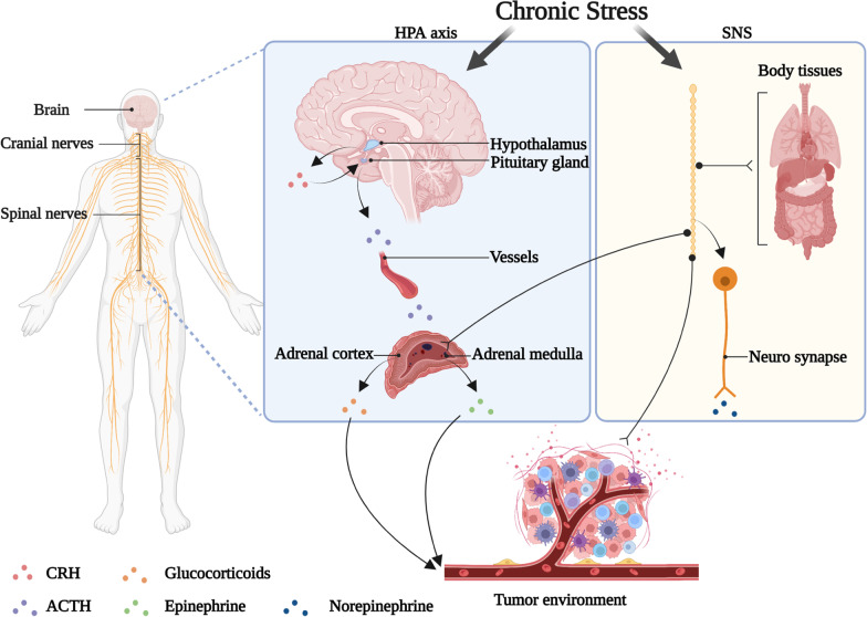
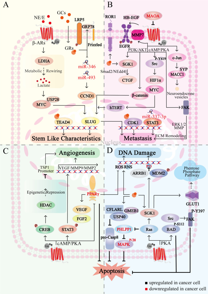
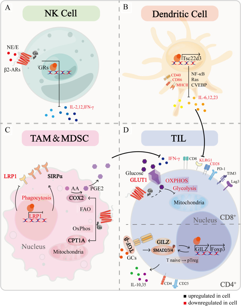
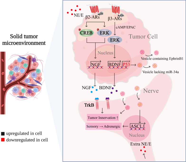
Chronic stress results in disturbances of body hormones through the neuroendocrine system. Cancer patients often experience recurrent anxiety and restlessness during disease progression and treatment, which aggravates disease progression and hinders treatment effects. Recent studies have shown that chronic stress-regulated neuroendocrine systems secret hormones to activate many signaling pathways related to tumor development in tumor cells. The activated neuroendocrine system acts not only on tumor cells but also modulates the survival and metabolic changes of surrounding non-cancerous cells. Current clinical evidences also suggest that chronic stress affects the outcome of cancer treatment. However, in clinic, there is lack of effective treatment for chronic stress in cancer patients. In this review, we discuss the main mechanisms by which chronic stress regulates the tumor microenvironment, including functional regulation of tumor cells by stress hormones (stem cell-like properties, metastasis, angiogenesis, DNA damage accumulation, and apoptotic resistance), metabolic reprogramming and immune escape, and peritumor neuromodulation. Based on the current clinical treatment framework for cancer and chronic stress, we also summarize pharmacological and non-pharmacological therapeutic approaches to provide some directions for cancer therapy.

Keywords: Cancer treatment; Chronic stress; Tumor development; Tumor microenvironment.


Conflict of interest statement

The authors declare that they have no known competing financial interests or personal relationships that could have appeared to influence the work reported in this paper.

Figures

Fig. 1
Fig. 1
Chronic stress regulates the tumor microenvironment through the neuroendocrine system. The nervous system is composed of cranial and spinal nerves. Under chronic stress, the hypothalamus releases CRH, which triggers ACTH secretion from pituitary to stimulate the secretion of glucocorticoids from the adrenal cortex. Chronic stress also activates the SNS, which directly innervates organs through sympathetic neurotransmitter norepinephrine (NE) release from neuro synapse, promoting the synthesis and secretion of epinephrine (E) from the adrenal medulla. NE generally reaches the TME through nerve fibers, and E/glucocorticoids (GCs) reach it through the blood
Fig. 2
Fig. 2
The biological mechanism of chronic stress affecting cancer cells. A Chronic stress promotes the stem cell characteristics in cancer cells. Chronic stress-induced E activates LDHA, promotes glycolysis, and leads to lactate secretion, this enhances the interaction between USP28 and MYC, which promotes stem cell-like-associated genes expression via SLUG. Chronic stress-induced GCs promote β-catenin expression through the interaction of GRP78 with LRP5. GCs also downregulate miR-346 and miR-493, which in turn upregulate Cyclin D1 and accelerate the cell cycle. In addition, GCs promote GRs-dependent nuclear accumulation and TEAD4 transcriptional activation, promoting the maintenance of CSCs. B Chronic stress accelerates cancer cell metastasis. Chronic stress-induced β-ARs signaling activates FAK via cAMP/PKA, which induces extracellular matrix remodeling via Erk1/2-MMP; Src is also activated by cAMP/PKA, and Y419 phosphorylation of Src amplifies HIF1α and MYC, further inducing hTERT overexpression in the nucleus. hTERT activates SLUG and in turn upregulates metastasis-related genes. β-ARs also promotes β-catenin expression and nuclear localization through PI3K/AKT and increases SLUG promoter activity. NE-induced downregulation of miR-337-3p activates STAT3. β-ARs activates MMP7 and releases HB-EGF to activate EGFR, whereas MAOA can target β2-ARs to reverse these processes. In addition, β-ARs promotes neuroendocrine phenotypic transformation and metastasis through MACC1 upregulation, which binds directly to SYP via c-Jun. GRs contribute to increased ROR1. In addition, GRs increase CTGF expression via PI3K/SGK/Nedd4l-Smad2 and promotes lung metastasis. GRs localize to the CDK1 promoter in nucleus and stimulate CDK1 through epigenetic regulation. C Chronic stress promotes angiogenesis. β-ARs activates CREB and STAT3 via cAMP/PKA, STAT3 translocates to the nucleus and stimulates VEGF and MMP2/9 transcription, CREB targets HDAC2 activation, which epistemically represses TSP1. In addition, PPARγ inhibits VEGF by suppressing ROS, while β-ARs signaling reverses the effect of PPARγ on VEGF/FGF2. D Chronic stress promotes DNA damage accumulation and anti-apoptosis. β-ARs mediates Src/FAK and BAD anti-apoptotic pathways by PKA, BAD-S112 phosphorylation and FAK-Y397 phosphorylation gain resistance to apoptosis. GCs stabilize CFLARL through GMEB1-USP40 interaction, inhibit pro-CASP8 activation, thereby inhibiting apoptosis. SGK1, a key downstream effector of Ras, promotes glucose-mediated carbon flux into multiple metabolic pathways to inhibit oxidation by GLUT1 activation; in addition, Ras blocks apoptosis by reducing PHLPP1, which activates p38 MAPK to promote apoptosis. Elevated NE/E and GCs increase MDM2 activity and decrease p53 function via SGK1 and ARRB1, β-ARs and GRs also stimulate the production of ROS and RNS, ultimately leading to DNA damage accumulation
Fig. 3
Fig. 3
The biological mechanism of chronic stress affecting immune cells. A NK cell: E/NE inhibits cytokines production, including IL-12, IFN-γ and IL-2, through DNA-dependent mechanisms activation and protein synthesis. B Dendritic cell: chronic stress downregulates maturation-related markers such as CD40, CD68, and MHC II receptors in DCs. GRs upregulate TSC22D3 expression, mediate immunosuppressive effects through NF-κB, Ras and CVEBP, reduce pro-inflammatory cytokines IL-6, 12, and 23 productions, and block antigen presentation, resulting in the inability of TILs to acquire mature phenotype, as evidenced by low KLRG-1. C TAM & MDSC: chronic stress mediates metabolic reprogramming of macrophages and promotes immunosuppression. β2-ARs increases oxidative phosphorylation and promotes CPT1A expression; β2-ARs also increases FAO, which increases PGE2 production through COX2 overexpression and inhibits IFN-γ production by CD8+ T cells. GCs-GRs inhibit LRP1 and increase SIRPα, leading to an imbalance in the LRP1/SIRPα axis and inhibiting the phagocytosis of tumor cells by macrophages. D TIL: chronic stress reshaped the TIL phenotype in the TME, CD4+ TIL exhibit PD-1+, FOXP3+, CD8+ TIL exhibit PD-1+, TIM3+, Lag3+, IFN-γ, CD28. β2-ARs decreased GLUT1 and glucose uptake, resulting in reduced glycolysis and oxidative phosphorylation. GCs-GRs induce GILZ expression, which synergistically induces FoxP3 expression with TGF-β and SMAD2/3/4, further enhancing TGF-β signaling and promoting the conversion of naive T cells to Treg cells. Treg cells can produce immunosuppressive cytokines, including IL-10 and IL-35
Fig. 4
Fig. 4
The biological mechanism of chronic stress affecting perineural nerve of tumor. Chronic stress-induced ADRB increases BDNF via ADRB3/cAMP/EpAC/JNK and NGF via ADRB2/CREB or ADRB2/ERK. BDNF and NGF bind to TrkB in peritumor sensory neurons, promoting innervation of the tumor microenvironment and producing a sustained accumulation of adrenergic signals, forming a feed-forward loop. Chronic stress-induced low p53 in tumor cells leads to miR-34a loss in exosomes, resulting in increased ASCL1 in peritumor sensory neurons, which redifferentiates peritumor sensory neurons into adrenergic neurons. In addition, tumor cells secrete exosomes containing EphrinB1 into peritumor sensory nerves, inducing tumor innervation

Similar articles

Cited by

References

    1. Yuan K, Zheng YB, Wang YJ, Sun YK, Gong YM, Huang YT, Chen X, Liu XX, Zhong Y, Su SZ, et al. A systematic review and meta-analysis on prevalence of and risk factors associated with depression, anxiety and insomnia in infectious diseases, including COVID-19: a call to action. Mol Psychiatry. 2022 doi: 10.1038/s41380-022-01638-z. - DOI - PMC - PubMed
    1. Russell G, Lightman S. The human stress response. Nat Rev Endocrinol. 2019;15(9):525–534. - PubMed
    1. Cole SW, Nagaraja AS, Lutgendorf SK, Green PA, Sood AK. Sympathetic nervous system regulation of the tumour microenvironment. Nat Rev Cancer. 2015;15(9):563–572. - PMC - PubMed
    1. Sloan EK, Capitanio JP, Tarara RP, Cole SW. Social temperament and lymph node innervation. Brain Behav Immun. 2008;22(5):717–726. - PMC - PubMed
    1. Cui B, Peng F, Lu J, He B, Su Q, Luo H, Deng Z, Jiang T, Su K, Huang Y, et al. Cancer and stress: NextGen strategies. Brain Behav Immun. 2021;93:368–383. - PubMed