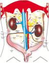
Structural organization of the neuronal pathways of the superior ovarian nerve in the rat
- PMID: 36707870
- PMCID: PMC9883865
- DOI: 10.1186/s13048-023-01109-1
Structural organization of the neuronal pathways of the superior ovarian nerve in the rat
Abstract
Background: In the rat, studies have shown that ovary innervation arrives via the superior ovarian nerve (SON) and the ovarian plexus nerve, which originates from the celiac plexus (CP). In the present study, we performed a neuroanatomical technique to investigate the anatomy of the SON between the ovary and the CP.
Results: We found that the SON fibers were concentrated on the lateral border of the suprarenal ganglion and projected towards, then inserted into the suspensory ligament. Then, it ran parallel to the long axis of the ligament to reach and innervate the ovaries. At this level, the SON was composed of two coiled nerve fibers, each between 10 and 15 µm in diameter. The SON was linked to three different ganglia: the suprarenal ganglia, the celiac ganglia, and the superior mesenteric ganglion.
Conclusions: The postganglionic fibers that project to the ovary via the SON emerge from the suprarenal ganglia. The trajectories on the right and left sides to each ovary are similar. The somas of ipsilateral and contralateral SON neurons are located in the prevertebral ganglia, mostly in the celiac ganglia.
Keywords: Celiac ganglion; Ovary; Superior ovarian nerve; Suprarenal ganglion; Suspensory ligament.
© 2023. The Author(s).
Conflict of interest statement
The authors declare no competing interests.
Figures


Similar articles
-
The extrinsic innervation of the abdominal organs in the female rat.Acta Anat (Basel). 1979;104(3):243-67. doi: 10.1159/000145073. Acta Anat (Basel). 1979. PMID: 90440
-
Anatomical organization and neural pathways of the ovarian plexus nerve in rats.J Ovarian Res. 2017 Mar 14;10(1):18. doi: 10.1186/s13048-017-0311-x. J Ovarian Res. 2017. PMID: 28292315 Free PMC article.
-
Neural activity between ovaries and the prevertebral celiac-superior mesenteric ganglia varies during the estrous cycle of the rat.Endocrine. 2005 Mar;26(2):147-52. doi: 10.1385/ENDO:26:2:147. Endocrine. 2005. PMID: 15888926
-
Anatomical localization of afferent and postganglionic sympathetic neurons innervating the rat ovary.Neurosci Lett. 1988 Feb 29;85(2):217-22. doi: 10.1016/0304-3940(88)90354-0. Neurosci Lett. 1988. PMID: 3374837
-
Physiology of mammalian prevertebral ganglia.Annu Rev Physiol. 1981;43:53-68. doi: 10.1146/annurev.ph.43.030181.000413. Annu Rev Physiol. 1981. PMID: 6260023 Review.
Cited by
-
Organization of the Subdiaphragmatic Vagus Nerve and Its Connection with the Celiac Plexus and the Ovaries in the Female Rat.Brain Sci. 2023 Jul 6;13(7):1032. doi: 10.3390/brainsci13071032. Brain Sci. 2023. PMID: 37508964 Free PMC article.
-
The role of the autonomic nervous system in polycystic ovary syndrome.Front Endocrinol (Lausanne). 2024 Jan 19;14:1295061. doi: 10.3389/fendo.2023.1295061. eCollection 2023. Front Endocrinol (Lausanne). 2024. PMID: 38313837 Free PMC article. Review.
-
Metabolic control of ovarian function through the sympathetic nervous system: role of leptin.Front Endocrinol (Lausanne). 2025 Feb 3;15:1484939. doi: 10.3389/fendo.2024.1484939. eCollection 2024. Front Endocrinol (Lausanne). 2025. PMID: 39963180 Free PMC article. Review.
References
-
- Kelly WA, Marler RJ, Weikel JH. Drug-induced mesovarial leiomyomas in the rat—a review and additional data. J Am Coll Toxicol. 1993;12(1):13–22. doi: 10.3109/10915819309140618. - DOI
-
- van der Schoot P, Emmen JM. Development, structure and function of the cranial suspensory ligaments of the mammalian gonads in a cross-species perspective; their possible role in effecting disturbed testicular descent. Hum Reprod Update. 1996;2(5):399–418. doi: 10.1093/humupd/2.5.399. - DOI - PubMed
MeSH terms
Grants and funding
LinkOut - more resources
Full Text Sources
Miscellaneous

