. 2023 Jan 27;11(1):21.
doi: 10.1186/s40478-023-01510-3.

Lack of a protective effect of the Tmem106b "protective SNP" in the Grn knockout mouse model for frontotemporal lobar degeneration

Affiliations

Lack of a protective effect of the Tmem106b "protective SNP" in the Grn knockout mouse model for frontotemporal lobar degeneration

Anne-Sophie Cabron et al. Acta Neuropathol Commun. .

Abstract

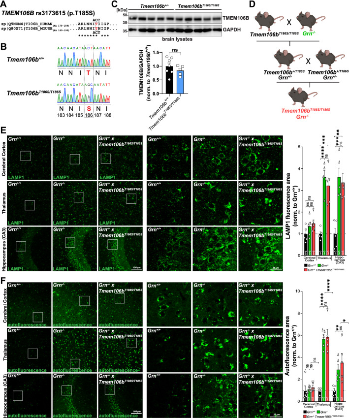
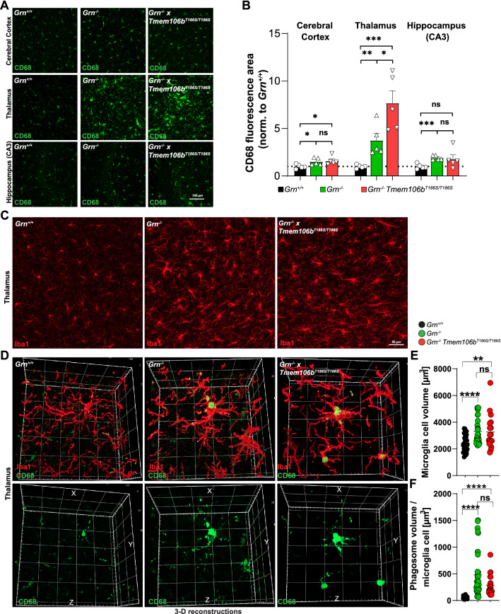
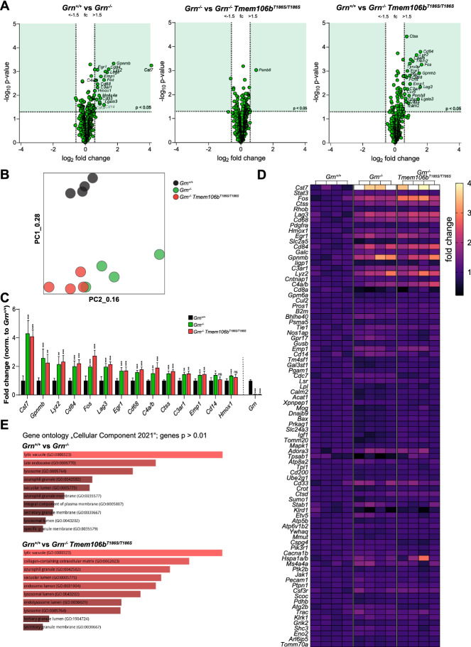
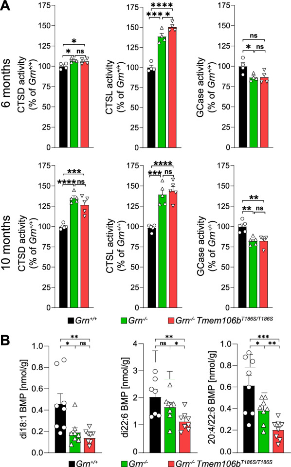
Genetic variants in TMEM106B are a common risk factor for frontotemporal lobar degeneration and the most important modifier of disease risk in patients with progranulin (GRN) mutations (FTLD-GRN). TMEM106B is encoding a lysosomal transmembrane protein of unknown molecular function. How it mediates its disease-modifying function remains enigmatic. Several TMEM106B single nucleotide polymorphisms (SNPs) are significantly associated with disease risk in FTLD-GRN carriers, of which all except one are within intronic sequences of TMEM106B. Of note, the non-coding SNPs are in high linkage disequilibrium with the coding SNP rs3173615 located in exon six of TMEM106B, resulting in a threonine to serine change at amino acid 185 in the minor allele, which is protective in FTLD-GRN carriers. To investigate the functional consequences of this variant in vivo, we generated and characterized a knockin mouse model harboring the Tmem106bT186S variant. We analyzed the effect of this protective variant on FTLD pathology by crossing Tmem106bT186S mice with Grn-/- knockout mice, a model for GRN-mediated FTLD. We did not observe the amelioration of any of the investigated Grn-/- knockout phenotypes, including transcriptomic changes, lipid alterations, or microgliosis in Tmem106bT186S/T186S × Grn-/- mice, indicating that the Tmem106bT186S variant is not protective in the Grn-/- knockout mouse model. These data suggest that effects of the associated SNPs not directly linked to the amino acid exchange in TMEM106B are critical for the modifying effect.

Keywords: FTLD; Progranulin; Protective SNP; TMEM106B.


Conflict of interest statement

MD participated in advisory board meetings of Arkuda Therapeutics and received a speaker honorarium from Takeda.

Figures

Fig. 1
Fig. 1
A Alignment of the protein sequence of human and mouse TMEM106B around the critical threonine (red) that is coded by the rs3173615 SNP in humans. T185 in humans corresponds to T186 in mice. The codon coding for the threonine is indicated. B Sequencing chromatogram of genomic tail DNA of a wildtype and a homozygous Tmem106bT186S/T186S mouse with the codons for threonine 186 in the wildtype mouse and serin 186 (red) in the knockin mouse after gene editing of the “ACT” codon to “AGC”. A third base in the following codon is edited in the wobble-position without affecting the coded amino acid asparagine. C Immunoblot of brain lysates from wildtype and Tmem106bT186S/T186S mice (n = 5–7, nine months old) with antibodies against TMEM106B and GAPDH as a loading control. The quantification of the TMEM106B signal is depicted (average of wildtype set as “1”). D Breeding scheme depicting the crossing of Tmem106bT186S/T186S and Grn−/− mice to obtain Grn−/− × Tmem106T186S/T186S. E Representative images of immunofluorescence stainings of the cerebral cortex, thalamus, and the hippocampus (CA3 subfield) of a wildtype, Grn−/−, and Grn−/− × Tmem106bT186S/T186S with an antibody against LAMP1 (green). The quantification of the LAMP1-positive area/section is provided (n = 5, age: 6 months). F Representative images of autofluorescence recorded with laser excitation at 594 nm of the cerebral cortex, thalamus, and the hippocampus (CA3 subfield) of a wildtype, Grn−/−, and Grn−/− × Tmem106bT186S/T186S. The quantification of the autofluorescence-positive area/section is provided (n = 5. Age: 6 months)
Fig. 2
Fig. 2
A Representative images of immunofluorescence stainings of the cerebral cortex, thalamus, and the hippocampus (CA3 subfield) of a wildtype, Grn−/−, and Grn−/− × Tmem106bT186S/T186S with an antibody against CD68 (green). B The quantification of the CD68-positive area/section is indicated (n = 5; Age: 6 months). C Representative images of immunofluorescence stainings of the thalamus of a wildtype, Grn−/−, and Grn−/− × Tmem106bT186S/T186S with an antibody against Iba1 (red). Age: 6 month. D Three-dimensional reconstructions from representative microglia cells of a wildtype, Grn−/−, and Grn−/− × T186S/T186S mouse stained for Iba1 (red) and CD68 (green) from 50 images in the Z-direction of the thalamus (age: 6 months). E Volumetric quantification of the microglia cell volume of individual cells depicted from 3D reconstructed images. F Volumetric quantification of the CD68-positive phagosome compartment in individual Iba1-positive microglia cells depicted from 3D reconstructed images. B, E, F *p < 0.05; **p < 0.01; ****p < 0.001; ns Not significant
Fig. 3
Fig. 3
A Volcano plot presentation of the differently expressed transcripts between wildtype vs. Grn−/− (left panel), Grn−/− vs. Grn−/− × Tmem106bT186S/T186S and wildtype vs Grn−/− × Tmem106bT186S/T186S mice (n = 4, each genotype) in RNA isolated from 6-month-old mice. The thresholds for transcripts reaching statistically significant differences (fold change (fc) < − 1.5 / > 1.5; p < 0.05) are indicated (dotted lines), and the quadrants with transcripts reaching the threshold are colored in green. Grn as the top-down-regulated transcript in Grn−/− and Grn−/− × Tmem106bT186S/T186S mice was excluded in the volcano-plot representation. B Principal component analysis (PCA) of the 12 total brain samples analyzed by Nanostring nCounter “Glia Profiling” panel. Every individual point represents one individual biological replicate: Wildtype = black, Grn−/− = green Grn−/− × Tmem106bT186S/T186S = red. Note that the wildtype group can be clearly separated from the other two groups, while there is an overlap between Grn−/− and Grn−/− × Tmem106bT186S/T186S. C Fold changes of the top 14 transcripts differentially expressed between wildtype and Grn−/− mice determined by NanoString nCounter analysis. Statistics indicate significant differences between wildtype and Grn−/− / wildtype and Grn−/− × Tmem106bT186S/T186S mice. For non of the selected genes, any significant differences were found between Grn−/− and wildtype and Grn−/− × Tmem106bT186S/T186S. *p < 0.05; **p < 0.01; ***p < 0.001; ns = not significant; (n = 4, each genotype). D Heatmap of 86 gene transcripts analyzed by NanoString nCounter analysis in wildtype, Grn−/−, Grn−/− × Tmem106bT186S/T186S mice (n = 4, each genotype) from total brain RNA isolated from 6-month-old mice. The expression-corrected and housekeeping gene-normalized RNA counts for each gene and sample were normalized to the mean value of the wildtype animals. Grn was excluded from this representation. E Pathway enrichment of differentially expressed genes (p < 0.01) between wildtype and Grn−/− mice (upper panel) and wildtype and Grn−/− × Tmem106bT186S/T186S mice (lower panel) by the “Enrichr” tool. The top-enriched terms of the gene ontology term “Cellular Component 2021” are depicted
Fig. 4
Fig. 4
A Specific enzymatic activity of the three lysosomal enzymes cathepsin D (CTSD), cathepsin L (CTSL), and β-Glucocerebrosidase (GCase) in brain lysates of 6-month-old wildtype, Grn−/−, Grn−/− × Tmem106bT186S/T186S mice (n = 4, each genotype) (upper panel) and 10-month-old mice (lower panel). B The levels of the BMP species with different fatty acid compositions (di18:1, di22:6, and 20:4/22:6) were quantified in lipid extracts of the brain from 6-month-old wildtype, Grn−/−, Grn−/− × Tmem106bT186S/T186S mice (n = 8, each genotype) by mass spectrometry

Similar articles

  • TMEM106B p.T185S regulates TMEM106B protein levels: implications for frontotemporal dementia.
    Nicholson AM, Finch NA, Wojtas A, Baker MC, Perkerson RB 3rd, Castanedes-Casey M, Rousseau L, Benussi L, Binetti G, Ghidoni R, Hsiung GY, Mackenzie IR, Finger E, Boeve BF, Ertekin-Taner N, Graff-Radford NR, Dickson DW, Rademakers R. Nicholson AM, et al. J Neurochem. 2013 Sep;126(6):781-91. doi: 10.1111/jnc.12329. Epub 2013 Jul 1. J Neurochem. 2013. PMID: 23742080 Free PMC article.
  • TMEM106B regulates progranulin levels and the penetrance of FTLD in GRN mutation carriers.
    Finch N, Carrasquillo MM, Baker M, Rutherford NJ, Coppola G, Dejesus-Hernandez M, Crook R, Hunter T, Ghidoni R, Benussi L, Crook J, Finger E, Hantanpaa KJ, Karydas AM, Sengdy P, Gonzalez J, Seeley WW, Johnson N, Beach TG, Mesulam M, Forloni G, Kertesz A, Knopman DS, Uitti R, White CL 3rd, Caselli R, Lippa C, Bigio EH, Wszolek ZK, Binetti G, Mackenzie IR, Miller BL, Boeve BF, Younkin SG, Dickson DW, Petersen RC, Graff-Radford NR, Geschwind DH, Rademakers R. Finch N, et al. Neurology. 2011 Feb 1;76(5):467-74. doi: 10.1212/WNL.0b013e31820a0e3b. Epub 2010 Dec 22. Neurology. 2011. PMID: 21178100 Free PMC article.
  • Loss of Tmem106b exacerbates FTLD pathologies and causes motor deficits in progranulin-deficient mice.
    Zhou X, Brooks M, Jiang P, Koga S, Zuberi AR, Baker MC, Parsons TM, Castanedes-Casey M, Phillips V, Librero AL, Kurti A, Fryer JD, Bu G, Lutz C, Dickson DW, Rademakers R. Zhou X, et al. EMBO Rep. 2020 Oct 5;21(10):e50197. doi: 10.15252/embr.202050197. Epub 2020 Aug 5. EMBO Rep. 2020. PMID: 32761777 Free PMC article.
  • What we know about TMEM106B in neurodegeneration.
    Nicholson AM, Rademakers R. Nicholson AM, et al. Acta Neuropathol. 2016 Nov;132(5):639-651. doi: 10.1007/s00401-016-1610-9. Epub 2016 Aug 20. Acta Neuropathol. 2016. PMID: 27543298 Free PMC article. Review.
  • TMEM106B a novel risk factor for frontotemporal lobar degeneration.
    van der Zee J, Van Broeckhoven C. van der Zee J, et al. J Mol Neurosci. 2011 Nov;45(3):516-21. doi: 10.1007/s12031-011-9555-x. Epub 2011 May 26. J Mol Neurosci. 2011. PMID: 21614538 Free PMC article. Review.

Cited by

References

    1. Ahmed Z, Sheng H, Xu YF, Lin WL, Innes AE, Gass J, Yu X, Wuertzer CA, Hou H, Chiba S, et al. Accelerated lipofuscinosis and ubiquitination in granulin knockout mice suggest a role for progranulin in successful aging. Am J Pathol. 2010;177:311–324. doi: 10.2353/ajpath.2010.090915. - DOI - PMC - PubMed
    1. Arrant AE, Roth JR, Boyle NR, Kashyap SN, Hoffmann MQ, Murchison CF, Ramos EM, Nana AL, Spina S, Grinberg LT, et al. Impaired beta-glucocerebrosidase activity and processing in frontotemporal dementia due to progranulin mutations. Acta Neuropathol Commun. 2019;7:218. doi: 10.1186/s40478-019-0872-6. - DOI - PMC - PubMed
    1. Baker M, Mackenzie IR, Pickering-Brown SM, Gass J, Rademakers R, Lindholm C, Snowden J, Adamson J, Sadovnick AD, Rollinson S, et al. Mutations in progranulin cause tau-negative frontotemporal dementia linked to chromosome 17. Nature. 2006;442:916–919. doi: 10.1038/nature05016. - DOI - PubMed
    1. Beel S, Moisse M, Damme M, De Muynck L, Robberecht W, Van Den Bosch L, Saftig P, Van Damme P. Progranulin functions as a cathepsin D chaperone to stimulate axonal outgrowth in vivo. Hum Mol Genet. 2017;26:2850–2863. doi: 10.1093/hmg/ddx162. - DOI - PMC - PubMed
    1. Bellenguez C, Kucukali F, Jansen IE, Kleineidam L, Moreno-Grau S, Amin N, Naj AC, Campos-Martin R, Grenier-Boley B, Andrade V, et al. New insights into the genetic etiology of Alzheimer's disease and related dementias. Nat Genet. 2022;54:412–436. doi: 10.1038/s41588-022-01024-z. - DOI - PMC - PubMed

Publication types

MeSH terms

LinkOut - more resources