The NLRP3 inflammasome - interleukin 1β axis in uveal melanoma
- PMID: 36707938
- PMCID: PMC9989921
- DOI: 10.1002/2211-5463.13566
The NLRP3 inflammasome - interleukin 1β axis in uveal melanoma
Abstract
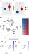
Uveal melanoma (UM) is the most common primary intraocular cancer in the adult population. Recent studies suggested that the NLRP3 inflammasome could be a therapeutic target for cutaneous melanoma (CM), but the role of NLRP3 in UM remains unknown. Here, we analyzed the NLRP3-IL-1β axis in 5 UM and 4 CM cell lines. Expression of NLRP3 mRNA in UM and CM was low, and expression in UM was lower than in CM (P < 0.001). NLRP3 protein levels were below detection limit for all cell lines. UM exhibited lower baseline IL-1β secretion than CM, especially when compared to the Hs294t cell line (P < 0.05). Bioinformatic analysis of human tumor samples showed that UM has significantly lower expression of NLRP3 and IL-1β compared with CM. In conclusion, our work shows evidence of extremely low NLRP3 expression and IL-1β secretion by melanoma cells and highlight differences between CM and UM.
Keywords: IL-1; NLRP3; inflammasome; melanoma; skin; uveal.
© 2023 The Authors. FEBS Open Bio published by John Wiley & Sons Ltd on behalf of Federation of European Biochemical Societies.
Conflict of interest statement
The authors declare no conflict of interest.
Figures


Similar articles
-
Forskolin attenuates the NLRP3 inflammasome activation and IL-1β secretion in human macrophages.Pediatr Res. 2019 Dec;86(6):692-698. doi: 10.1038/s41390-019-0418-4. Epub 2019 May 13. Pediatr Res. 2019. PMID: 31086288
-
Sendai Virus V Protein Inhibits the Secretion of Interleukin-1β by Preventing NLRP3 Inflammasome Assembly.J Virol. 2018 Sep 12;92(19):e00842-18. doi: 10.1128/JVI.00842-18. Print 2018 Oct 1. J Virol. 2018. PMID: 30021903 Free PMC article.
-
Reduced NLRP3 Gene Expression Limits the IL-1β Cleavage via Inflammasome in Monocytes from Severely Injured Trauma Patients.Mediators Inflamm. 2018 May 9;2018:1752836. doi: 10.1155/2018/1752836. eCollection 2018. Mediators Inflamm. 2018. PMID: 29861655 Free PMC article.
-
The NLRP3 inflammasome: activation and regulation.Trends Biochem Sci. 2023 Apr;48(4):331-344. doi: 10.1016/j.tibs.2022.10.002. Epub 2022 Nov 4. Trends Biochem Sci. 2023. PMID: 36336552 Free PMC article. Review.
-
[Advances in mechanisms for NLRP3 inflammasomes regulation].Yao Xue Xue Bao. 2016 Oct;51(10):1505-12. Yao Xue Xue Bao. 2016. PMID: 29924571 Review. Chinese.
Cited by
-
miR-22 negatively regulating NOD-like receptor protein 3 gene in the proliferation, invasion, and migration of malignant melanoma cells.Postepy Dermatol Alergol. 2024 Jun;41(3):284-291. doi: 10.5114/ada.2024.140521. Epub 2024 Apr 25. Postepy Dermatol Alergol. 2024. PMID: 39027690 Free PMC article.
-
Inflammasome activation in melanoma progression: the latest update concerning pathological role and therapeutic value.Arch Dermatol Res. 2025 Jan 16;317(1):258. doi: 10.1007/s00403-025-03802-1. Arch Dermatol Res. 2025. PMID: 39820618 Review.
References
-
- Jager MJ, Shields CL, Cebulla CM, Abdel‐Rahman MH, Grossniklaus HE, Stern MH, Carvajal RD, Belfort RN, Jia R, Shields JA et al. (2020) Uveal melanoma. Nat Rev Dis Primers 6, 18–20. - PubMed
-
- Shields CL, Furuta M, Thangappan A, Nagori S, Mashayekhi A, Lally DR, Kelly CC, Rudich DS, Nagori AV, Wakade OA et al. (2009) Metastasis of uveal melanoma millimeter‐by‐millimeter in 8033 consecutive eyes. Arch Ophthalmol 127, 989–998. - PubMed
-
- Collaborative Ocular Melanoma Study Group (2005) Development of metastatic disease after enrollment in the COMS trials for treatment of choroidal melanoma. Arch Ophthalmol 123, 1639–1643. - PubMed
Publication types
MeSH terms
Substances
Grants and funding
LinkOut - more resources
Full Text Sources
Medical

