

Non-specific and specific DNA binding modes of bacterial histone, HU, separately regulate distinct physiological processes through different mechanisms
- PMID: 36708073
- PMCID: PMC10120378
- DOI: 10.1111/mmi.15033
Non-specific and specific DNA binding modes of bacterial histone, HU, separately regulate distinct physiological processes through different mechanisms
Abstract
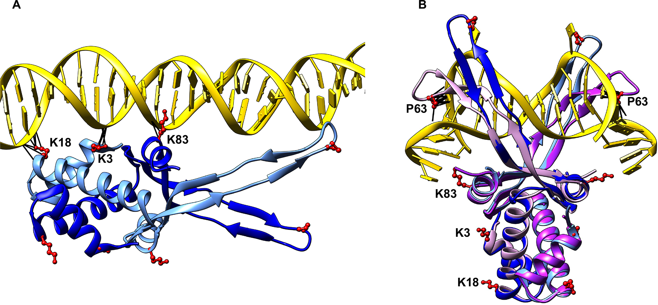
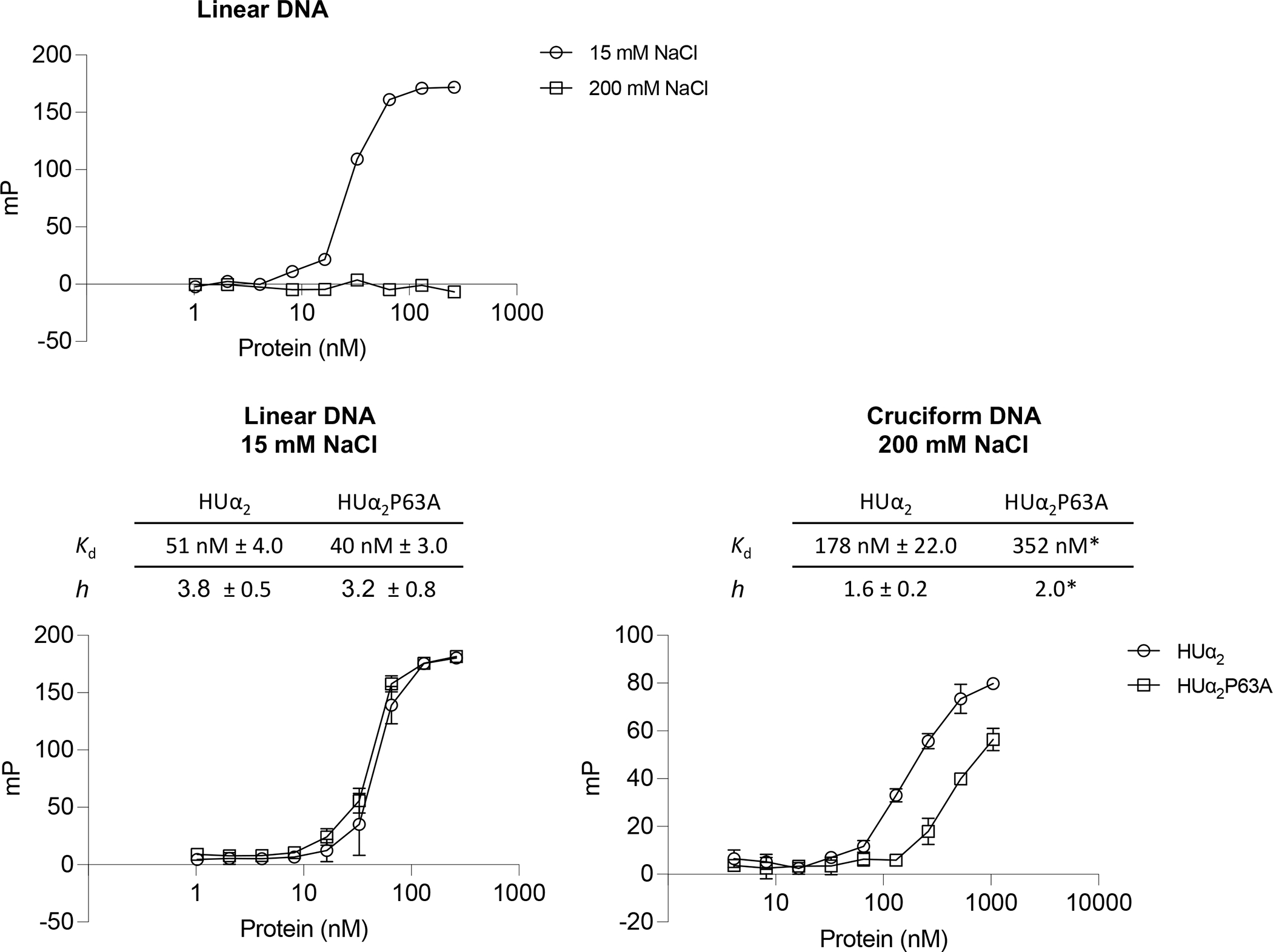
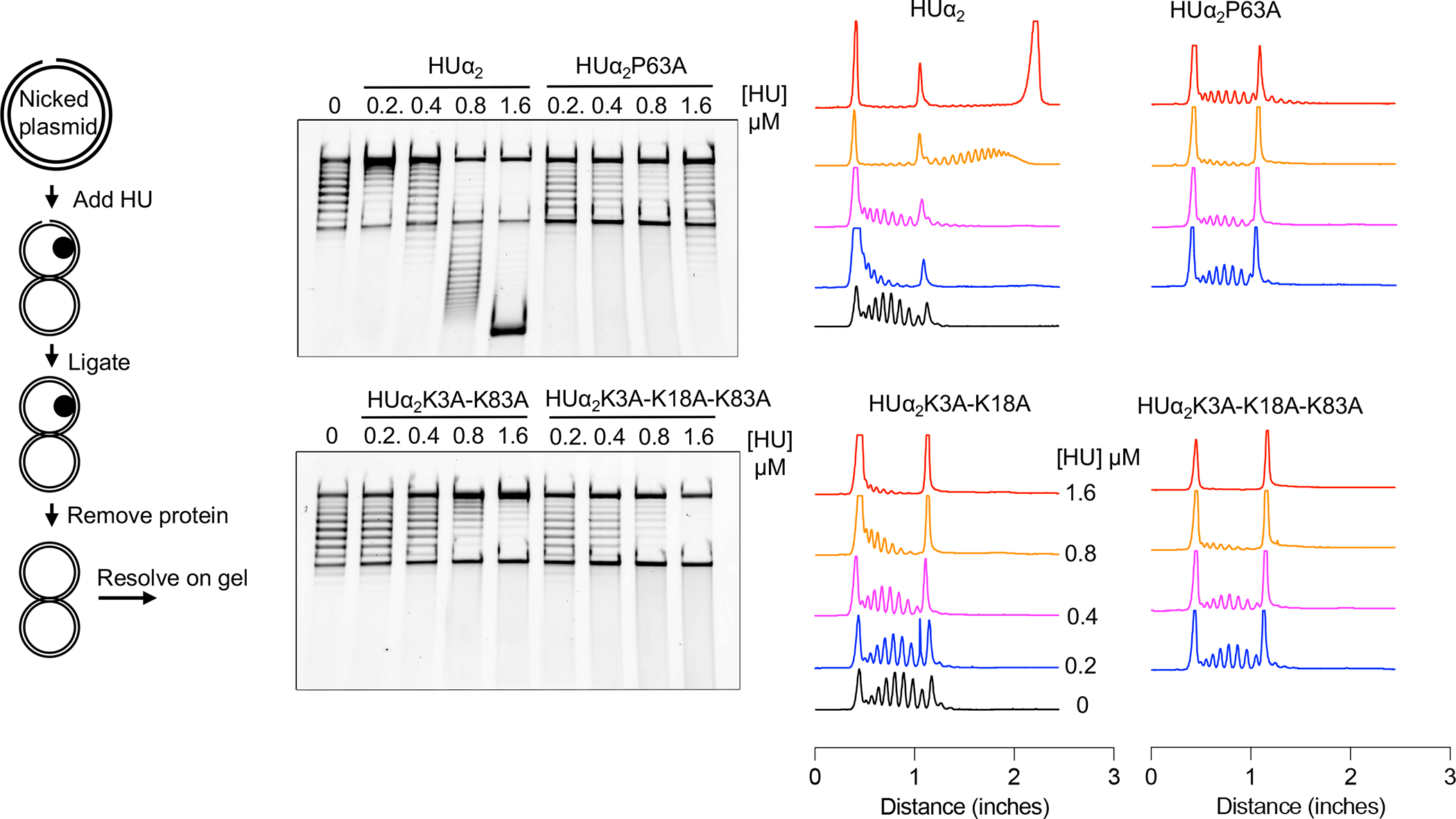
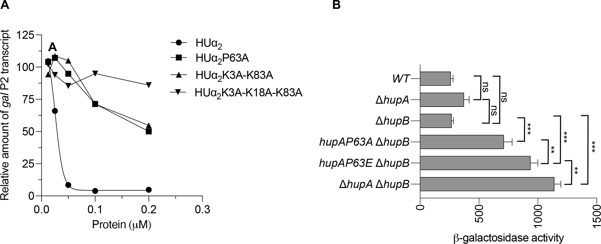
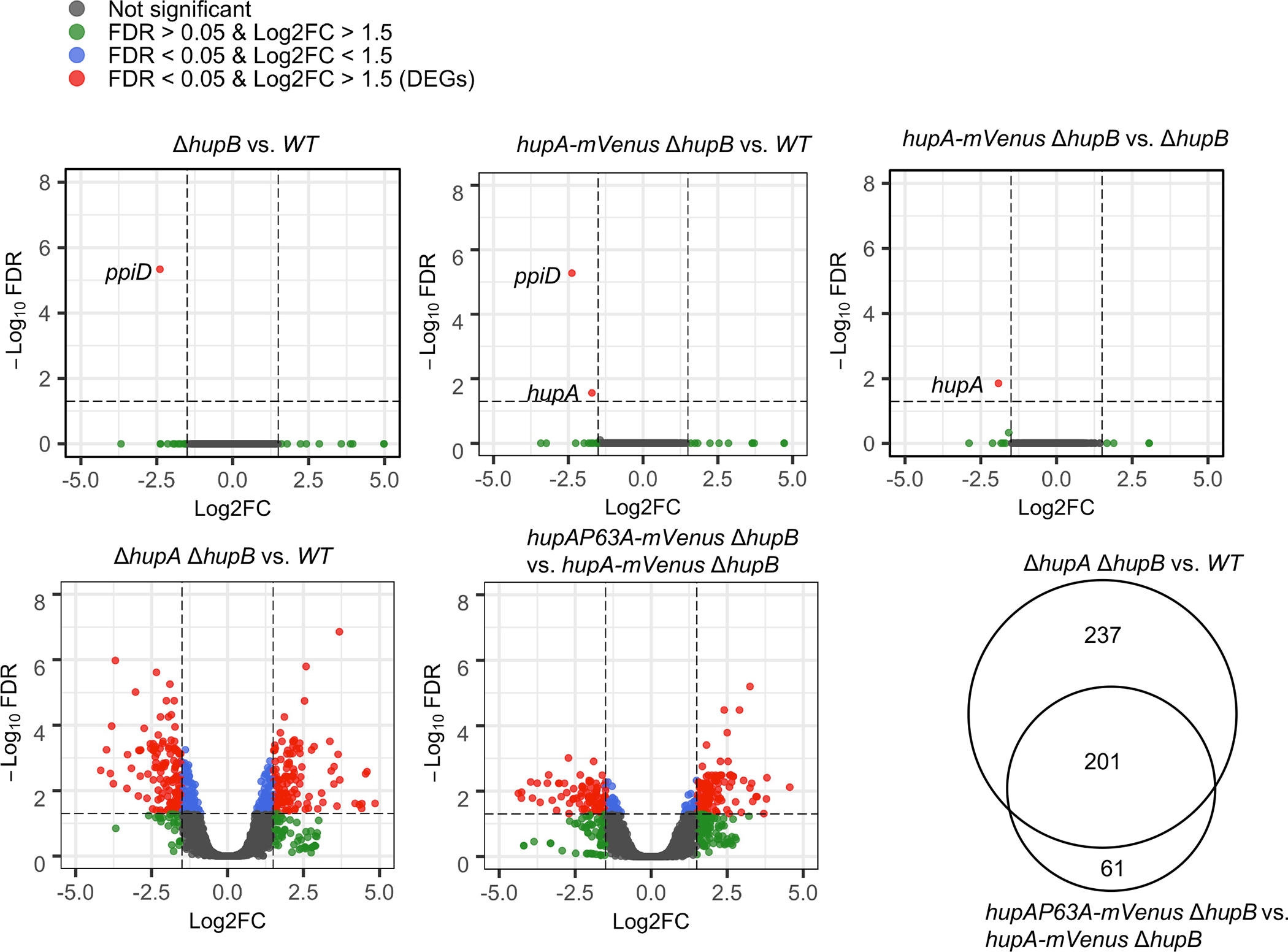
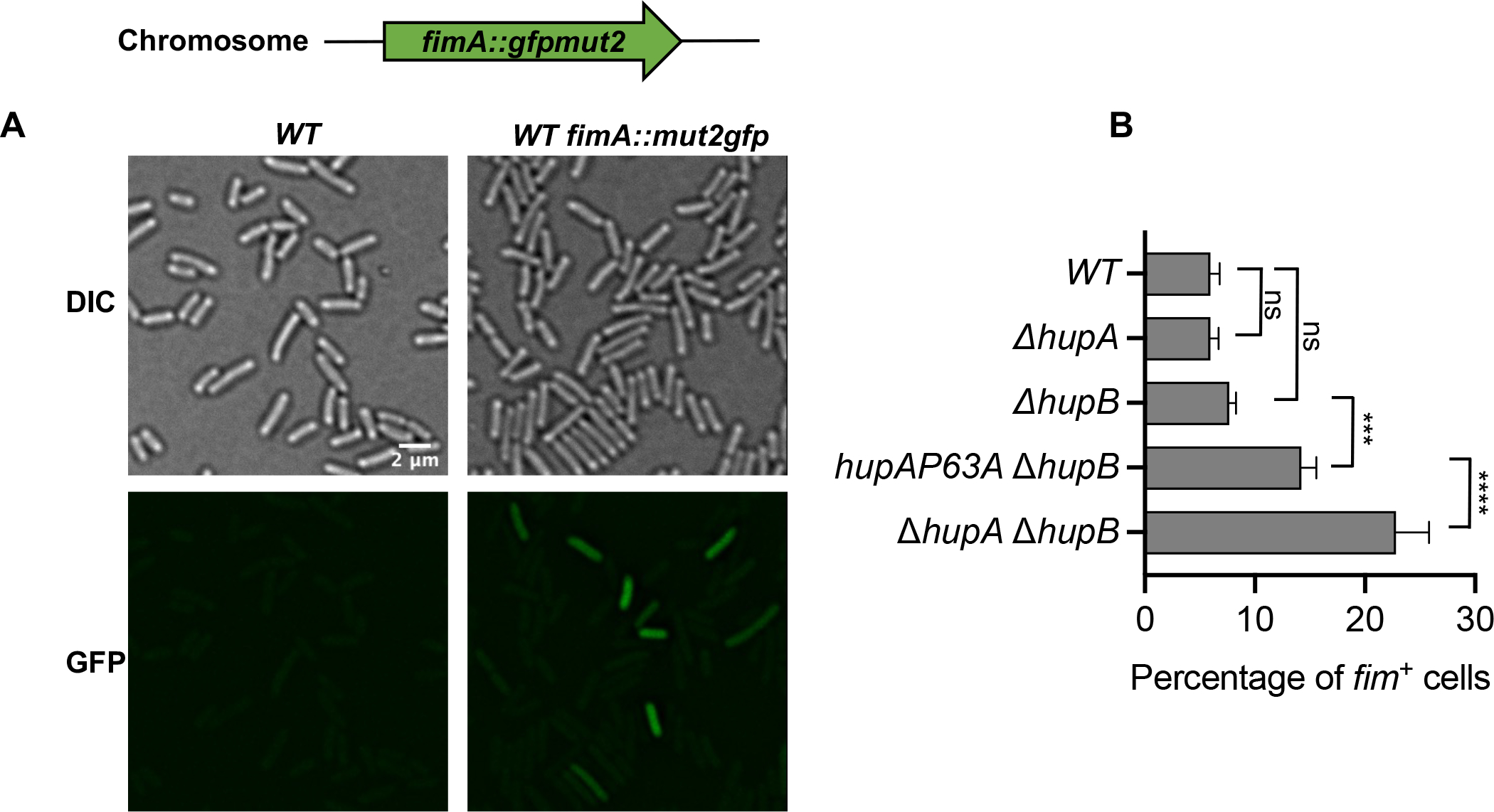
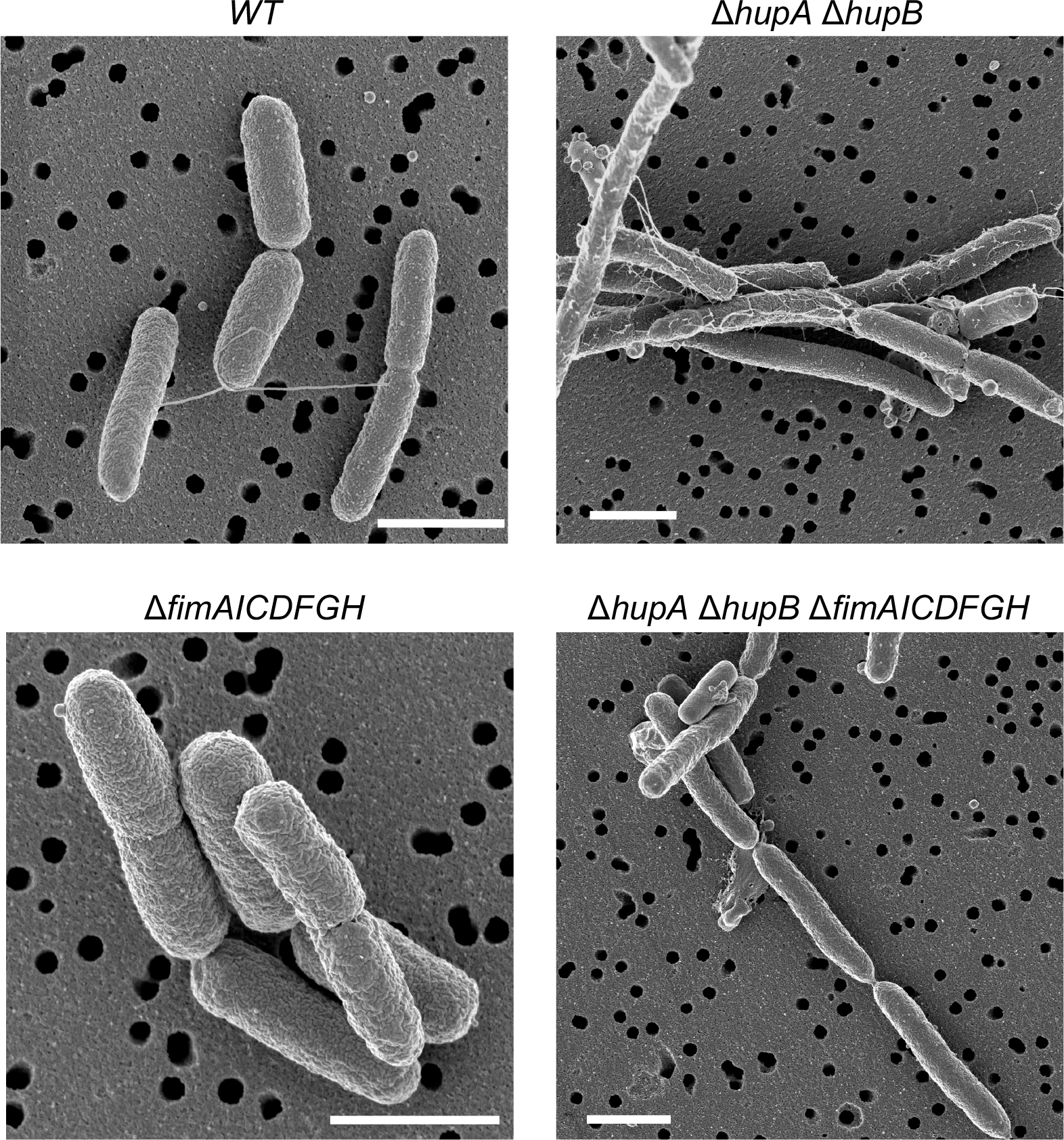
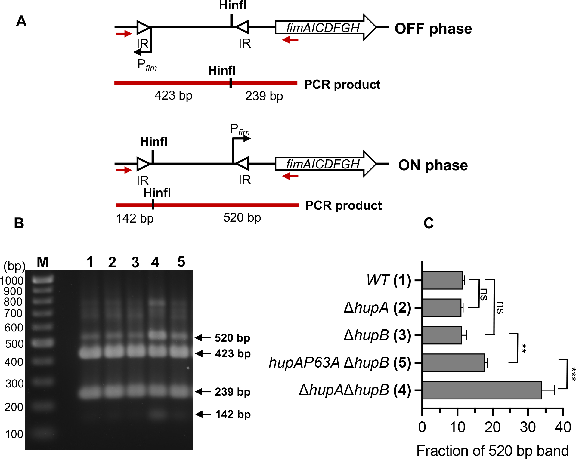
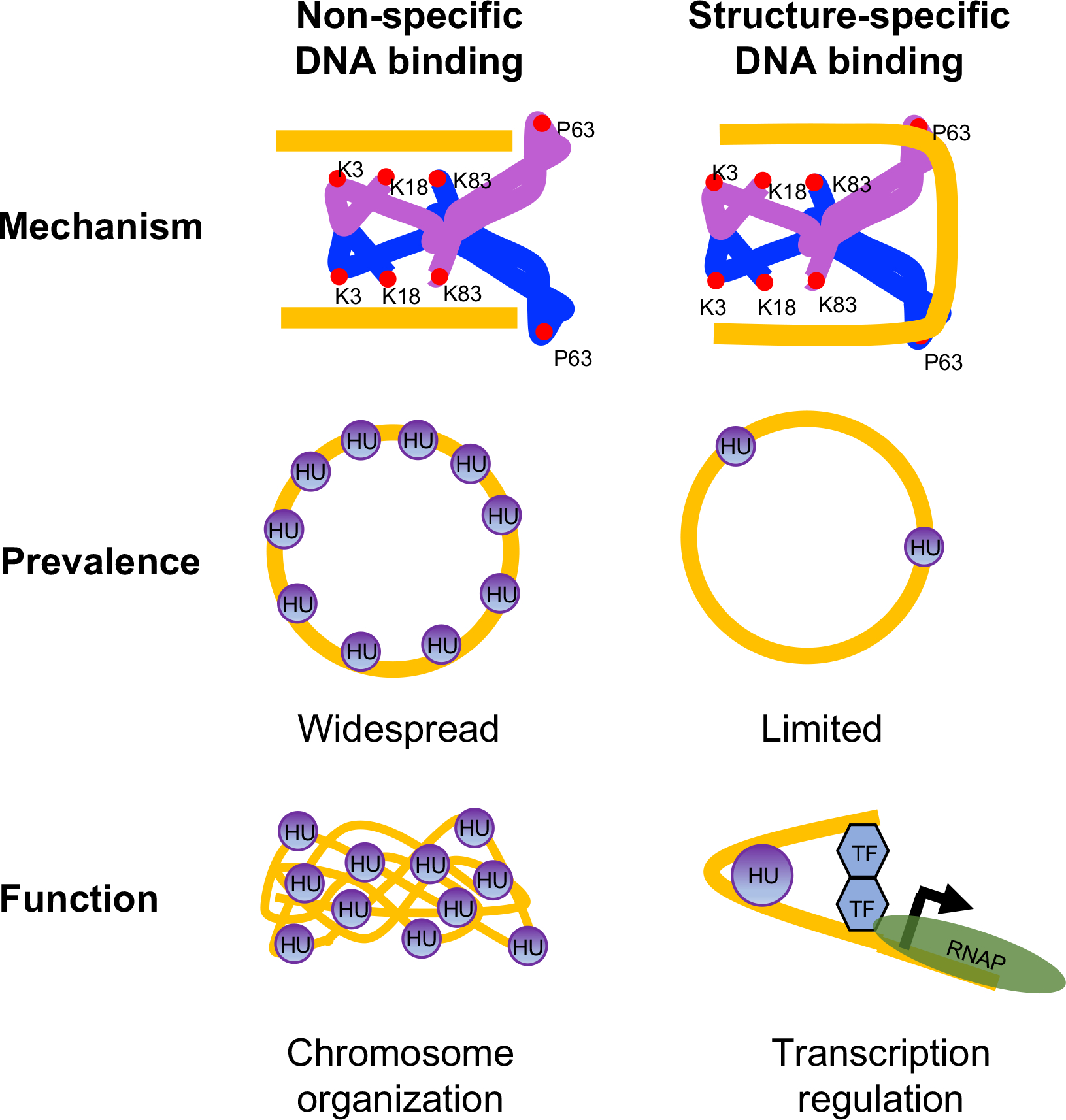
The histone-like protein HU plays a diverse role in bacterial physiology from the maintenance of chromosome structure to the regulation of gene transcription. HU binds DNA in a sequence-non-specific manner via two distinct binding modes: (i) random binding to any DNA through ionic bonds between surface-exposed lysine residues (K3, K18, and K83) and phosphate backbone (non-specific); (ii) preferential binding to contorted DNA of given structures containing a pair of kinks (structure-specific) through conserved proline residues (P63) that induce and/or stabilize the kinks. First, we show here that the P63-mediated structure-specific binding also requires the three lysine residues, which are needed for a non-specific binding. Second, we demonstrate that substituting P63 to alanine in HU had no impact on non-specific binding but caused differential transcription of diverse genes previously shown to be regulated by HU, such as those associated with the organonitrogen compound biosynthetic process, galactose metabolism, ribosome biogenesis, and cell adhesion. The structure-specific binding also helps create DNA supercoiling, which, in turn, may influence directly or indirectly the transcription of other genes. Our previous and current studies show that non-specific and structure-specific HU binding appear to have separate functions- nucleoid architecture and transcription regulation- which may be true in other DNA-binding proteins.
Keywords: HU; non-specific binding; nucleoid architecture; structure-specific binding; transcription regulation.
Published 2023. This article is a U.S. Government work and is in the public domain in the USA.
Conflict of interest statement
Figures
Similar articles
-
Single-molecule tracking reveals that the nucleoid-associated protein HU plays a dual role in maintaining proper nucleoid volume through differential interactions with chromosomal DNA.Mol Microbiol. 2021 Jan;115(1):12-27. doi: 10.1111/mmi.14572. Epub 2020 Aug 3. Mol Microbiol. 2021. PMID: 32640056 Free PMC article.
-
HU multimerization shift controls nucleoid compaction.Sci Adv. 2016 Jul 29;2(7):e1600650. doi: 10.1126/sciadv.1600650. eCollection 2016 Jul. Sci Adv. 2016. PMID: 27482541 Free PMC article.
-
Comparison of histone-like HU protein DNA-binding properties and HU/IHF protein sequence alignment.PLoS One. 2017 Nov 13;12(11):e0188037. doi: 10.1371/journal.pone.0188037. eCollection 2017. PLoS One. 2017. PMID: 29131864 Free PMC article.
-
Functional evolution of bacterial histone-like HU proteins.Curr Issues Mol Biol. 2011;13(1):1-12. Epub 2010 May 20. Curr Issues Mol Biol. 2011. PMID: 20484776 Review.
-
Nucleoid-Associated Protein HU: A Lilliputian in Gene Regulation of Bacterial Virulence.Front Cell Infect Microbiol. 2019 May 10;9:159. doi: 10.3389/fcimb.2019.00159. eCollection 2019. Front Cell Infect Microbiol. 2019. PMID: 31134164 Free PMC article. Review.
Cited by
-
Stress-induced nucleoid remodeling in Deinococcus radiodurans is associated with major changes in Heat Unstable (HU) protein dynamics.Nucleic Acids Res. 2024 Jun 24;52(11):6406-6423. doi: 10.1093/nar/gkae379. Nucleic Acids Res. 2024. PMID: 38742631 Free PMC article.
-
Replicating Chromosomes in Whole-Cell Models of Bacteria.Methods Mol Biol. 2024;2819:625-653. doi: 10.1007/978-1-0716-3930-6_29. Methods Mol Biol. 2024. PMID: 39028527
-
One Earth: The Equilibrium between the Human and the Bacterial Worlds.Int J Mol Sci. 2023 Oct 10;24(20):15047. doi: 10.3390/ijms242015047. Int J Mol Sci. 2023. PMID: 37894729 Free PMC article. Review.
-
Synergistic collaboration between AMPs and non-direct antimicrobial cationic peptides.Nat Commun. 2024 Aug 25;15(1):7319. doi: 10.1038/s41467-024-51730-x. Nat Commun. 2024. PMID: 39183339 Free PMC article.
-
Dynamics of chromosome organization in a minimal bacterial cell.Front Cell Dev Biol. 2023 Aug 9;11:1214962. doi: 10.3389/fcell.2023.1214962. eCollection 2023. Front Cell Dev Biol. 2023. PMID: 37621774 Free PMC article.
References
-
- Balandina A, Claret L, Hengge-Aronis R, and Rouviere-Yaniv J (2001). The Escherichia coli histone-like protein HU regulates rpoS translation. Molecular microbiology 39, 1069–1079. - PubMed
-
- Bettridge K, Verma S, Weng X, Adhya S, and Xiao J (2020). Single-molecule tracking reveals that the nucleoid-associated protein HU plays a dual role in maintaining proper nucleoid volume through differential interactions with chromosomal DNA. Molecular microbiology. 10.1111/mmi.14572. - DOI - PMC - PubMed
Publication types
MeSH terms
Substances
Grants and funding
LinkOut - more resources
Full Text Sources
