. 2023 Feb 8;31(2):260-272.e7.
doi: 10.1016/j.chom.2023.01.002. Epub 2023 Jan 27.

Structure of the Inmazeb cocktail and resistance to Ebola virus escape

Affiliations

Structure of the Inmazeb cocktail and resistance to Ebola virus escape

Vamseedhar Rayaprolu et al. Cell Host Microbe. .

Abstract

Monoclonal antibodies can provide important pre- or post-exposure protection against infectious disease for those not yet vaccinated or in individuals that fail to mount a protective immune response after vaccination. Inmazeb (REGN-EB3), a three-antibody cocktail against Ebola virus, lessened disease and improved survival in a controlled trial. Here, we present the cryo-EM structure at 3.1 Å of the Ebola virus glycoprotein, determined without symmetry averaging, in a simultaneous complex with the antibodies in the Inmazeb cocktail. This structure allows the modeling of previously disordered portions of the glycoprotein glycan cap, maps the non-overlapping epitopes of Inmazeb, and illuminates the basis for complementary activities and residues critical for resistance to escape by these and other clinically relevant antibodies. We further provide direct evidence that Inmazeb protects against the rapid emergence of escape mutants, whereas monotherapies even against conserved epitopes do not, supporting the benefit of a cocktail versus a monotherapy approach.

Keywords: Ebola; antiviral; cocktail; cryo-EM; escape; structure; therapeutics.


Conflict of interest statement

Declaration of interests The Regeneron employees have stock and/or options in the company. C.A.K. is an officer of the company. The antibodies are patented: WO/2016/123019.

Figures
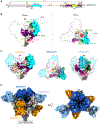
Figure 1.
Figure 1.. Structural overview of REGN-EB3 bound to EBOV GP
(A) Schematic representation of EBOV GP ectodomain. The mucin-like domain (MLD) and transmembrane (TM) domains are deleted. Thermolysin (ThL) and furin cleavage sites are indicated. A disulfide bond links the N-terminal portion of GP1 (light gray) to GP2 (dark gray). The glycan cap, internal fusion loop (IFL), and heptad repeats 1 (HR1) are colored cyan, purple, and yellow, respectively. Hash-marked regions in the schematic correspond to disordered regions in the structure. (B) Surface representation of side and top views of the EBOV GP trimer in the complex. The dotted line indicates the approximate region of the MLD. The β1-β2 loop (pink), receptor-binding site (RBS; brick red), ThL cut site (red), glycan cap (cyan), mucin-like domain (MLD; white), internal fusion loop (IFL; purple), heptad repeat 1 (HR1; yellow), and glycans (green) are shown. (C) Surface representation of EBOV GP trimer with each of the three different antibody footprints illustrated as a black outline in three separate views The dotted line and the red triangle indicated the 3-fold axis of the GP trimer. (D) The 3.1 Å cryo-EM map of the EBOV GP bound to eight Fabs of the REGN-EB3 cocktail. GP1 and GP2 are light and dark gray, respectively. REGN3470, REGN3471, and REGN3479 are colored light blue, dark blue, and orange, respectively. The number of copies of each antibody bound simultaneously to the GP trimer is labeled. See also Figures S1, S2, and S6 and Tables S1 and S2.
Figure 2.
Figure 2.. REGN3479 targets a quaternary epitope on the GP trimer
(A) Surface representation of the GP trimer with the REGN3479 epitope footprint colored in orange, glycan moieties are in green, and the thermolysin cleavage site is in red. (B) REGN3479 residues (orange) interact extensively with the Asn563 glycan (green). (C) REGN3479 interactions (orange) with the fusion loop (purple). LC residue Phe32 wedges into the fusion loop tip. (D) Overlay GP (residues 547–554) in the unbound (gray, PDB ID:5JQ3) and REGN3479-bound form (purple) with the HR1 shown as a gray (unbound) and yellow (bound), respectively, ribbon diagram. Antibody binding introduces an additional helical turn in the HR1 N terminus (purple). (E) The β1/β2 loop shifts by 7 Å in the unbound (gray) to bound (pink) shows an ~7 Å shift mediated by REGN3479 binding. See also Table S3.
Figure 3.
Figure 3.. REGN3471 binds both the head and glycan cap of GP
(A) Top view of REGN3471 footprint shown in blue, receptor-binding site (RBS) in brick red. Residues that overlap between the REGN3471 footprint and RBS shown in teal. (B) REGN3471 CDRL1 forms hydrogen bonds with residues in the hydrophilic, receptor-binding crest of GP. (C) A salt-bridge interaction between Lys 272 of GP and Glu 61 of REGN3471 enhances antibody binding affinity. The inset shows the overlay of GP in unbound (gray, PDB ID:5JQ3) and bound (cyan) forms. Steric clashes with Ser62 of the Fab displace the GP Thr 270 by ~9 Å. Thr 270 loop movement is not observed with mAb114 binding. (D) Electrostatic maps of GP bound to REGN3471 (left) or mAb114 (right). Antibody residues unique to REGN3471 or mAb114 are labeled. Several hydrophobic residues in REGN3471 occupy the hydrophilic crest of GP. Although the resolution of the mAb114 complex is low, stronger and more favorable hydrophilic residues of mAb114 relative to REGN3471 interact with the GP crest to form high-affinity salt bridges and also participate in other interactions. See also Table S3.
Figure 4.
Figure 4.. REGN3470 stabilizes the β17/β18 loop in the glycan cap
(A) Cryo-EM map of REGN3470 bound to EBOV GP. Inset compares the β17/18 loop in REGN3470 bound to GP and two previously published structures (PDB ID:3CSY and 5JQ3). The unambiguous density of this loop is observed in the REGN3470 complex. (B) Comparison of unbound GP (PDB ID: 5JQ3, gray) with GP bound to REGN3470 shows the movement of the loop upon REGN3470 binding. See also Table S3.
Figure 5.
Figure 5.. Glycan cap removal abolishes REGN3470 GP binding and reduces REGN3471 GP binding
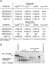
(A) Ligand binding properties of REGN-EB3 cocktail antibodies. Summary of equilibrium dissociation constants (KD) for the interaction of surface-captured anti-EBOV antibody with recombinant EBOV GP or GPCL trimer protein, respectively. ka, association rate constant; kd, dissociation rate constant; KD, equilibrium dissociation constant; t½, dissociative half-life. (B) Time-dependent thermolysin cleavage of GP in the absence and presence of REGN3479. See Figures S3 and S4.
Figure 6.
Figure 6.. REGN-EB3 prevents rapid viral escape
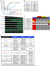
(A) Neutralization curves of fully replicative VSV-EBOV-GP by individual REGN-EB3 components, mAb114, a two-mAb combination (REGN3479 + mAb114), or the three-mAb REGN-EB3. (B) IC50 values (in M). (C) Images of representative plates. (D) Selection of escape variants to individual mAbs and combinations. To identify escape variants, VSV-EBOV-GP was propagated under antibody pressure to select for the resistant virus. The maximal antibody concentration under which virus replication was observed for each passage is indicated: ≥10 μg/mL (red), 1–10 μg/mL (orange), and <1 μg/mL (white). (E) Variants identified through RNA-seq under greater than ≥10 μg/mL of antibody selection. Values in parentheses are the relative frequency of reads encoding a given variant among all reads in that position. N.D., not determined, as no viral replication was observed at high mAb concentration (≥ 10 μg/mL). Complete: at least one previous passage at high antibody concentration (≥10 μg/mL). See also Figures S4 and S5.

References

    1. Pratt C. (2020). Two Ebola virus variants circulating during the 2020 Equateur Province outbreak. https://virological.org/t/two-ebola-virus-variants-circulating-during-th....
    1. Mulangu S, Dodd LE, Davey RT Jr., Tshiani Mbaya O, Proschan M, Mukadi D, Lusakibanza Manzo M, Nzolo D, Tshomba Oloma A, Ibanda A, et al. (2019). A randomized, controlled trial of Ebola virus disease therapeutics. N. Engl. J. Med 381, 2293–2303. 10.1056/NEJMoa1910993. - DOI - PMC - PubMed
    1. Center for Drug Evaluation and Research (2020). FDA approves treatment for Ebola virus (U.S. Food and Drug Administration; ). https://www.fda.gov/drugs/news-events-human-drugs/fda-approves-treatment....
    1. Sanchez A, Yang ZY, Xu L, Nabel GJ, Crews T, and Peters CJ (1998). Biochemical analysis of the secreted and virion glycoproteins of Ebola virus. J. Virol 72, 6442–6447. 10.1128/JVI.72.8.6442-6447.1998. - DOI - PMC - PubMed
    1. Lee JE, Fusco ML, Hessell AJ, Oswald WB, Burton DR, and Saphire EO (2008). Structure of the Ebola virus glycoprotein bound to an antibody from a human survivor. Nature 454, 177–182. 10.1038/nature07082. - DOI - PMC - PubMed

Substances