

doi: 10.1016/j.jid.2022.12.021.
Epub 2023 Jan 26.
Characterizing Dermal Transcriptional Change in the Progression from Sun-Protected Skin to Actinic Keratosis
Affiliations
- PMID: 36708948
- PMCID: PMC10293087
- DOI: 10.1016/j.jid.2022.12.021
Item in Clipboard
Characterizing Dermal Transcriptional Change in the Progression from Sun-Protected Skin to Actinic Keratosis
J Invest Dermatol.
2023 Jul.
No abstract available
Conflict of interest statement
Conflict of Interest Statement
The authors have no conflicts of interest to declare.
Figures
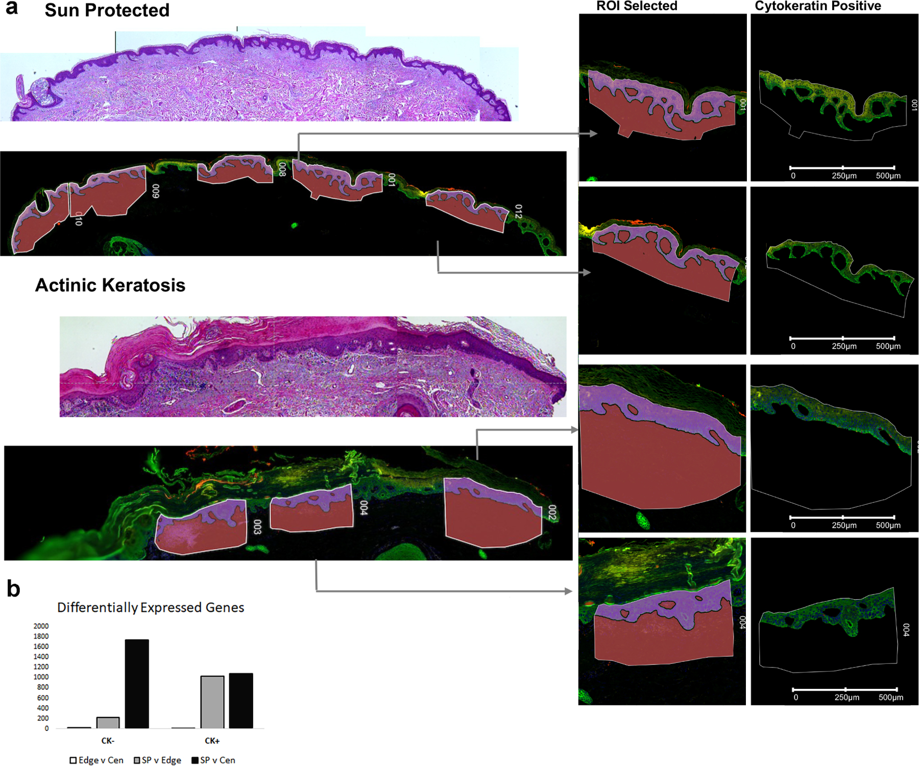
a: Digital Spatial Profiler identified regions of interest (ROI) highlights examples of areas of patient tissue sampled for transcriptional profiling. b: Differentially expressed genes identified using a pairwise FDR adjusted p-value of less than 0.05 and an overall t-test statistic of 0.05 highlights greater number DEGs (y-axis) in the cytokeratin negative compartment (CK-) comparing sun protected normal skin (SP) with actinic keratosis center (Cen). CK+ = cytokeratin positive compartment, Edge = actinic keratosis edge.
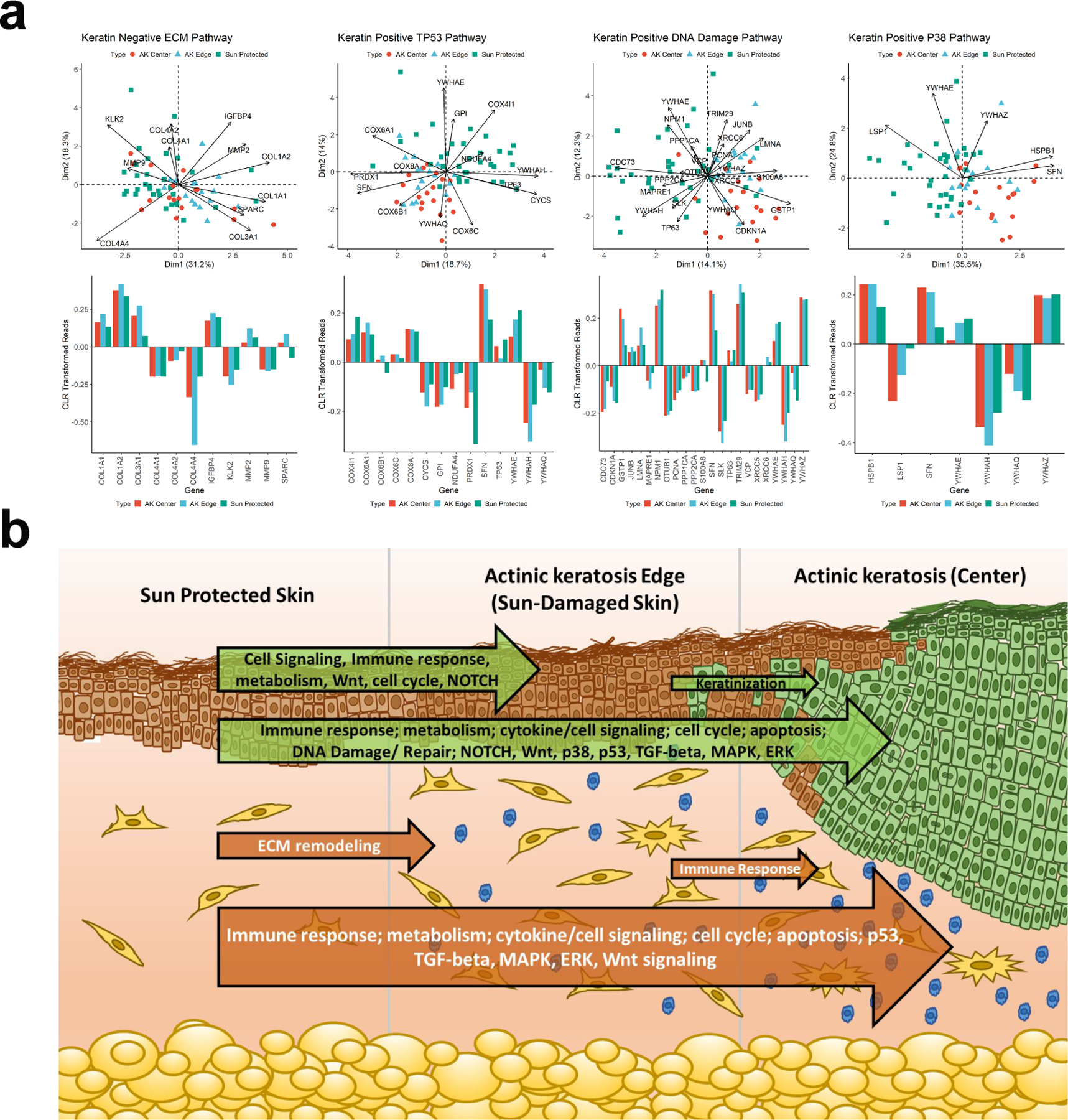
a: Biplots showing centered log-ratio (CLR) transformed reads comparing sun-protected and actinic keratosis samples. Individual samples are shown with three color and symbol combinations and the mean values are larger symbols on the PCA plane with the CLR standardized gene expression plotted. For CK-negative samples the SP and AK edge cluster together, and for CK-positive samples AK edge and center cluster together relative to the SP samples. The bar charts help interpret the directionality of the projections which can be inferred from the PCA plot but is simplified by the bar graphs. b: Graphical representation of transcriptional change moving through sun protected skin to actinic keratosis. Major pathways (identified using methodology described in Ben-Ari Fuchs et al., 2016) changing comparing each of the three compartments are indicated within the arrows representing the overall extent of transcriptional change, green arrows represent transcriptional change within the cytokeratin positive compartment while orange arrows represent the dermal (cytokeratin negative) compartment.
Similar articles
-
Quantitative fluorescence in situ hybridization measurement of telomere length in skin with/without sun exposure or actinic keratosis.Hum Pathol. 2014 Mar;45(3):473-80. doi: 10.1016/j.humpath.2013.10.009. Epub 2013 Oct 19. Hum Pathol. 2014. PMID: 24411948
-
Molecular discrimination of cutaneous squamous cell carcinoma from actinic keratosis and normal skin.Mod Pathol. 2011 Jul;24(7):963-73. doi: 10.1038/modpathol.2011.39. Epub 2011 Apr 1. Mod Pathol. 2011. PMID: 21743436
-
Gene expression patterns of normal human skin, actinic keratosis, and squamous cell carcinoma: a spectrum of disease progression.Arch Dermatol. 2010 Mar;146(3):288-93. doi: 10.1001/archdermatol.2009.378. Arch Dermatol. 2010. PMID: 20231500
-
Actinic keratoses.Cancer Treat Res. 2009;146:227-39. doi: 10.1007/978-0-387-78574-5_20. Cancer Treat Res. 2009. PMID: 19415207 Review. No abstract available.
-
Topical therapies for skin cancer and actinic keratosis.Eur J Pharm Sci. 2015 Sep 18;77:279-89. doi: 10.1016/j.ejps.2015.06.013. Epub 2015 Jun 16. Eur J Pharm Sci. 2015. PMID: 26091570 Review.
References
-
- Kim YS, Shin S, Jung SH, Park YM, Park GS, Lee SH, et al. Genomic Progression of Precancerous Actinic Keratosis to Squamous Cell Carcinoma. J Invest Dermatol 2021. - PubMed
Publication types
MeSH terms
Grants and funding
LinkOut - more resources
Full Text Sources
Medical
Molecular Biology Databases
