

Developing a metabolic clearance rate framework as a translational analysis approach for hyperpolarized 13C magnetic resonance imaging
- PMID: 36709217
- PMCID: PMC9884306
- DOI: 10.1038/s41598-023-28643-8
Developing a metabolic clearance rate framework as a translational analysis approach for hyperpolarized 13C magnetic resonance imaging
Abstract
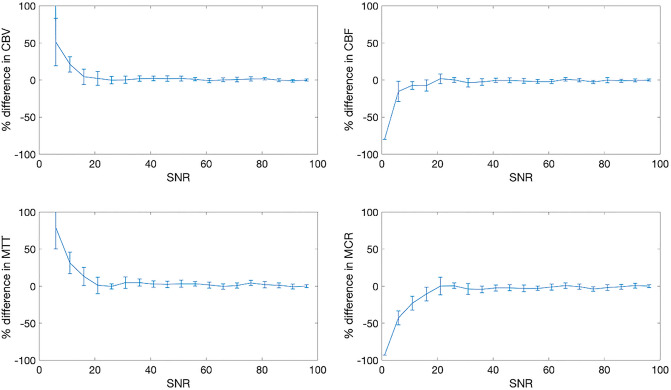
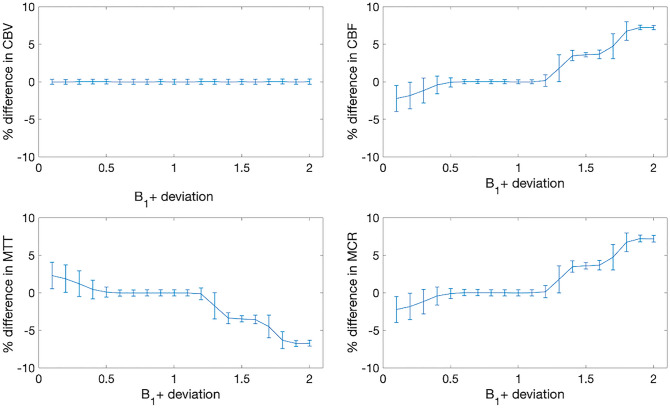
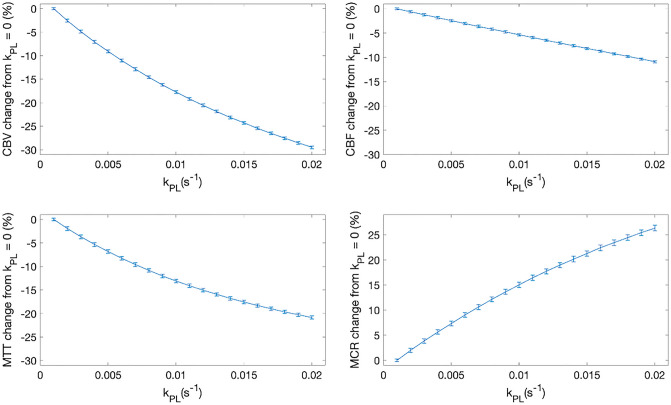
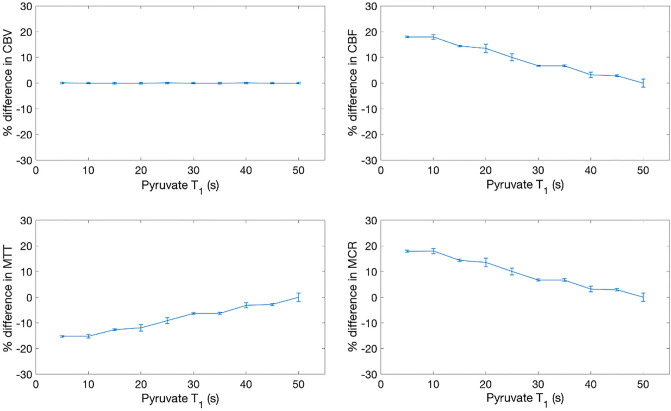
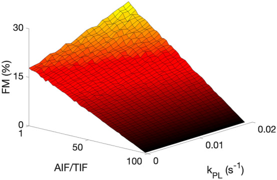
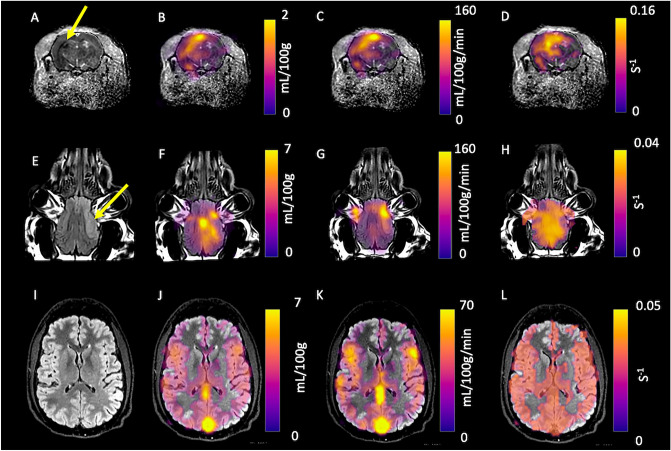
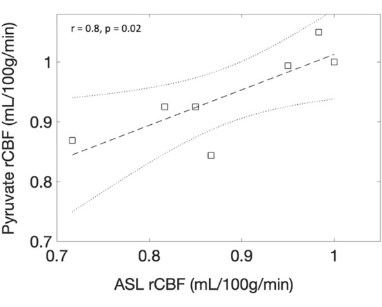
Hyperpolarized carbon-13 magnetic resonance imaging is a promising technique for in vivo metabolic interrogation of alterations between health and disease. This study introduces a formalism for quantifying the metabolic information in hyperpolarized imaging. This study investigated a novel perfusion formalism and metabolic clearance rate (MCR) model in pre-clinical stroke and in the healthy human brain. Simulations showed that the proposed model was robust to perturbations in T1, transmit B1, and kPL. A significant difference in ipsilateral vs contralateral pyruvate derived cerebral blood flow (CBF) was detected in rats (140 ± 2 vs 89 ± 6 mL/100 g/min, p < 0.01, respectively) and pigs (139 ± 12 vs 95 ± 5 mL/100 g/min, p = 0.04, respectively), along with an increase in fractional metabolism (26 ± 5 vs 4 ± 2%, p < 0.01, respectively) in the rodent brain. In addition, a significant increase in ipsilateral vs contralateral MCR (0.034 ± 0.007 vs 0.017 ± 0.02/s, p = 0.03, respectively) and a decrease in mean transit time (31 ± 8 vs 60 ± 2 s, p = 0.04, respectively) was observed in the porcine brain. In conclusion, MCR mapping is a simple and robust approach to the post-processing of hyperpolarized magnetic resonance imaging.
© 2023. The Author(s).
Conflict of interest statement
The authors declare no competing interests.
Figures
Similar articles
-
Metabolic MRI with hyperpolarized [1-13C]pyruvate separates benign oligemia from infarcting penumbra in porcine stroke.J Cereb Blood Flow Metab. 2021 Nov;41(11):2916-2927. doi: 10.1177/0271678X211018317. Epub 2021 May 20. J Cereb Blood Flow Metab. 2021. PMID: 34013807 Free PMC article.
-
Quantifying normal human brain metabolism using hyperpolarized [1-13C]pyruvate and magnetic resonance imaging.Neuroimage. 2019 Apr 1;189:171-179. doi: 10.1016/j.neuroimage.2019.01.027. Epub 2019 Jan 11. Neuroimage. 2019. PMID: 30639333 Free PMC article.
-
Assessing the effect of anesthetic gas mixtures on hyperpolarized 13 C pyruvate metabolism in the rat brain.Magn Reson Med. 2022 Sep;88(3):1324-1332. doi: 10.1002/mrm.29274. Epub 2022 Apr 25. Magn Reson Med. 2022. PMID: 35468245 Free PMC article.
-
Assessing Therapeutic Efficacy in Real-time by Hyperpolarized Magnetic Resonance Metabolic Imaging.Cells. 2019 Apr 11;8(4):340. doi: 10.3390/cells8040340. Cells. 2019. PMID: 30978984 Free PMC article. Review.
-
Hyperpolarized MRI - An Update and Future Perspectives.Semin Nucl Med. 2022 May;52(3):374-381. doi: 10.1053/j.semnuclmed.2021.09.001. Epub 2021 Nov 14. Semin Nucl Med. 2022. PMID: 34785033 Review.
Cited by
-
2-deoxy-D-glucose chemical exchange-sensitive spin-lock MRI of cerebral glucose metabolism after transient focal stroke in the rat.J Cereb Blood Flow Metab. 2025 Jul 8:271678X251355049. doi: 10.1177/0271678X251355049. Online ahead of print. J Cereb Blood Flow Metab. 2025. PMID: 40626496 Free PMC article.
-
Investigating cerebral perfusion with high resolution hyperpolarized [1-13 C]pyruvate MRI.Magn Reson Med. 2023 Dec;90(6):2233-2241. doi: 10.1002/mrm.29844. Epub 2023 Sep 4. Magn Reson Med. 2023. PMID: 37665726 Free PMC article.
References
-
- Hurd RE, Yen Y-F, Chen A, Ardenkjaer-Larsen JH. Hyperpolarized 13C metabolic imaging using dissolution dynamic nuclear polarization. J. Magn. Reson. Imaging. 2012;36:1314–1328. - PubMed
Publication types
MeSH terms
Substances
Grants and funding
LinkOut - more resources
Full Text Sources
Medical
