. 2023 Jan 28;13(1):1611.
doi: 10.1038/s41598-023-28814-7.

Expression of Chrna9 is regulated by Tbx3 in undifferentiated pluripotent stem cells

Affiliations

Expression of Chrna9 is regulated by Tbx3 in undifferentiated pluripotent stem cells

Takashi Yazawa et al. Sci Rep. .

Abstract

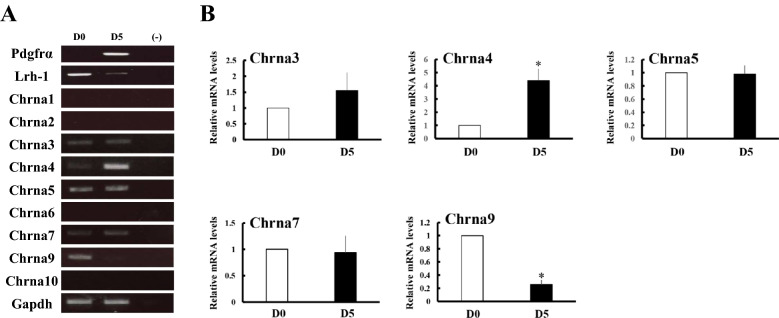
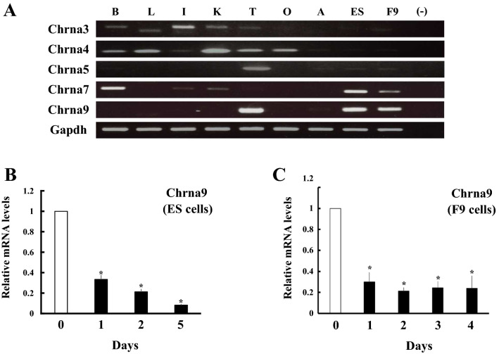
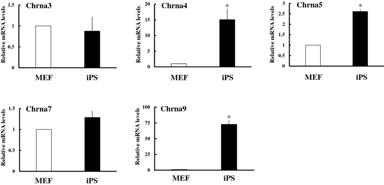
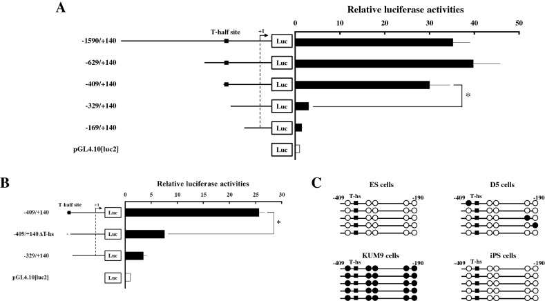
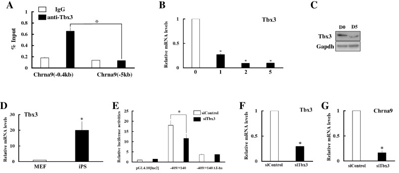
It was reported that nicotinic acetylcholine receptor (nAChR)-mediated signaling pathways affect the proliferation and differentiation of pluripotent stem cells. However, detail expression profiles of nAChR genes were unrevealed in these cells. In this study, we comprehensively investigated the gene expression of α subunit of nAChRs (Chrna) during differentiation and induction of pluripotent stem cells. Mouse embryonic stem (ES) cells expressed multiple Chrna genes (Chrna3-5, 7 and 9) in undifferentiated status. Among them, Chrna9 was markedly down-regulated upon the differentiation into mesenchymal cell lineage. In mouse tissues and cells, Chrna9 was mainly expressed in testes, ES cells and embryonal F9 teratocarcinoma stem cells. Expression of Chrna9 gene was acutely reduced during differentiation of ES and F9 cells within 24 h. In contrast, Chrna9 expression was increased in induced pluripotent stem cells established from mouse embryonic fibroblast. It was shown by the reporter assays that T element-like sequence in the promoter region of Chrna9 gene is important for its activities in ES cells. Chrna9 was markedly reduced by siRNA-mediated knockdown of Tbx3, a pluripotency-related transcription factor of the T-box gene family. These results indicate that Chrna9 is a nAChR gene that are transcriptionally regulated by Tbx3 in undifferentiated pluripotent cells.


Conflict of interest statement

The authors declare no competing interests.

Figures

Figure 1
Figure 1
Expression of Chrna genes during differentiation of ES cells into mesenchymal cell lineage. ES cells were cultured on collagen IV-coated dishes with differentiation media for 5 days. (A) RT-PCR analyses of each gene in ES cells (D0) and differentiated cells (D5). (B) Expression of each Chrna gene was analyzed in ES cells (D0) and differentiated cells (D5) by qPCR and normalized to 36B4 expression. Data represent the mean ± SEM of four independent experiments. Differences between groups are indicated by *P < 0.05.
Figure 2
Figure 2
Expression of Chrna9 in murine tissues and pluripotent stem cells. (A) Expression of Chrna genes in murine tissues and pluripotent stem cells. mRNA expression of each gene was analyzed in brain (lane B), liver (lane L), intestine (lane I), kidney (lane K), testis (lane T), ovary (lane O), adrenal (lane A), ES cells (lane ESC) and F9 cells (lane F9) by RT-PCR. (B) Expression of Chrna9 during differentiation of ES cells into mesenchymal stem cells. Expression of Chrna9 gene was analyzed by qPCR and normalized to 36B4 expression. Data represent the mean ± SEM of four independent experiments. *P < 0.05 vs. day 0. (C) Expression of Chrna9 during differentiation of F9 cells into parietal endodermal cells. Expression of Chrna9 gene was analyzed by qPCR and normalized to 36B4 expression. Data represent the mean ± SEM of four independent experiments. *P < 0.05 vs. day 0.
Figure 3
Figure 3
Comparison of Chrna gene expression between MEF and MEF-derived iPS cells. Expression of each Chrna gene was analyzed in MEF and iPS cells by qPCR and normalized to 36B4 expression. Data represent the mean ± SEM of four independent experiments. Differences between groups are indicated by *P < 0.05.
Figure 4
Figure 4
Analyses of mouse Chrna9 promoter in ES cells. (A) 5′-deletion analysis of the mouse Chrna9 promoter region. Progressive deletions of the upstream region are illustrated schematically in the left panel. Each vector was transfected by lipofection into ES cells. At 24 h after transfection, luciferase assays were performed using the cell lysates. Relative luciferase activities are shown. Data are the mean ± SEM values of at least four independent experiments; and *, P < 0.05. (B) Effects of deletion in the T-half site within the Chrna9 promoter region. The constructs used are drawn schematically. Transfection and luciferase assays were performed as described in A. Data are the mean ± SEM values of at least four independent experiments; and *, P < 0.05. (C) Methylation analysis of the promoter region (− 409 to − 190) of Chrna9 gene in undifferentiated ES, differentiated (D5), KUM9 and iPS cells. Each circle denotes cytosine bases in CpG dinucleotides, and filled and open circles represent methylated and unmethylated cytosines, respectively.
Figure 5
Figure 5
Involvement of Tbx3 in the transcriptional regulation of Chrna9 in pluripotent stem cells. (A) ChIP assays showing that Tbx3 occupied its binding site on the promoter region of Chrna9. Assays were performed on ES cell extracts using control IgG or anti-TBX3 antibody. Recovered chromatin was subjected to qPCR analysis using primers encompassing proximal Tbx3 binding site (− 0.4 kb) or 5 kb from the transcription start site (− 5 kb). P < 0.05 between the indicated groups. (B) Expression of Tbx3 during differentiation of ES cells into mesenchymal cells. Expression of Chrna9 gene was analyzed by qPCR and normalized to 36B4 expression. Data represent the mean ± SEM of four independent experiments. *P < 0.05 vs. day 0. (C) Western blot analyses were conducted in ES cells before (D0) and after differentiation (D5), using antibodies against Tbx3 and Gapdh. (D) Expression of Tbx3 was analyzed in MEF and iPS cells by qPCR and normalized to 36B4 expression. Data represent the mean ± SEM of four independent experiments. Differences between groups are indicated by *P < 0.05. (E) Effects of siRNA-mediated Tbx3 knockdown on the promoter activities of Chrna9 gene. Each reporter construct was co-transfected to ES cells with control siRNA (white boxes) or Tbx3 siRNA (black boxes). To investigate the effects of Tbx3 knockdown, luciferase assays were performed using the cell lysates at 48 h after transfection. Relative luciferase activities are shown. Data are the mean ± SEM values of at least four independent experiments; and *, P < 0.05. (F, G) Effects of siRNA-mediated Tbx3 knockdown on the expression of Tbx3 (F) and Chrna9 (G) in ES cells. Expression of each gene was analyzed by qPCR and normalized to 36B4 expression. Data represent the mean ± SEM of four independent experiments. Differences between groups are indicated by *P < 0.05.

References

    1. Niwa H. The principles that govern transcription factor network functions in stem cells. Development (Cambridge, England) 2018;145(6):157420. - PubMed
    1. Nichols J, Zevnik B, Anastassiadis K, et al. Formation of pluripotent stem cells in the mammalian embryo depends on the POU transcription factor Oct4. Cell. 1998;95(3):379–391. - PubMed
    1. Niwa H, Miyazaki J, Smith AG. Quantitative expression of Oct-3/4 defines differentiation, dedifferentiation or self-renewal of ES cells. Nat. Genet. 2000;24(4):372–376. - PubMed
    1. Avilion AA, Nicolis SK, Pevny LH, Perez L, Vivian N, Lovell-Badge R. Multipotent cell lineages in early mouse development depend on SOX2 function. Genes Dev. 2003;17(1):126–140. - PMC - PubMed
    1. Mitsui K, Tokuzawa Y, Itoh H, et al. The homeoprotein Nanog is required for maintenance of pluripotency in mouse epiblast and ES cells. Cell. 2003;113(5):631–642. - PubMed

Publication types