. 2023 Jan 28;14(1):67.
doi: 10.1038/s41419-023-05606-5.

A first-in-class HBO1 inhibitor WM-3835 inhibits castration-resistant prostate cancer cell growth in vitro and in vivo

Affiliations

A first-in-class HBO1 inhibitor WM-3835 inhibits castration-resistant prostate cancer cell growth in vitro and in vivo

Yuan-Yuan Mi et al. Cell Death Dis. .

Abstract

The prognosis and overall survival of castration-resistant prostate cancer (CRPC) patients are poor. The search for novel and efficient anti-CRPC agents is therefore extremely important. WM-3835 is a cell-permeable, potent and first-in-class HBO1 (KAT7 or MYST2) inhibitor. Here in primary human prostate cancer cells-derived from CRPC patients, WM-3835 potently inhibited cell viability, proliferation, cell cycle progression and in vitro cell migration. The HBO1 inhibitor provoked apoptosis in the prostate cancer cells. It failed to induce significant cytotoxicity and apoptosis in primary human prostate epithelial cells. shRNA-induced silencing of HBO1 resulted in robust anti-prostate cancer cell activity as well, and adding WM-3835 failed to induce further cytotoxicity in the primary prostate cancer cells. Conversely, ectopic overexpression of HBO1 further augmented primary prostate cancer cell proliferation and migration. WM-3835 inhibited H3-H4 acetylation and downregulated several pro-cancerous genes (CCR2, MYLK, VEGFR2, and OCIAD2) in primary CRPC cells. Importantly, HBO1 mRNA and protein levels are significantly elevated in CRPC tissues and cells. In vivo, daily intraperitoneal injection of WM-3835 potently inhibited pPC-1 xenograft growth in nude mice, and no apparent toxicities detected. Moreover, intratumoral injection of HBO1 shRNA adeno-associated virus (AAV) suppressed the growth of primary prostate cancer xenografts in nude mice. H3-H4 histone acetylation and HBO1-dependent genes (CCR2, MYLK, VEGFR2, and OCIAD2) were remarkably decreased in WM-3835-treated or HBO1-silenced xenograft tissues. Together, targeting HBO1 by WM-3835 robustly inhibits CRPC cell growth.


Conflict of interest statement

The authors declare no competing interests.

Figures

Fig. 1
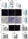
Fig. 1. WM-3835 inhibits primary CRPC cell viability, cell cycle progression, proliferation and migration in vitro.
The primary human prostate cancer cells derived from a CRPC patient, “pPC-1”, were treated with WM-3835 (at designated concentrations) or vehicle control (“Veh”) for designated hours, cell viability (CCK-8 assay, A), colony formation (B), cell death (by testing medium LDH releasing, C) and proliferation (by testing EdU incorporation, D and E), as well as cell cycle distribution (F) and in vitro cell migration (G) were tested. The primary human prostate cancer cells derived from three other CRPC patients, “pPC-2/pPC-3/pPC-4” (HJ), or the primary human prostate epithelial cells (“pEpi1” and “pEpi2”, derived from two patients) (K) were treated WM-3835 (10 μM) or vehicle control (“Veh”) for designated hours, cell viability (H, K), proliferation (I), and migration (J) were examined similarly. Data were expressed as the mean ± standard deviation (SD, n = 5). *P < 0.05 versus “Veh” group. “n. s.” stands for non-statistical difference (P > 0.05). Scale bar = 100 μm.
Fig. 2
Fig. 2. WM-3835 provokes apoptosis in primary CRPC cells.
The primary human prostate cancer cells derived from a CRPC patient, “pPC-1”, were treated with WM-3835 (10 μM) or vehicle control (“Veh”) for designated hours, caspase-PARP activation was measured (AC); Cell apoptosis was tested by nuclear TUNEL staining assay (D). pPC-1 cells were pretreated with zDEVD-fmk (45 μM), zVAD-fmk (45 μM), or DMSO (0.1%) for 1 h, followed by WM-3835 (10 μM) treatment for another 72 h, cell death was tested by LDH releasing assay (E). The primary human prostate cancer cells derived from three other CRPC patients, “pPC-2/pPC-3/pPC-4” (F, G), or the primary human prostate epithelial cells (“pEpi1” and “pEpi2”, derived from two patients) (H) were treated WM-3835 (10 μM) or vehicle control (“Veh”) for designated hours, caspase-3 activity (F), and apoptosis (G, H) were tested using the similar methods. Data were expressed as the mean ± standard deviation (SD, n = 5). *P < 0.05 versus “Veh” group. #P < 0.05 versus “DMSO” treatment (E). “n. s.” stands for non-statistical difference (P > 0.05). Scale bar = 100 μm.
Fig. 3
Fig. 3. HBO1 silencing inhibits primary CRPC cell viability, proliferation and migration in vitro.
The primary pPC-1 cells were infected with the HBO1-shRNA-expressing lentivirus (“shHBO1”) or the scramble control shRNA-expressing lentivirus (“shC”), and stable cells were formed after selection and verification. The shHBO1 pPC-1 cells were further treated with WM-3835 (10 μM) or vehicle control (“Veh”) for designated hours, expression of listed mRNAs and proteins was measured (AC); Cell viability, proliferation, in vitro migration and apoptosis were tested by CCK-8 (D), the nuclear EdU incorporation staining (E), “Transwell” (F) and nuclear TUNEL staining (G) assays, respectively. The primary human prostate cancer cells derived from three other CRPC patients, pPC-2/pPC-3/pPC-4 (HK), or the primary human prostate epithelial cells (“pEpi1” and “pEpi2”, derived from two patients) (L, M), were stably transduced with shHBO1 or shC, HBO1 mRNA expression was measured (H, L); After culturing for indicated time periods, cell viability (I, M), cell proliferation (J) and in vitro cell migration (K) were measured similarly. Data were expressed as the mean ± standard deviation (SD, n = 5). *P < 0.05 versus “shC” group. “n. s.” stands for non-statistical difference (P > 0.05). Scale bar = 100 μm.
Fig. 4
Fig. 4. Ectopic overexpression of HBO1 exerts pro-cancerous activity in primary CRPC cells.
The primary pPC-1 cells were transduced with a lentiviral HBO1-expressing construct, and two stable selections, oeHBO1-Slc-1 and oeHBO1-Slc-2, were formed after selection and overexpression verification. Control pPC-1 cells were stably transduced with the empty vector (“Vec”); Expression of HBO1 mRNA and listed proteins was tested (AC). Cells were cultivated for the designated hours, and cell proliferation and in vitro cell migration were tested by nuclear EdU incorporation (D) and “Transwell” (E) assays, respectively. The primary human prostate cancer cells derived from three other CRPC patients, pPC-2/pPC-3/pPC-4 (FH), or the primary human prostate epithelial cells (“pEpi1” and “pEpi2”, derived from two patients) (I, J), were stably transduced with the lentiviral HBO1-expressing construct (“oeHBO1”) or the empty vector (“Vec”), HBO1 mRNA expression was measured (F, I). After culturing for indicated time periods, cell proliferation (G), in vitro migration (H) and cell viability (J) were measured similarly. Data were expressed as the mean ± standard deviation (SD, n = 5). *P < 0.05 versus “Vec” group. “n. s.” stands for non-statistical difference (P > 0.05). Scale bar = 100 μm.
Fig. 5
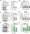
Fig. 5. WM-3835 inhibits H3-H4 acetylation and expression of several pro-cancerous genes in primary CRPC cells.
The primary pPC-1 cells were treated with WM-3835 (10 μM) or vehicle control (“Veh”) for designated hours, expression of listed proteins and mRNAs were tested (A and B). The stable primary pPC-1 cells with HBO1 shRNA (“shHBO1”) or the scramble control shRNA (“shC”) were further treated with or without WM-3835 (10 μM) for designated hours, expression of listed proteins and mRNAs were tested (C and D); The primary pPC-1 cells, with the lentiviral HBO1-expressing construct (oeHBO1-Slc-1 and oeHBO1-Slc-2, two stable selections) or the empty vector (“Vec”) were established, expression of listed proteins and mRNAs were tested (E and F); Data were expressed as the mean ± standard deviation (SD, n = 5). *P < 0.05 versus “Veh”/“shC”/“Vec” group. “n. s.” stands for non-statistical difference (P > 0.05).
Fig. 6
Fig. 6. HBO1 overexpression in CRPC cells and tissues.
Expression of HBO1 mRNA (A, C) and listed proteins (B, D) in primary human prostate epithelial cells (“pEpi1”), the listed primary CRPC cells (“pPC-1/pPC-2/pPC-3/pPC-4”), the CRPC tumor tissues (“T”, derived from 10 different CRPC patients, n = 10) or tumor-surrounding normal prostate tissues (“N”) were shown. Data were expressed as the mean ± standard deviation (SD). *P < 0.05 versus “pEpi1” cells or “N” tissues.
Fig. 7
Fig. 7. WM-3835 injection hinders pPC-1 xenograft growth in nude mice.
The pPC-1 xenograft-bearing nude mice were intraperitoneally (i.p.) injected with WM-3835 (at 5 mg/kg body weight, daily for 14 days) or the vehicle control (“Veh”), tumor volumes (A) and mice body weights (D) were recorded every seven days (“Day-1” to “Day-42”), daily tumor growth (in mm3 per day, B) and tumor weights (at Day-42, C) were measured as well. Expression of listed genes and proteins in the listed pPC-1 xenograft tissue lysates was tested (EH). Data were expressed as the mean ± standard deviation (SD). *P < 0.05 versus “Veh” group. “n. s.” stands for non-statistical difference (P > 0.05).
Fig. 8
Fig. 8. HBO1 silencing hinders pPC-1 xenograft growth in nude mice.
The pPC-1 xenograft-bearing nude mice were intratumorally injected with AAV-packed HBO1 shRNA (“aav-shHBO1”) or AAV-packed scramble control shRNA (“aav-shC”). AAV was injected daily for 10 consecutive days. Tumor volumes (A) and mice body weights (D) were recorded every seven days (“Day-1” to “Day-42”), daily tumor growth (in mm3 per day, B) and tumor weights (at Day-42, C) were measured as well. Expression of listed genes and proteins in the listed pPC-1 xenograft lysates were tested (E, H). Data were expressed as the mean ± standard deviation (SD). *P < 0.05 versus “aav-shC” group.

Similar articles

Cited by

References

    1. Sung H, Ferlay J, Siegel RL, Laversanne M, Soerjomataram I, Jemal A, et al. Global Cancer Statistics 2020: GLOBOCAN estimates of incidence and mortality worldwide for 36 cancers in 185 countries. CA Cancer J Clin. 2021;71:209–49. doi: 10.3322/caac.21660. - DOI - PubMed
    1. Siegel RL, Miller KD, Fuchs HE, Jemal A. Cancer Statistics, 2021. CA Cancer J Clin. 2021;71:7–33. - PubMed
    1. Sandhu S, Moore CM, Chiong E, Beltran H, Bristow RG, Williams SG. Prostate cancer. Lancet. 2021;398:1075–90. - PubMed
    1. Attard G, Parker C, Eeles RA, Schroder F, Tomlins SA, Tannock I, et al. Prostate cancer. Lancet. 2016;387:70–82. - PubMed
    1. Ku SY, Gleave ME, Beltran H. Towards precision oncology in advanced prostate cancer. Nat Rev Urol. 2019;16:645–54. - PMC - PubMed

Publication types