

Proteomics identifies novel biomarkers of synovial joint disease in a canine model of mucopolysaccharidosis I
- PMID: 36709534
- PMCID: PMC9918716
- DOI: 10.1016/j.ymgme.2023.107371
Proteomics identifies novel biomarkers of synovial joint disease in a canine model of mucopolysaccharidosis I
Abstract
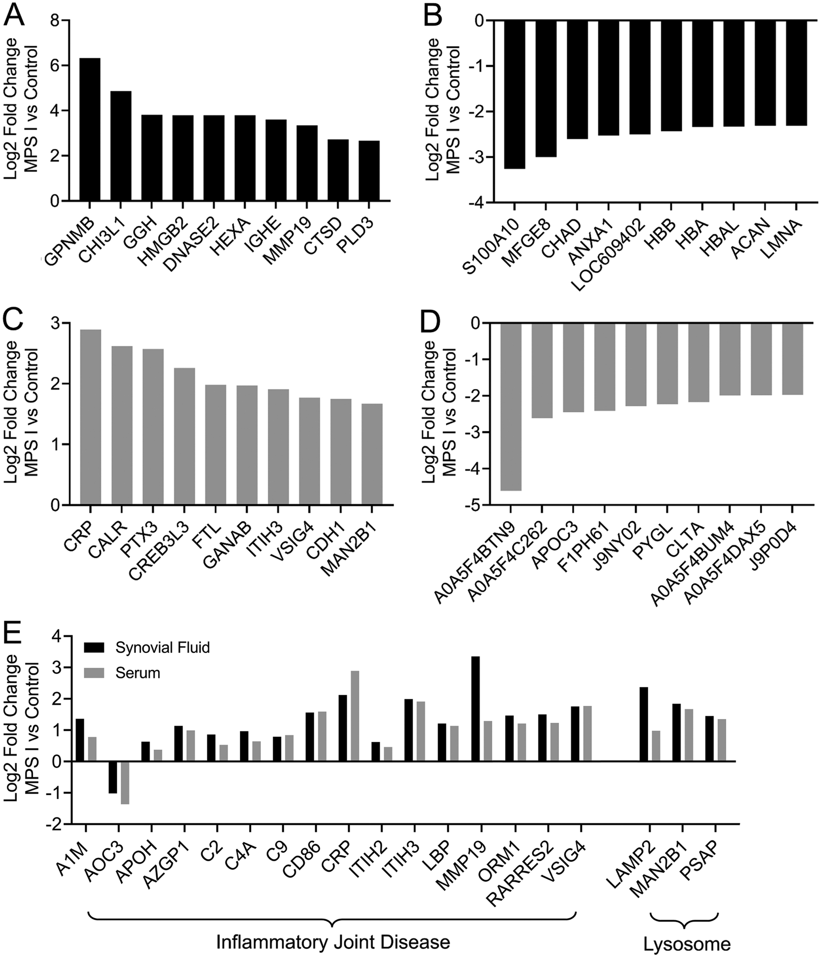
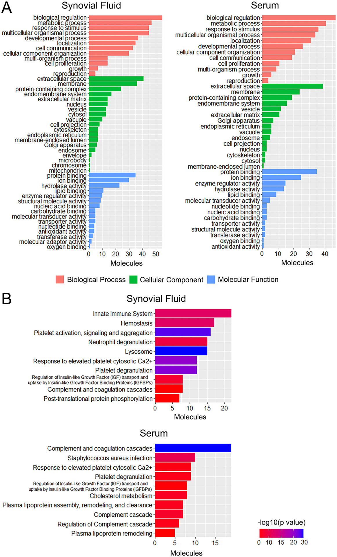
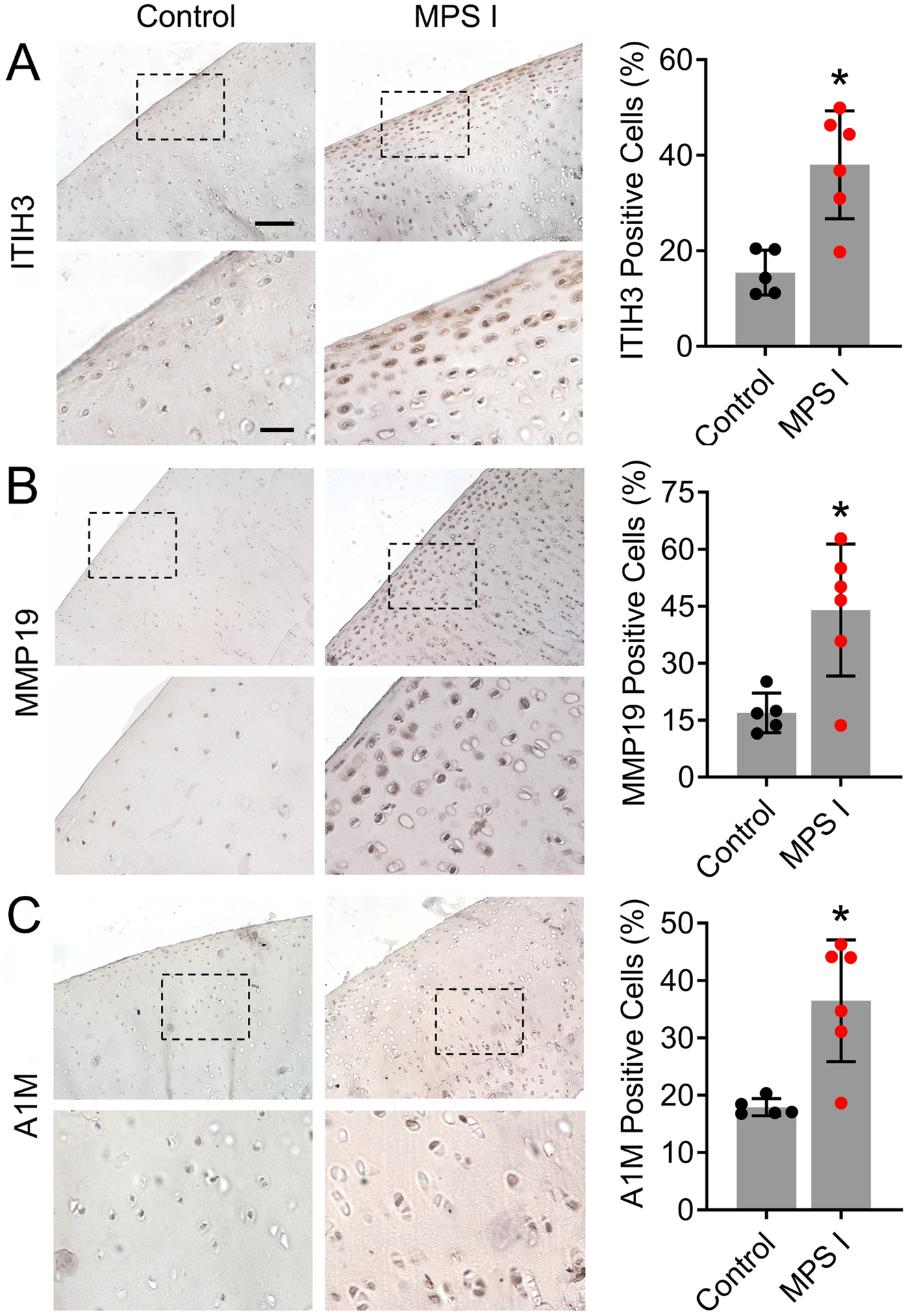
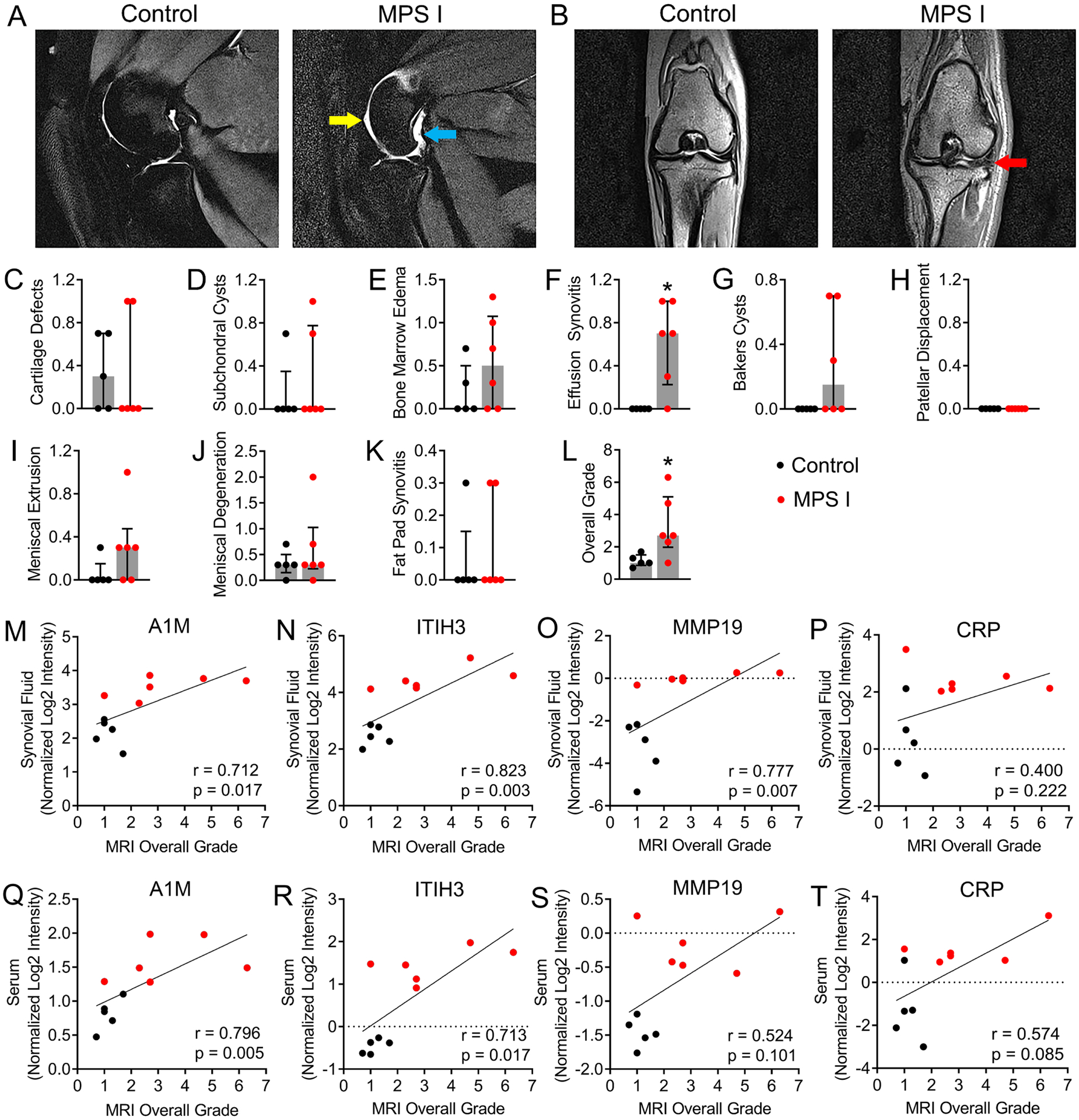
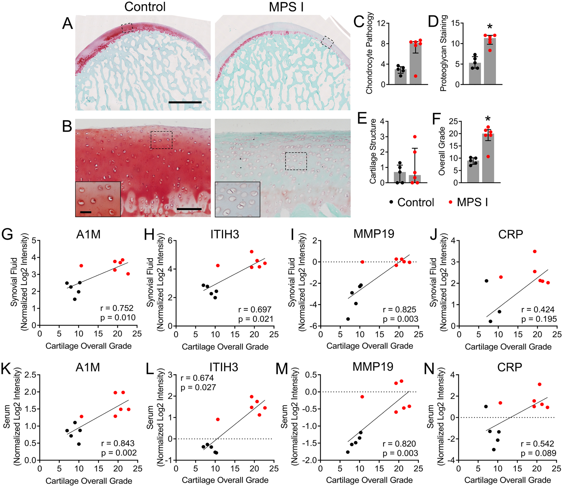
Mucopolysaccharidosis I is a lysosomal storage disorder characterized by deficient alpha-L-iduronidase activity, leading to abnormal accumulation of glycosaminoglycans in cells and tissues. Synovial joint disease is prevalent and significantly reduces patient quality of life. There is a critical need for improved understanding of joint disease pathophysiology in MPS I, including specific biomarkers to predict and monitor joint disease progression, and response to treatment. The objective of this study was to leverage the naturally-occurring MPS I canine model and undertake an unbiased proteomic screen to identify systemic biomarkers predictive of local joint disease in MPS I. Synovial fluid and serum samples were collected from MPS I and healthy dogs at 12 months-of-age, and protein abundance characterized using liquid chromatography tandem mass spectrometry. Stifle joints were evaluated postmortem using magnetic resonance imaging (MRI) and histology. Proteomics identified 40 proteins for which abundance was significantly correlated between serum and synovial fluid, including markers of inflammatory joint disease and lysosomal dysfunction. Elevated expression of three biomarker candidates, matrix metalloproteinase 19, inter-alpha-trypsin inhibitor heavy-chain 3 and alpha-1-microglobulin, was confirmed in MPS I cartilage, and serum abundance of these molecules was found to correlate with MRI and histological degenerative grades. The candidate biomarkers identified have the potential to improve patient care by facilitating minimally-invasive, specific assessment of joint disease progression and response to therapeutic intervention.
Keywords: Cartilage; Dog; Inflammation; Lysosomal storage disease; Serum; Synovial fluid.
Copyright © 2023 Elsevier Inc. All rights reserved.
Conflict of interest statement
Declaration of Competing Interest LJS: Scientific Advisory Board, National MPS Society; Scientific Advisory Board, JOR Spine. GRD: Co-founder and CEO, Mechano-Therapeutics LLC. WM: Book royalties, “Diagnostic MRI in Dogs and Cats”, Taylor and Francis. RG: Consultant, Acorn Biolabs; Scientific Advisory Board, JOR Spine. MLC, CRS, CZ, ZJ, YKL, SYJ, LAS, HF: No relevant disclosures.
Figures

Similar articles
-
Intra-articular enzyme replacement therapy with rhIDUA is safe, well-tolerated, and reduces articular GAG storage in the canine model of mucopolysaccharidosis type I.Mol Genet Metab. 2014 Aug;112(4):286-93. doi: 10.1016/j.ymgme.2014.05.015. Epub 2014 Jun 6. Mol Genet Metab. 2014. PMID: 24951454 Free PMC article.
-
Characterization of joint disease in mucopolysaccharidosis type I mice.Int J Exp Pathol. 2013 Oct;94(5):305-11. doi: 10.1111/iep.12033. Epub 2013 Jun 21. Int J Exp Pathol. 2013. PMID: 23786352 Free PMC article.
-
Effects of lithium administration on vertebral bone disease in mucopolysaccharidosis I dogs.Bone. 2022 Jan;154:116237. doi: 10.1016/j.bone.2021.116237. Epub 2021 Oct 22. Bone. 2022. PMID: 34695616 Free PMC article.
-
Enzyme replacement therapy in mucopolysaccharidosis type I: progress and emerging difficulties.J Inherit Metab Dis. 2001 Apr;24(2):245-50. doi: 10.1023/a:1010379320378. J Inherit Metab Dis. 2001. PMID: 11405343 Review.
-
Biomarkers and proteomic analysis of osteoarthritis.Matrix Biol. 2014 Oct;39:56-66. doi: 10.1016/j.matbio.2014.08.012. Epub 2014 Aug 30. Matrix Biol. 2014. PMID: 25179675 Review.
Cited by
-
Evaluation of tendon and ligament microstructure and mechanical properties in a canine model of mucopolysaccharidosis I.J Orthop Res. 2024 Jul;42(7):1409-1419. doi: 10.1002/jor.25813. Epub 2024 Feb 18. J Orthop Res. 2024. PMID: 38368531 Free PMC article.
-
The function of the inter-alpha-trypsin inhibitors in the development of disease.Front Med (Lausanne). 2024 Aug 1;11:1432224. doi: 10.3389/fmed.2024.1432224. eCollection 2024. Front Med (Lausanne). 2024. PMID: 39149600 Free PMC article. Review.
-
Application of tandem mass spectrometry in the screening and diagnosis of mucopolysaccharidoses.Orphanet J Rare Dis. 2024 Apr 29;19(1):179. doi: 10.1186/s13023-024-03195-w. Orphanet J Rare Dis. 2024. PMID: 38685110 Free PMC article. Review.
References
-
- Neufeld EF, Muenzer J, The Mucopolysaccharidoses, in: Scriver CR, Beaudet AL, Sly WS, Valle D (Eds.), The metabolic and molecular bases of inherited disease, McGraw-Hill, New York, 2001, pp. 3421–3452.
-
- White KK, Orthopaedic aspects of mucopolysaccharidoses Rheumatology 50 (2011) V26–V33. - PubMed
-
- Muenzer J, The mucopolysaccharidoses: a heterogeneous group of disorders with variable pediatric presentations J Pediatr 144 (2004) S27–34. - PubMed
Publication types
MeSH terms
Substances
Grants and funding
LinkOut - more resources
Full Text Sources
Medical
Molecular Biology Databases
