. 2023 Feb:116:28-48.
doi: 10.1016/j.matbio.2023.01.003. Epub 2023 Jan 26.

Mechanical loading is required for initiation of extracellular matrix deposition at the developing murine myotendinous junction

Affiliations

Mechanical loading is required for initiation of extracellular matrix deposition at the developing murine myotendinous junction

Sarah N Lipp et al. Matrix Biol. 2023 Feb.

Abstract

The myotendinous junction (MTJ) contributes to the generation of motion by connecting muscle to tendon. At the adult MTJ, a specialized extracellular matrix (ECM) is thought to contribute to the mechanical integrity of the muscle-tendon interface, but the factors that influence MTJ formation during mammalian development are unclear. Here, we combined 3D imaging and proteomics with murine models in which muscle contractility and patterning are disrupted to resolve morphological and compositional changes in the ECM during MTJ development. We found that MTJ-specific ECM deposition can be initiated via static loading due to growth; however, it required cyclic loading to develop a mature morphology. Furthermore, the MTJ can mature without the tendon terminating into cartilage. Based on these results, we describe a model wherein MTJ development depends on mechanical loading but not insertion into an enthesis.

Keywords: ECM; MTJ; Muscular dysgenesis (mdg); Musculoskeletal development; Myotendinous junction; Tbx3; Type XXII collagen; Ulnar-mammary syndrome.


Conflict of interest statement

Declaration of Competing Interest The authors declare no competing interests.

Figures
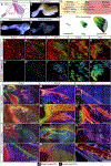
Figure 1:
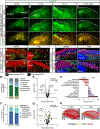
Figure 1:. The ECM undergoes distinct morphological and compositional changes during MTJ development.
(A) The insertion of the lateral triceps (pink) and long triceps (purple) into the olecranon (elbow) were either (B) wholemount, (C) decellularized (decell) in SDS, or (D) SeeDB-cleared. Representative E18.5 limbs shown. (E) Samples were stained with markers used to differentially visualize muscle, MTJ, and tendon, then imaged. (F) Schematic of changes in MTJ morphology as assessed by COL22A1 staining. (G-K’) In decellularized tissue, COL22A1 (green, depth projection) was identified at the putative MTJ starting at E13.5, and the morphology changed from thin, aligned structures at E13.5, to jagged protrusions at E18.5, then a highly interdigitated interface at P21. WGA (blue) marked general architecture by labeling proteoglycans and glycoproteins that contain sialic acid and n-acetylglucosamine [40]. (L-N’) TNC (green) and THBS4 (red) were broadly distributed at E14.5 and became enriched in the tendon of the long (open arrowheads) and lateral triceps (closed arrowheads) at E18.5 and P21. (O-Q’) POSTN (green) was found in muscle (LAMA2, blue), tendon, and cartilage during development, but was restricted to the MTJ (arrowheads) at P21. PRELP (red) was found in the cartilage at E14.5 and 18.5, as well as the tendon at P21. G-K’: 63× decell, 3D rendering, z = 64 μm; L-Q: 10× cryosections; L’-Q’: 63× cryosections. Scale bars: 1 mm (B-D, L-Q); 10 μm (G-K’); 100 μm (L’-Q’). t = tendon; m = muscle. Representative images from n = 3 biological replicates.
Figure 2:
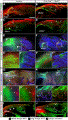
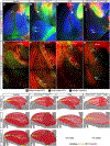
Figure 2:. Muscle contraction was required for continuing maturation of the MTJ.
(A-D) Distinct contraction of the triceps was observed in ScxGFP (green) forelimbs at E13.5 and E14.5 in response to acetylcholine, but not at E12.5 or in the mdg E14.5 limb. (E-F) The distribution of COL22A1 (green) was reduced at the termination of the long (open arrowheads) triceps in mdg compared to control forelimbs at E14.5 (WGA, marks general architecture of proteoglycans and glycoproteins, red). (G-H) By E18.5, COL22A1 was further reduced in the mdg mouse compared to control. (I-J) LAM+ muscle basement membranes (red) terminated in THBS4+ (green) MTJs in control, but not mdg limbs. WGA (blue). (K-L) EMILIN1+ (red) long triceps tendons were reduced in size in the mdg limb. (M) Proteomic analysis of E18.5 triceps muscles revealed proteins from the cytoskeletal and nuclear compartment were significantly higher in mdg compared to controls (two-tailed t-test, cytoskeletal: p = 0.017; nuclear: p = 0.043; Table S1). (N) Volcano plot of the proteome in the mdg triceps versus control. Grey lines indicate ≥ 2-fold change and p < 0.05 (two-tailed t-test). (O) Selected gene ontology (GO) terms generated by analysis of proteins upregulated in, or exclusive to, the different genotypes. (P) The distribution of matrisome components was similar when muscle motility was disrupted (two-tailed t-test, all ns). (Q) Volcano plot of matrisome components. Grey lines indicate ≥ 2-fold change and p < 0.05 (two-tailed t-test). (R) Schematic comparing differences in the limb structure between E18.5 control and mdg triceps. Cartilage (grey), tendon (yellow), MTJ (green), muscle (red). E-L: 63× decell, 3D rendering, z = 110 μm. Scale bars: 500 μm (A,D); 100 μm (E-L). A-L: representative images from n ≥ 3 biological replicates. M-R: average of n = 3 biological replicates.
Figure 3:
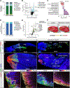
Figure 3:. Knockout of Tbx3 resulted in formation of an ectopic MTJ and tendon in E14.5 forelimbs.
(A, B) Muscle fibers are outlined by COL5 (green) and WGA (red, marks general architecture of proteoglycans and glycoproteins), and LAM+ (blue) blood vessels in the lateral triceps (dotted line) were morphologically normal, but abnormally oriented, in Prrx1CreTg/+;Tbx3fl/fl forelimbs. (A’, B’) Schematic comparing difference in orientation between E14.5 control and Prrx1CreTg/+;Tbx3fl/fl lateral triceps. Cartilage (grey), tendon (yellow), MTJ (green), muscle (red). (C) Comparison of Pearson correlation coefficients revealed a high degree of similarity between the matrisome of Prrx1CreTg/+;Tbx3fl/fl (Tbx3 KO) forelimbs and controls. (D) Volcano plot of matrisome components. Grey lines indicate ≥ 2-fold change and p < 0.05 (two-tailed t-test); Table S2. (E-F’) The ectopic insertion of the LAMA2+ lateral triceps muscle (blue) was TNC+ (green) and THBS4+ (red). (G-L) COL5, ELN, EMILIN1 (green), and WGA (red) were enriched in tendons of the long and lateral triceps in the controls, as well as the ectopic insertion. (M-N”) COL22A1+ (green or depth projection) fibers marked the MTJ of the control lateral and long triceps insertions at the olecranon, as well as the ectopic insertion. Red = WGA. (O, P) SOX9+ (red) nuclei and ScxGFP+ (green) cells did not co-localize at the ectopic insertion of the MY32+ (blue) lateral triceps muscle, but were localized in control entheses. A, B: 10× decell, z projection, z = 19.2 μm, inset 2.5×; E, F: 10×, cryosections; E’, F’: 63× cryosections; G-N’: 25× decell, 3D rendering, z = 110 μm; N”: 3D rendering 90° rotation of ectopic insertion z = 175 μm; O, P: 10× wholemount, z projection, z = 458 μm. A-B, E-P: representative images from n = 3 biological replicates; scale bars: 100 μm. C-D: average of n = 3 biological replicates.
Figure 4:
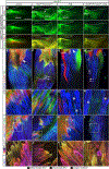
Figure 4:. The MTJ of the ectopic insertion was disorganized and the ectopic tendon did not terminate in an enthesis at E18.5.
(A-D) THBS4+ (green) MTJs were present at the interface between LAM+ myofibers (red, A, B) and EMILIN1+ tendons (red, C, D) in the long and the lateral triceps in both control and Prrx1CreTg/+;Tbx3fl/fl limbs. (E-F”) SOX9+ (red) nuclei co-localized with ScxGFP+ (green) cells at the enthesis of the control elbow (E’, F’); however, neither the control lateral triceps tendon nor the ectopic tendon contained SOX9+ nuclei (E”, F”). (G-H”) SOX9+ nuclei (red) co-localized with TNC (green) and neurofilament+ (blue) neurons in both the control and Prrx1CreTg/+;Tbx3fl/fl triceps. SOX9+ nuclei and TNC co-localized (*) at the enthesis of the control elbow; however, the TNC+ ectopic tendon did not contain SOX9+ nuclei. (I, J) COL2A1 (red) was present in the cartilage of the humerus, but not in either the control or Prrx1CreTg/+;Tbx3fl/fl forelimbs in the area of the ectopic insertion marked by COL22A1 (green). A-D, I J: 10× decell, 3D rendering, z = 290 μm (A, B), z = 680 μm (C-D), z = 450 μm (I, J); E, F: 10× wholemount, z-projection, z = 40 μm; E-F”: 63× wholemount, z-projection, z = 25 μm; G,H: 10× SeeDB-cleared, z-projection, z = 804 μm; G’-H” 63× 3D rendering, z = 86 μm. Scale bars: 500 μm (A-J); 10 μm (E, F); 100 μm (G, H). Representative images from n = 3 biological replicates.
Figure 5:
Figure 5:. The ectopic MTJ was mature by P21.
(A) Proteins from the cytoskeletal and matrisome fractions were significantly different in the P21 Prrx1CreTg/+;Tbx3fl/fl triceps unit compared with controls (two-tailed t-test: matrisome: p = 0.020; cytoskeletal: p < 0.0001; nuclear: p = 0.049; Table S4). (B) Volcano plot of all proteins revealed a reduction in slow-twitch muscle-related proteins. Grey lines indicate ≥ 2-fold change and p < 0.05 (two-tailed t-test). (C) Selected GO analysis terms for proteins upregulated, or exclusive to, the different genotypes. (D) The distribution of matrisome components was significantly different with more proteoglycan content in Prrx1CreTg/+;Tbx3fl/fl triceps (two-tailed t-test, proteoglycans: p = 0.027; all others: ns). (E) Volcano plot of matrisome components in control and Prrx1CreTg/+;Tbx3fl/fl triceps. Grey lines indicated ≥ 2-fold change and p < 0.05 (two-tailed t-test). (F) Schematic comparing difference in organization between P21 control and Prrx1CreTg/+;Tbx3fl/fl lateral triceps. Cartilage (grey), tendon (yellow), MTJ (green), muscle (red). Arrows indicate ectopic insertion. (G-H) COL22A1 (green) and THBS4 (red) were present in the MTJ of the control and ectopic lateral triceps. (I-J”) LAMA2+ (blue) muscle terminated in THBS4+ (green) and ScxGFP+ (red) tendons in control and ectopic lateral triceps MTJs. (I’-J’) LAMA2+ muscle fibers in both phenotypes terminated in morphologically mature MTJs. G, H: 10× decell, 3D rendering, z = 701 μm; I, J: 10× wholemount, z-projection, z = 1587 μm; I’-J”: 63×, z-projection, z = 79 μm. Scale bars: 500 μm (G-J); 100 μm (I’ -J”). A-F: average of n = 3 biological replicates. G-J”: representative images from n = 3 biological replicates.
Figure 6:
Figure 6:. Muscle contraction was critical for the fetal maturation of ectopic lateral triceps muscle and MTJ.
(A-D) Exposure of E18.5 limbs to 10 μm acetylcholine induced the contraction of the lateral triceps in control and Prrx1CreTg/+;Tbx3fl/fl, but not in mdg or Prrx1CreTg/+;Tbx3fl/fl;mdg limbs. Arrow indicates ectopic insertion. Dashed line indicates edge of tissue, solid line outlines the ectopic lateral triceps. (E-H) The lateral triceps was absent by E18.5 in the Prrx1CreTg/+;Tbx3fl/fl;mdg and TNC was not observed at the location of the putative ectopic insertion. (E’-H’) Striated muscle fibers were observed in the control and Prrx1CreTg/+;Tbx3fl/fl lateral triceps, but myofibers in the mdg and Prrx1CreTg/+;Tbx3fl/fl;mdg had reduced striations and contained vacuoles (*). (I-L) THBS4+ (green)/ScxGFP+ (red) tendons were present in the triceps of control, Prrx1CreTg/+;Tbx3fl/fl, and mdg, but not Prrx1CreTg/+;Tbx3fl/fl;mdg limbs. Blue = LAMA2 (muscle). (I’-L’) MTJs at the end of mdg myofibers were less mature, with fewer invaginations, than controls. E-H: 10× wholemount, z-projection, z = 547 μm; E’-H’, I-L’: 63× wholemount, z-projection, z = 44 μm (E’-H’) z = 39 μm (I, I’, K, K’, L, L’), z = 80 μm (J, J’). Scale bars: 500 μm (A-D); 100 μm (E-J); 10 μm (E’-J’). Representative images from n = 3 biological replicates.
Figure 7:
Figure 7:. COL22A1 MTJ formation at the ectopic insertion depended on muscle contraction.
(A-D) ScxGFP (green) and TNC (red) marked the long and lateral triceps tendon in E14.5 control, mdg, and Tbx3 limbs. MY32 muscle (blue) did not terminate in ScxGFP+ or TNC+ tissue in the Prrx1CreTg/+;Tbx3fl/fl;mdg limbs. (E-H) At E14.5, COL22A1 (green) was present in the lateral and long triceps MTJ in both control and Prrx1CreTg/+;Tbx3fl/fl. COL22A1 was reduced in the mdg and absent in the ectopic insertion in Prrx1CreTg/+;Tbx3fl/fl;mdg limbs. (I) In the control lateral triceps, the organization of the COL22A1+ (green) MTJ developed from fibers (embryonic time point) to a cap-like structure with jagged protrusions (fetal), before maturing into caps with slight shallow serrated invaginations with a greater frequency, lower amplitude (adult). (J) When muscles were mispatterned (Prrx1CreTg/+;Tbx3fl/fl), the MTJ formed at the embryonic time points at the normal insertion of the long triceps and the ectopic insertion of the lateral triceps. (K) When muscle contraction was disrupted (mdg), the MTJ failed to mature. At embryonic time points, there was decreased COL22A1 in the MTJ, and by the fetal time point, the MTJ failed to mature, and tendon ECM decreased in abundance. (L) When mispatterned lateral triceps did not contract (Prrx1CreTg/+; Tbx3fl/fl;mdg), tendon and MTJ did not form in the embryonic limb, and by the fetal time point, the lateral triceps regressed. Cartilage (grey), tendon (yellow), MTJ (green), muscle (red). A-D: 10× wholemount, z-projection, z = 88 μm; E-H: 25× decell, 3D rendering, z = 78 μm. Scale bars: 100 μm. Representative images from n = 3 biological replicates.

Similar articles

Cited by

References

    1. Narayanan N, Calve S, Extracellular matrix at the muscle – tendon interface: functional roles, techniques to explore and implications for regenerative medicine, Connect Tissue Res 62(1) (2021) 53–71. - PMC - PubMed
    1. Tonti OR, Larson H, Lipp SN, Luetkemeyer CM, Makam M, Vargas D, Wilcox SM, Calve S, Tissue-specific parameters for the design of ECM-mimetic biomaterials, Acta Biomater 132 (2021) 83–102. - PMC - PubMed
    1. Jacobson KR, Lipp S, Acuna A, Leng Y, Bu Y, Calve S, Comparative Analysis of the Extracellular Matrix Proteome Across the Myotendinous Junction, J Proteome Res. 19 (2020) 3955–3967. - PMC - PubMed
    1. Koch M, Schulze J, Hansen U, Ashwodt T, Keene DR, Brunken WJ, Burgeson RE, Bruckner P, Bruckner-Tuderman L, A novel marker of tissue junctions, collagen XXII, J Biol Chem 279(21) (2004) 22514–22521. - PMC - PubMed
    1. Kim M, Franke V, Brandt B, Lowenstein ED, Schöwel V, Spuler S, Akalin A, Birchmeier C, Single-nucleus transcriptomics reveals functional compartmentalization in syncytial skeletal muscle cells, Nature Communications 11(1) (2020) 6375. - PMC - PubMed

Publication types

LinkOut - more resources